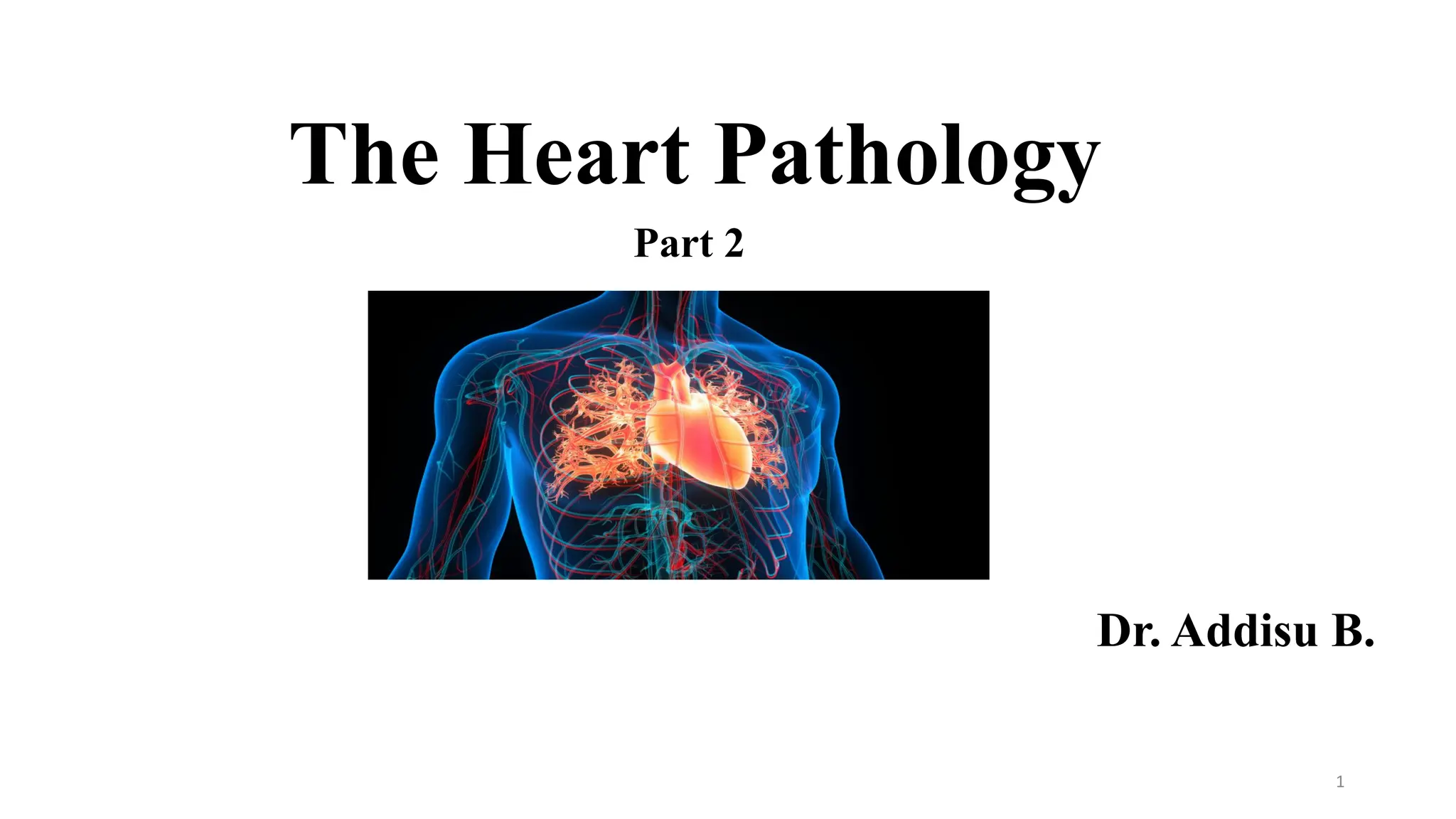
Myocarditis
- A groupof inflammatory processes of the myocardium that result
in injury to the cardiac myocytes.
4.
The chief causesof myocarditis
Viral infections - the most common cause of myocarditis eg.
Coxsackie groups A & B, influenza, echovirus, EBV, HIV
Bacterial infection eg diphteria, leptospirosis, meningococcus
Parasitic infections eg chagas disease, toxoplasmosis
Ionising radiation
Drugs eg doxorubucin
5.
Morphology
- The heartmay be of normal size, but more commonly it is dilated.
- The myocardium is flabby and pale.
- In cases of myocarditis caused by bacteria, frank abscesses may be
visible
6.
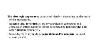
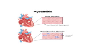
The histologic appearancevaries considerably, depending on the cause
of the myocarditis.
- In acute viral myocarditis, the myocardium is edematous and
contains an inflammatory infiltrate dominated by lymphocytes and
other mononuclear cells .
- Some degree of myocyte degeneration and/or necrosis is almost
always present.
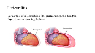
• Primary pericarditisis uncommon. It is typically due to viral
infection (often with concurrent myocarditis), although bacteria,
fungi, or parasites may also be involved
• In most cases, pericarditis is secondary to acute MI or cardiac
surgery (so-called “Dressler’s syndrome”), radiation to the
mediastinum, or processes involving other thoracic structures (e.g.,
pneumonia or pleuritis).
• Uremia is the most common systemic disorder associated with
pericarditis.
10.
• Pericarditis can:-
(1) Cause immediate hemodynamic complications if it elicits a large
effusion (resulting in cardiac tamponade),
(2) Resolve without significant sequelae, or
3) Progress to a chronic fibrosing proces
Clinical features
• Pericarditisclassically manifests with atypical chest pain (not related
to exertion and worse in recumbency), and a prominent friction rub.
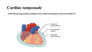
• When associated with significant fluid accumulation, acute pericarditis
can cause cardiac tamponade, which leads to declining cardiac
output and consequent shock
13.
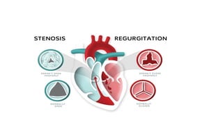
VALVULAR HEART DISEASES
-Valvular involvement by disease causes stenosis, insufficiency (
regurgitation or incompetence) or both.
- Stenosis - the failure of a valve to open completely impending forward
flow.
- Regurgitation – results from failure a valve to close completely
allowing reversed flow.
15.
- Abnormalities offlow often produce abnormal heart sounds known as
murmurs
- Diseases of the heart valves include a diverse group of acquired and
congenital lesions.
- Some of these occur in isolation, and others occur in association with
other heart diseases
16.
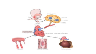
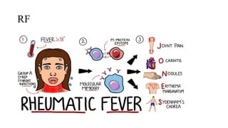
Rheumatic Fever andHeart Disease
- Rheumatic fever is an acute, immunologically mediated, multisystem
inflammatory disease that follows an episode of group A streptococcal
pharyngitis after an interval of a few weeks.
- The most important consequence of RF are chronic valvular
deformities characterized principally by deforming fibrotic valvular
disease (particularly mitral stenosis)- chronic rheumatic heart disease
- It produces permanent dysfunction & severe cardiac problems decades
later.
17.
Pathogenesis
- It isstrongly suspected that acute rheumatic fever is a
hypersensitivity reaction induced by group A streptococci.
- It is proposed that antibodies directed against the M proteins of
group A streptococci cross-react with normal proteins present in the
heart, joints, and other tissues.
19.
- Only minorityof infected patients develop RF, indicating genetic
susceptibility influences the hypersensitivity reaction.
- The chronic sequelae result from progressive fibrosis due to both
healing of the acute inflammatory lesions & the turbulence induced by
ongoing valvular deformity.
20.
MORPHOLOGY
- In acuterheumatic fever, inflammatory infiltrates may occur
in a wide range of sites, including synovium, joints, skin, and
(most importantly) the heart.
- Areas of fibrosis eventually develop at sites of inflammation.
- Fibrosis is particularly common in cardiac tissues, where it is
responsible for the valvular deformities seen in chronic
rheumatic heart disease.
21.
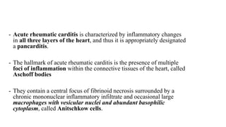
- Acute rheumaticcarditis is characterized by inflammatory changes
in all three layers of the heart, and thus it is appropriately designated
a pancarditis.
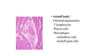
- The hallmark of acute rheumatic carditis is the presence of multiple
foci of inflammation within the connective tissues of the heart, called
Aschoff bodies
- They contain a central focus of fibrinoid necrosis surrounded by a
chronic mononuclear inflammatory infiltrate and occasional large
macrophages with vesicular nuclei and abundant basophilic
cytoplasm, called Anitschkow cells.
• Chronic rheumaticheart disease is characterized by organization of
the acute inflammation & subsequent fibrosis.
• The major anatomic changes of the valves in chronic RHD are leaflet
thickening, commissural fusion (resulting in ‘fish mouth’ shape in
mitral valve)& shorting & thickening & fusion of the tendinous cords.
24.
• The mitralvalve is abnormal in approximately 95% of cases of
chronic rheumatic heart disease, and combined aortic and mitral valve
disease is present in about 25% of patients.
• Right-sided valvular disease is relatively uncommon.
25.
Clinical Features
- Acuterheumatic fever occurs anywhere from 10 days to 6 weeks after an
episode of pharyngitis caused by group A streptococci in about 3% of
patients.
- The peak incidence is between the ages of 5 and 15, although younger
children and adults may also develop the disease.
- Although pharyngeal cultures for streptococci are negative by the time the
illness begins, antibodies to one or more streptococcal enzymes, such as
streptolysin O and DNAse B, are present in the sera of most patients.
26.
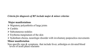
Criteria for diagnosisof RF include major & minor criterias
Major manifestations
Migratory polyarthritis of large joints
Carditis
Subcutaneous nodules
Erythema marginatum of the skin
Sydenham chorea, neurologic disorder with involuntary purposeless movements
Minor manifestation
Non specific sign & symptoms that include fever, arthralgia or elevated blood
levels of acute phase reactants
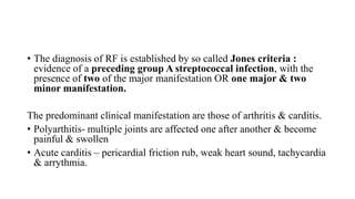
• The diagnosisof RF is established by so called Jones criteria :
evidence of a preceding group A streptococcal infection, with the
presence of two of the major manifestation OR one major & two
minor manifestation.
The predominant clinical manifestation are those of arthritis & carditis.
• Polyarthitis- multiple joints are affected one after another & become
painful & swollen
• Acute carditis – pericardial friction rub, weak heart sound, tachycardia
& arrythmia.
30.
- Chronic rheumaticcarditis usually does not cause clinical
manifestations for years or even decades after the initial episode of
rheumatic fever.
- The signs and symptoms of valvular disease depend on which cardiac
valve or valves are involved.
31.
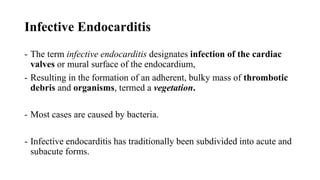
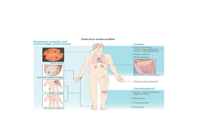
Infective Endocarditis
- Theterm infective endocarditis designates infection of the cardiac
valves or mural surface of the endocardium,
- Resulting in the formation of an adherent, bulky mass of thrombotic
debris and organisms, termed a vegetation.
- Most cases are caused by bacteria.
- Infective endocarditis has traditionally been subdivided into acute and
subacute forms.
33.
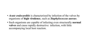
• Acute endocarditisis characterized by infection of the valves by
organisms of high virulence, such as Staphylococcus aureus.
• Such organisms are capable of infecting even structurally normal
valves and cause rapidly destructive infection, with little
accompanying local host reaction.
34.
- Subacute endocarditis,in contrast, is typically associated with
infection of previously abnormal valves by organisms of lower
virulence, such as α-hemolytic streptococci.
- The resultant infections tend to progress somewhat more slowly and
are often accompanied by the development of a local inflammatory
reaction and granulation tissue in the affected valve.
35.
Etiology and Pathogenesis
-Infection occurs when organisms are implanted on the
endocardial surface during episodes of bacteremia.
- The portal of entry of the agent into the blood stream may be
an obvious infection or procedures (surgical or dental).
36.
Conditions that increasethe risk of infective endocarditis include
- increased hemodynamic trauma to the endocardial surface, such as
high pressure shunts within the heart (e.g., small ventricular septal
defects) or chronic valvular diseases (e.g., chronic rheumatic heart
disease, degenerative calcific aortic stenosis, mitral valve prolapse),
prosthetic heart valves.
Host factors such as neutropenia, immunodeficiency, malignancy,
diabetes mellitus & intravenous drug abuse
37.
- Endocarditis ofabnormal valves is caused most commonly by
streptococcus viridans (50-60%)
- S. aureus attacks both healthy & deformed valves & are responsible
for 20% to 30% of cases.
- Other causes include bacteria such as enterococci, HACEK group
(Haemophilus, Actinobacillus, Cardiobacterium, Eikenella &
Kingella)
38.
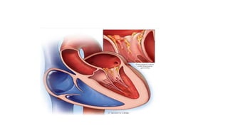
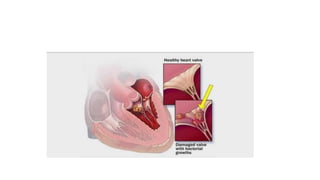
MORPHOLOGY
- The hallmarkof infective endocarditis is the presence of valvular
vegetations containing bacteria or other organisms.
- The vegetation is friable bulky, potentially destructive containing
fibrin, inflammatory cells & bacteria .
- The aortic and mitral valves are the most common sites of infection
- The valves of the right side of the heart may also be involved,
particularly in cases of endocarditis occurring in intravenous drug
abusers .
40.
• Systemic embolimay occur at any time because of the friable nature
of the vegetations, and they may cause infarcts in the brain, kidneys,
myocardium, and other tissues.
• Because the embolic fragments contain large numbers of virulent
organisms, abscesses often develop at the sites of such emboli
41.
Clinical Features
- Theonset of infective endocarditis may be gradual or explosive,
depending on the organism responsible for the infection.
- Low-grade fever, malaise, and weight loss are characteristic of cases
caused by organisms of low virulence
- While more acute cases, in contrast, typically present as high fevers,
shaking chills, and other evidence of overt septicemia.
43.
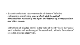
- Systemic emboliare very common in all forms of infective
endocarditis, manifesting as neurologic deficits, retinal
abnormalities, necrosis of the digits, and infarcts of the myocardium
and other viscera.
- Entrapment of infected emboli in the walls of blood vessels may cause
local infection and weakening of the vessel wall, with the formation of
so-called mycotic aneurysms.
44.
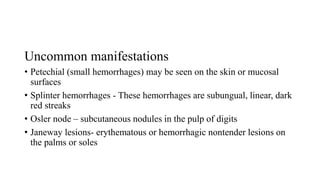
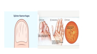
Uncommon manifestations
• Petechial(small hemorrhages) may be seen on the skin or mucosal
surfaces
• Splinter hemorrhages - These hemorrhages are subungual, linear, dark
red streaks
• Osler node – subcutaneous nodules in the pulp of digits
• Janeway lesions- erythematous or hemorrhagic nontender lesions on
the palms or soles
46.
• Blood culturesshould be done the moment the possibility of
endocarditis is considered
47.
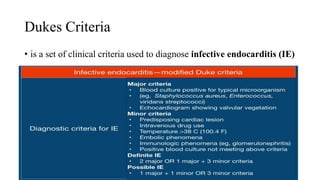
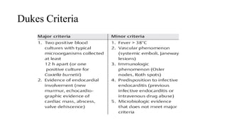
Dukes Criteria
• isa set of clinical criteria used to diagnose infective endocarditis (IE)


![CASE_PRESENTATION_ON_subdural_hematoma(SDH)[1 FINAL PPT]-1.pptx](https://cdn.slidesharecdn.com/ss_thumbnails/casepresentationonsubduralhematomasdh1finalppt-1-260129172522-d405d375-thumbnail.jpg?width=640&height=640&fit=bounds)


