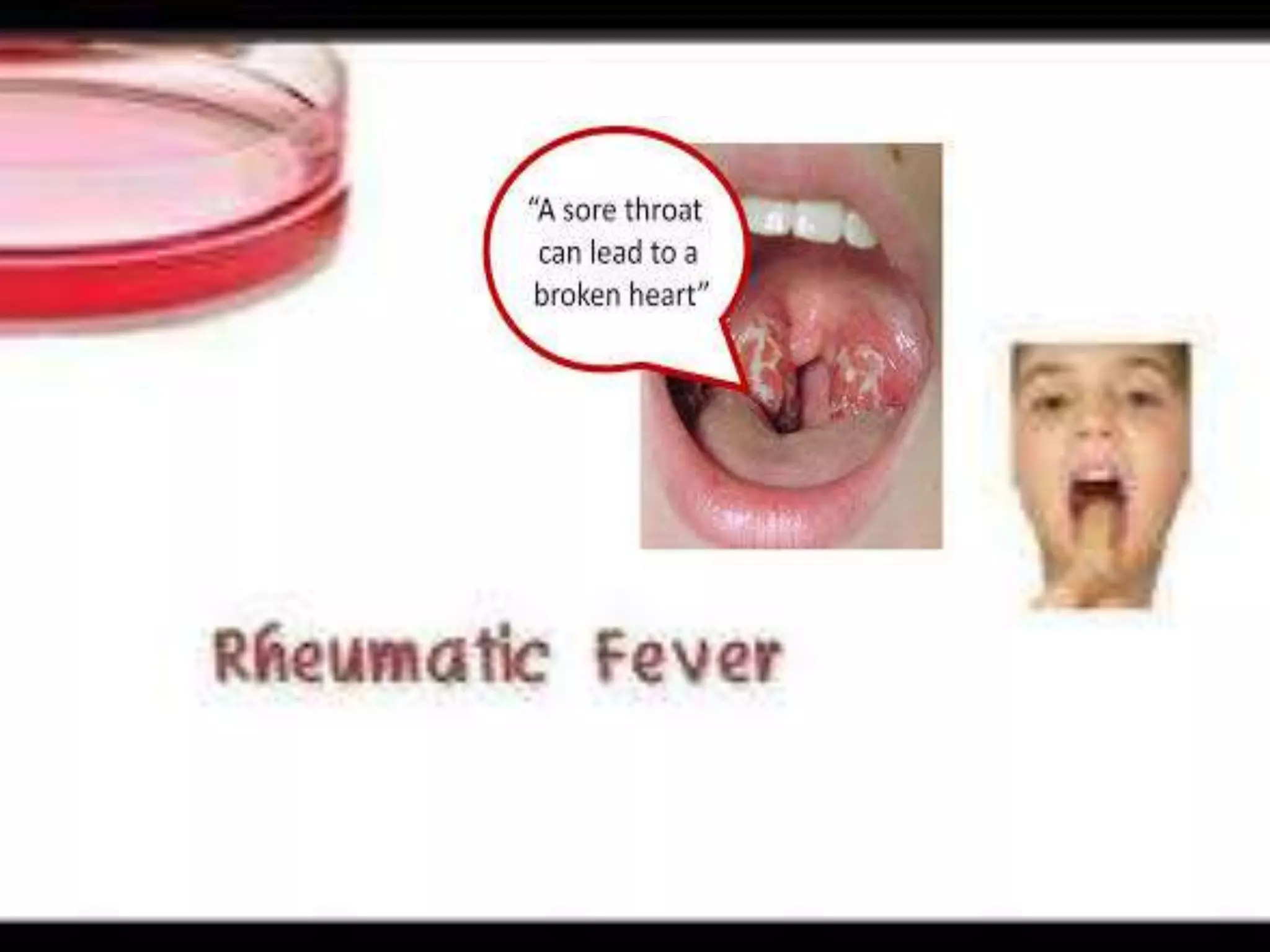
OBJECTIVE:
 Student will define rheumatic fever and
its causes.
 Student will explain its pathophysiology
 Relate its sign and symptoms with
pathophysiology
 Student will conceptualize diagnostic
measures and management of
rheumatic fever.
 Student will explain its complications and
prophylaxis .
OVERVIEW:
Rheumatic fever
 caused by group-A beta-hemolytic
streptococcus.
 It causes strep throat.It is an
inflammatory disorder.
It occurs in children of (6-15 yrs)
Family history of rheumatic fever is +
Initial attack of streptococcus occurs
1-5 weaks after streptococcal infection.
 Affects the valves of the heart.
 There is an autoimmune
response.
 Affects connective and
endothelial tissue in
heart,joint,skin as epitopes in
tisues of these areas is similar to
streptococal epitopes.
Definition:
Rheumatic fever is an autoimmune
disease that occurs as a sequelae of group A
beta-hemolytic streptococcal infection.It is
characterized by inflammatory lesions of
connective tissue and endothelial tissue
ANATOMY OF CONNECTIVE AND ENDOTHELIAL TISSUES:
 Endothelial tissue:
 It is a thin layer
of
simple squamous
cells called
endothelial cells
 It lines entire
circulatory
system from
heart to smallest
capillaries
 Connective tissue:
It IS a material made
up of fibers forming a
framework
 and support structure
for body tissues and
organs.
 It surrounds many
organs.
Cartilage and bone
It is found every-
Where in body
 Etiology:
 Underdiagnosed and undertreated strep
throat.
 Poor sanitation,poor housing.
 Incidence more during winter and early
spring.
 Family history of rheumatic fever.
PATHOPHYSIOLOGY:
Group-a streptococci
streptococcal pharyngitis
activation of T-cells synthesis of antistrpto-
by streptococcal antigen coccal antibodies by
B-cells
Autoimmune reaction results in cross reactivity between
epitopes of organism and host as epitopes in the cell wall
membrane,A,B,C regions of streptococcal M protein are
immunologically similar to human molecules.
Due to molecular mimicry between groupA streptococcal antigens
And host tissues antistreptococcal antibodies and t-cells
reacts with Strep like antigens in heart,joints,valves and
skin neurons and causes inflammation of heart
wall,valves and joints
Rheumatic
fever
 Joints:
 painful
 feel tender when touched
 Warm
 Swollen and red
 May contain fluid.
 Ankles, knees, elbows, and wrists are usually
affected. The shoulders, hips, and small joints of the
hands and feet also may be affected.
 Arthralgia.
 Fleeting and
fliting pain
 Arthritis.
 polyarthritis.
 HEART:
 valves are commonly affected
And developes new, larger, or different murmurs.
 Mitral valve is most commonly damaged.
 Pancarditis
CNS:
 Jerky, uncontrollable movements,
usually of both arms and legs and
particularly of the face, feet, and
hands, called Sydenham chorea.
 Skin:
 develops(redness of the skin or mucous
membrane) involving pink
rings Erythema marginatum
.
Sign and symptoms:
AS PER JONES CRITERIA:
 MAJOR
MANIFESTATIONS
 Polyarthritis:tenderness
swelling and pain in many
joints.
 Chorea-Involuntary movements:
 Erythema marginatum-pink macular rash
on trunk and extremities
 Carditis-Inflammation of heart
 MINOR MANIFESTATIONS:
 A temperature
Of 101°F or above.
 Arthralgia:Pain in joint
without inflammation and
tenderness.
 COMPLICATIONS:
 MITRAL STENOSIS:Mitral stenosis is a narrowing
of the mitral valve opening.Due to
inflammation. Mitral stenosis restricts
blood flow from the left atrium to the left
ventricle.
MITRAL INSUFFICIENCY:
Mitral insufficiency (MI), mitral regurgitation or mitral
incompetence is a disorderof the heart in which the
mitral valve does not close properly when the heart
pumps out blood. It is the abnormal leaking of blood
Backwards from the left ventricle, through the mitral
valve, into the left atrium,when the left ventricle
contracts
 COMPLICATIONS:
 AORTIC STENOSIS:Aortic stenosis is a narrowing
of the aortic valve .Due to inflammation.
Aortic stenosis restricts blood flow from
the left ventricle to all body parts.
 AORTIC insufficiency: Aortic insufficiency is
a heart valve disease in which the aortic valve
does not close tightly. This allows blood to flow
from the aorta into the left ventricle.
 Rheumatic endocardiatis:
Inflammation of endocardium.
 Rheumatic pericardiatis:
Inflammation of pericardium.
 Rheumatic myoardiatis:
Inflammation of endocardium.
 DIAGNOSTIC MEASURES:
by jones criteria
 Physical examination-A heart
murmur may
suggest ARF
 History Collection-
 Antistreptolysin O titre (ASOT) – this blood test looks for
evidence of antibodies produced by the immune
system in response to the streptococcal infection
 Erythrocyte sedimentation rate (ESR) –
 C- reactive protein (CRP) – which tests the level of C
reactive protein (CRP) in your blood. CRP is
produced by the liver. If there's more CRP in
the blood than usual, there's inflammation in
the body.
 Chest x-ray-To check size of heart
 ECG:To check electrical activity of heart.
Echocardiogram-To check valve damage and
chamber size
 Treatment:
 Medical management:
 Antibiotics- benzathine penicillin –single dose-IM
or 10day course of oral pencillin v.
 Oral erythromycin is given to those who
are allergic to pencillin.
 Anti-inflammatory drugs-aspirin 4-6 weaks for
carditis.
 Corticosteroids used in severe cases to control
cardiac inflammation.
 Neurological agents:Phenobarbital,diazepam to
control chorea.
 Complete bed rest during acute phase.
 NURSING MANAGEMENT:
 NURSING ASSESMENT:
 Moniter pulse.
 Ausultate heart for heart murmurs.
 Observe for involuntary movements.
 Asses pain level using pain scale.
 Asses temperature.
 Observe for rash on skin.
 Observe joints to check inflammation.
 Observe for pale skin.
 Nursing diagnosis:
 Decreased cardiac output realated to carditis.
 Pain related to arthritis
 Risk for injury related to chorea.
 Nursing INTERVENTIONs:
 For improving cardiac output:
 Monitir BP and intake output.
 Explain child family regarding complete bed
rest.(Aproximately 2 weaks).
 Observe for pallor skin and cynosis
 Reduce physical stress and activities.
 Administer medications as prescribed.
 Relieving pain-
 Asses pain level.
 Non-pharmacological pain management.
 Anti-inflammatory drugs as prescibed.
 Administer analgesics.
 Give comfortable possitioning.
 Protecting child from chorea:
 Use side rails
 Assist with feeding and other activities.
 Avoid unnecessary procedures.
 Limit activities.
 Administer medications as prescribed.
 FAMILY EDUCATION:
 Teach appropriate administration of all medicines
 Instruct additional prophylaxis for endocarditis before
dental procedures and surgery as indicated.
 Encourage to keep follow up with health care
Provider
 Encourage other family members to be screened for
streptococcus .
 Explain child and family need for complete rest
during acute phase (aproximately 2 weeks).
 PROPHYLAXIS:
 prophylaxis of rheumatic fever
For all individuals who have had an initial attack of
rheumatic fever, whether or not they have rheumatic
heart disease is manditory to prevent infection of the
upper respiratory tract by group A streptococci.
 Regular intramuscular injection of repository
penicillin (benzathine benzylpenicillin) 1,200,000
units of benzylbenzathine penicillin given every
three weeks to prevent recurrences of rheumatic
fever.
 For patients allergic to penicillin, an oral
sulfonamide is recommended . And those who
cannot take penicillin or sulfadiazine, erythromycin
in a dose of 250 mg twice daily may be used.
Patient Duration
No carditis/RHD To 18 years and at
least five years
after the last attack
Documented
carditis
At least to 25
years and often
longer
Chronic carditis For life
With artificial
valves
For life
*:
DURATION
The general principles for secondary
prophylaxis are:

Rheumatic fever

  • 2.
    OBJECTIVE:  Student willdefine rheumatic fever and its causes.  Student will explain its pathophysiology  Relate its sign and symptoms with pathophysiology  Student will conceptualize diagnostic measures and management of rheumatic fever.  Student will explain its complications and prophylaxis .
  • 3.
    OVERVIEW: Rheumatic fever  causedby group-A beta-hemolytic streptococcus.  It causes strep throat.It is an inflammatory disorder. It occurs in children of (6-15 yrs) Family history of rheumatic fever is + Initial attack of streptococcus occurs 1-5 weaks after streptococcal infection.  Affects the valves of the heart.
  • 4.
     There isan autoimmune response.  Affects connective and endothelial tissue in heart,joint,skin as epitopes in tisues of these areas is similar to streptococal epitopes.
  • 5.
    Definition: Rheumatic fever isan autoimmune disease that occurs as a sequelae of group A beta-hemolytic streptococcal infection.It is characterized by inflammatory lesions of connective tissue and endothelial tissue
  • 6.
    ANATOMY OF CONNECTIVEAND ENDOTHELIAL TISSUES:  Endothelial tissue:  It is a thin layer of simple squamous cells called endothelial cells  It lines entire circulatory system from heart to smallest capillaries
  • 7.
     Connective tissue: ItIS a material made up of fibers forming a framework  and support structure for body tissues and organs.  It surrounds many organs. Cartilage and bone It is found every- Where in body
  • 8.
     Etiology:  Underdiagnosedand undertreated strep throat.  Poor sanitation,poor housing.  Incidence more during winter and early spring.  Family history of rheumatic fever.
  • 9.
    PATHOPHYSIOLOGY: Group-a streptococci streptococcal pharyngitis activationof T-cells synthesis of antistrpto- by streptococcal antigen coccal antibodies by B-cells Autoimmune reaction results in cross reactivity between epitopes of organism and host as epitopes in the cell wall membrane,A,B,C regions of streptococcal M protein are immunologically similar to human molecules. Due to molecular mimicry between groupA streptococcal antigens
  • 10.
    And host tissuesantistreptococcal antibodies and t-cells reacts with Strep like antigens in heart,joints,valves and skin neurons and causes inflammation of heart wall,valves and joints
  • 11.
  • 12.
     Joints:  painful feel tender when touched  Warm  Swollen and red  May contain fluid.  Ankles, knees, elbows, and wrists are usually affected. The shoulders, hips, and small joints of the hands and feet also may be affected.  Arthralgia.  Fleeting and fliting pain  Arthritis.  polyarthritis.
  • 13.
     HEART:  valvesare commonly affected And developes new, larger, or different murmurs.  Mitral valve is most commonly damaged.  Pancarditis
  • 14.
    CNS:  Jerky, uncontrollablemovements, usually of both arms and legs and particularly of the face, feet, and hands, called Sydenham chorea.  Skin:  develops(redness of the skin or mucous membrane) involving pink rings Erythema marginatum .
  • 15.
    Sign and symptoms: ASPER JONES CRITERIA:  MAJOR MANIFESTATIONS  Polyarthritis:tenderness swelling and pain in many joints.
  • 16.
  • 17.
     Erythema marginatum-pinkmacular rash on trunk and extremities  Carditis-Inflammation of heart
  • 18.
     MINOR MANIFESTATIONS: A temperature Of 101°F or above.  Arthralgia:Pain in joint without inflammation and tenderness.
  • 19.
     COMPLICATIONS:  MITRALSTENOSIS:Mitral stenosis is a narrowing of the mitral valve opening.Due to inflammation. Mitral stenosis restricts blood flow from the left atrium to the left ventricle.
  • 20.
    MITRAL INSUFFICIENCY: Mitral insufficiency(MI), mitral regurgitation or mitral incompetence is a disorderof the heart in which the mitral valve does not close properly when the heart pumps out blood. It is the abnormal leaking of blood Backwards from the left ventricle, through the mitral valve, into the left atrium,when the left ventricle contracts
  • 21.
     COMPLICATIONS:  AORTICSTENOSIS:Aortic stenosis is a narrowing of the aortic valve .Due to inflammation. Aortic stenosis restricts blood flow from the left ventricle to all body parts.
  • 22.
     AORTIC insufficiency:Aortic insufficiency is a heart valve disease in which the aortic valve does not close tightly. This allows blood to flow from the aorta into the left ventricle.
  • 23.
     Rheumatic endocardiatis: Inflammationof endocardium.  Rheumatic pericardiatis: Inflammation of pericardium.
  • 24.
  • 25.
     DIAGNOSTIC MEASURES: byjones criteria  Physical examination-A heart murmur may suggest ARF  History Collection-
  • 26.
     Antistreptolysin Otitre (ASOT) – this blood test looks for evidence of antibodies produced by the immune system in response to the streptococcal infection  Erythrocyte sedimentation rate (ESR) –
  • 27.
     C- reactiveprotein (CRP) – which tests the level of C reactive protein (CRP) in your blood. CRP is produced by the liver. If there's more CRP in the blood than usual, there's inflammation in the body.  Chest x-ray-To check size of heart  ECG:To check electrical activity of heart.
  • 28.
    Echocardiogram-To check valvedamage and chamber size
  • 29.
     Treatment:  Medicalmanagement:  Antibiotics- benzathine penicillin –single dose-IM or 10day course of oral pencillin v.  Oral erythromycin is given to those who are allergic to pencillin.  Anti-inflammatory drugs-aspirin 4-6 weaks for carditis.  Corticosteroids used in severe cases to control cardiac inflammation.  Neurological agents:Phenobarbital,diazepam to control chorea.  Complete bed rest during acute phase.
  • 30.
     NURSING MANAGEMENT: NURSING ASSESMENT:  Moniter pulse.  Ausultate heart for heart murmurs.  Observe for involuntary movements.  Asses pain level using pain scale.  Asses temperature.  Observe for rash on skin.  Observe joints to check inflammation.  Observe for pale skin.
  • 31.
     Nursing diagnosis: Decreased cardiac output realated to carditis.  Pain related to arthritis  Risk for injury related to chorea.  Nursing INTERVENTIONs:  For improving cardiac output:  Monitir BP and intake output.  Explain child family regarding complete bed rest.(Aproximately 2 weaks).  Observe for pallor skin and cynosis  Reduce physical stress and activities.  Administer medications as prescribed.
  • 32.
     Relieving pain- Asses pain level.  Non-pharmacological pain management.  Anti-inflammatory drugs as prescibed.  Administer analgesics.  Give comfortable possitioning.  Protecting child from chorea:  Use side rails  Assist with feeding and other activities.  Avoid unnecessary procedures.  Limit activities.  Administer medications as prescribed.
  • 33.
     FAMILY EDUCATION: Teach appropriate administration of all medicines  Instruct additional prophylaxis for endocarditis before dental procedures and surgery as indicated.  Encourage to keep follow up with health care Provider  Encourage other family members to be screened for streptococcus .  Explain child and family need for complete rest during acute phase (aproximately 2 weeks).
  • 34.
     PROPHYLAXIS:  prophylaxisof rheumatic fever For all individuals who have had an initial attack of rheumatic fever, whether or not they have rheumatic heart disease is manditory to prevent infection of the upper respiratory tract by group A streptococci.  Regular intramuscular injection of repository penicillin (benzathine benzylpenicillin) 1,200,000 units of benzylbenzathine penicillin given every three weeks to prevent recurrences of rheumatic fever.  For patients allergic to penicillin, an oral sulfonamide is recommended . And those who cannot take penicillin or sulfadiazine, erythromycin in a dose of 250 mg twice daily may be used.
  • 35.
    Patient Duration No carditis/RHDTo 18 years and at least five years after the last attack Documented carditis At least to 25 years and often longer Chronic carditis For life With artificial valves For life *: DURATION The general principles for secondary prophylaxis are: