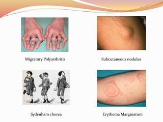
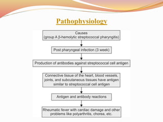
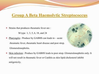
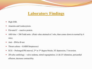
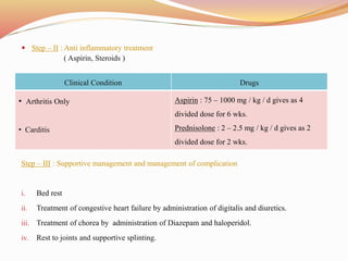
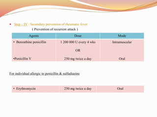
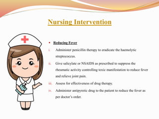
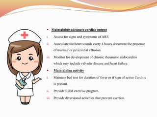
Introduction
Acute rheumatic fever is a systematic disease of childhood, often recurrent that
follows group A beta haemolytic streptococcus infection. It is a delayed non -
suppurative squeal to URII with GABH streptococci.
It is a diffuse inflammatory disease of connective tissue, primarily involving heart,
blood vessels, joints, subcutaneous tissue and central nervous system.
Rheumatic fever can develop if strep throat and scarlet fever infections are not
treated properly. Early diagnosis of these infections and treatment with antibiotics
are key to preventing rheumatic fever.
Epidemiology
 Ages : - 5 – 15 yrs. are most susceptible.
 Rare : - < 3 yrs.
 Girls > Boys
 Common in 3rd world countries.
 Environmental factor :- over crowding, poor sanitation, poverty.
 Incidence more during fall, winter and early spring.
Clinical manifestations
 Migratory polyarthritis : It means the arthritis appears in one or a few joints,
resolves but then appears in others, thus seeming to
move from one joint to another.
• Erythema marginatum : It is a serpiginous, flat or slightly raised, non – scar,
and painless rash. The rash usually appears on the
trunk and proximal extremities but not on the face.
• Sydenham chorea : Rapid and irregular jerking movement that may begin in
the hands but often become generalized, involving the feet
and face.
• Subcutaneous nodules.
• Heart murmur
 lethargy or fatigue
 Nose bleeds
 stomach pain
 red, hot, swollen joints
 shortness of breath
 fever
 sweating
 vomiting
 a decrease in attention span
 outbursts of crying or inappropriate laughter
 Chest pain
 Rapid fluttering or pounding chest palpitations
Migratory Polyarthritis Subcutaneous nodules
Sydenham chorea Erythema Marginatum
Pathophysiology
Group A Beta Haemolytic Streptococcus
 Strains that produces rheumatic fever are :
M type 1, 3, 5, 6, 18, and 24
 Pharyngitis : Produce by GABHS can leads to – acute
rheumatic fever, rheumatic heart disease and post strep.
Glomerulonephritis.
 Skin infection : Produce by GABHS leads to post strep. Glomerulonephritis only. It
will not result in rheumatic fever or Carditis as skin lipid cholesterol inhibit
antigenicity.
Diagnostic procedure
There is no specific laboratory test available for diagnosis of rheumatic fever……
 Look for a rash or skin nodules.
 Listen to their heart to check for abnormalities.
 Perform movement tests to determine their nervous system dysfunction.
 Examine their joints for inflammation.
 Test their throat and sometimes blood for evidence of strep bacteria.
 Perform an electrocardiogram (ECG or EKG), which measures the electric waves of
their heart.
 Perform an echocardiogram, which uses sound waves to produce images of their
heart.
Laboratory Findings
 High ESR.
 Anaemia and Leukocytosis.
 Elevated C – reactive protein.
 ASO titre > 200 Todd units : (Peak value attained at 3 wks, than comes down to normal by 6
wks).
 Anti – DNAs B test
 Throat culture – GABH Streptococci
 ECG – Prolonged PR interval, 2nd or 3rd degree blocks, ST depression, T inversion.
 2D echo cardiology – valve oedema, mitral regurgitation, LA & LV dilatation, pericardial
effusion, decrease contractility.
 Diagnosis of rheumatic fever is based on Modified Jones Criteria.
Jones Criteria (Revised) For Guidance In The
Diagnosis of Rheumatic Fever
 The presence of two major criteria, or of one major and two minor criteria, indicate a high
probability of acute rheumatic fever, if supported of evidence of Group A streptococcal
infection.
Major Manifestation Minor Manifestation Supporting evidence
of strep. infection
Clinical Laboratory
• Carditis
• Polyarthritis
• Chorea
• Erythema
marginatum
• Subcutaneous
nodules
• Previous
rheumatic fever or
rheumatic heart
disease, arthralgia
fever.
• Acute phase
reactant : Erythrocyte
sedimentation rate, C
– reactive protein,
Leukocytosis,
Prolonged P – R
interval.
• Increased titer of
anti – streptococcal
antibodies ASO (anti
– streptolysin O)
• Other – Positive
throat culture for
Group A beta
streptococcus recent
scarlet fever.
Complications of RF
 Congestive heart failure
 Cardiomegaly
 Chronic valve disease
 Rheumatic activity (recurrence)
 Pulmonary hypertension
Treatment
 Step – I : Primary Prevention
( Eradication of streptococci )
For individual allergic to penicillin
Agents Dose Mode Duration
• Benzathine penicillin G
• Penicillin V (Phenoxymethyl
penicillin
6 00 000 U for Pt. 27 kg and
1 200 000 U for Pt. > 27 kg
OR
Children – 250 mg
Adult & Adolescence – 500
mg
Intramuscular
Oral
Once
10 days
• Erythromycin : Estolate
• Ethylsuccinate
20 – 40 / kg / d
OR
40 mg / kg / d
Oral
Oral
10 days
10 days
 Step – II : Anti inflammatory treatment
( Aspirin, Steroids )
Step – III : Supportive management and management of complication
i. Bed rest
ii. Treatment of congestive heart failure by administration of digitalis and diuretics.
iii. Treatment of chorea by administration of Diazepam and haloperidol.
iv. Rest to joints and supportive splinting.
Clinical Condition Drugs
• Arthritis Only
• Carditis
Aspirin : 75 – 1000 mg / kg / d gives as 4
divided dose for 6 wks.
Prednisolone : 2 – 2.5 mg / kg / d gives as 2
divided dose for 2 wks.
 Step – IV : Secondary prevention of rheumatic fever
( Prevention of recurrent attack )
For individual allergic to penicillin & sulfadiazine
Agents Dose Mode
• Benzathine penicillin
•Penicillin V
1 200 000 U every 4 wks
OR
250 mg twice a day
Intramuscular
Oral
• Erythromycin 250 mg twice a day Oral
Nursing Management
 Nursing Assessment
 Subjective Data
i. Important health history.
ii. Functional health pattern.
iii. Complete past and present medical
history.
iv. Complete past and present surgical
history.
 Objective Data
i. General
ii. Integumentary
iii. Cardiovascular
iv. Neurological
v. Musculoskeletal
Nursing Intervention
 Reducing Fever
i. Administer penicillin therapy to eradicate the haemolytic
streptococcus.
ii. Give salicylate or NSAIDS as prescribed to suppress the
rheumatic activity controlling toxic manifestation to reduce fever
and relieve joint pain.
iii. Assess for effectiveness of drug therapy.
iv. Administer antipyretic drug to the patient to reduce the fever as
per doctor’s order.
 Maintaining adequate cardiac output
i. Assess for signs and symptoms of ARF.
ii. Auscultate the heart sounds every 4 hours document the presence
of murmur or pericardial effusion.
iii. Monitor for development of chronic rheumatic endocarditis
which may include valvular disease and heart failure.
 Maintaining activity
i. Maintain bed rest for duration of fever or if sign of active Carditis
is present.
ii. Provide ROM exercise program.
iii. Provide diversional activities that prevent exertion.
iv. Promote energy conservation. Provide rest periods between
activities to help pace the child’s energies and provide for
maximum comfort; if the child has chorea, inform visitors that the
child cannot control these movements, which are as upsetting to
the child as they are to others.
v. Prevent injury. Protect the child from injury by keeping the side
rails up and padding them; do not leave a child with chorea
unattended in a wheelchair, and use all appropriate safety
measures.
vi. Provide comfort and reduce pain. Position the child to reduce joint
pain; warm baths and gentle range-of-motion exercises help to
alleviate some of the joint discomforts; use pain indicator scales
with children so they are able to express the level of their pain.
Patient Education and Health Maintenance
i. Counsel about need for good nutrition.
ii. Counsel on hygiene practices ( hand washing ).
iii. Counsel about importance of adequate rest.
iv. Instruct patient to seek treatment immediately should sore throat or
fever occur.
v. Support patient in long term antibiotic therapy to prevent relapse.
vi. Instruct patient with valvular disease to use prophylaxis penicillin
therapy before certain procedures and surgery.
vii. Patient with previous history of ARF should be taught about the disease
process, possible sequelae and continues needs for prophylactic therapy.
viii. Teach about monthly injections of penicillin or erythromycin 1 or 2
times a day as prescribed by doctor.
Nursing Diagnosis
i. Acute pain or unpleasant sensory and emotional experience arising from actual or potential
tissue damage, sudden or slow onset of any intensity from mild to severe related to
inflammation and arthralgia as evidenced by verbal description of pain, warmth at affected
joints, edema and redness.
ii. Hyperthermia or body temperature elevated above the normal range related to illness or
inflammatory condition as evidence by hot, flushed skin, chills, tachycardia, tachypnea and
increased body temperature above normal range.
iii. Insufficient physiological or physiological energy to endure or complete required or desired
activity (activity intolerance) related to decrease cardiac output and muscle weakness as
evidence by prolonged bed, imposed activity restriction and imbalance oxygen supply and
demand.
iv. Risk of infection or at increased risk for being invaded by pathogenic organisms related to
chronic recurrence of disease and low immunity power as evidenced by high body
temperature, moist and clammy skin.
Rheumatic fever

Rheumatic fever

  • 2.
    Introduction Acute rheumatic feveris a systematic disease of childhood, often recurrent that follows group A beta haemolytic streptococcus infection. It is a delayed non - suppurative squeal to URII with GABH streptococci. It is a diffuse inflammatory disease of connective tissue, primarily involving heart, blood vessels, joints, subcutaneous tissue and central nervous system. Rheumatic fever can develop if strep throat and scarlet fever infections are not treated properly. Early diagnosis of these infections and treatment with antibiotics are key to preventing rheumatic fever.
  • 3.
    Epidemiology  Ages :- 5 – 15 yrs. are most susceptible.  Rare : - < 3 yrs.  Girls > Boys  Common in 3rd world countries.  Environmental factor :- over crowding, poor sanitation, poverty.  Incidence more during fall, winter and early spring.
  • 4.
    Clinical manifestations  Migratorypolyarthritis : It means the arthritis appears in one or a few joints, resolves but then appears in others, thus seeming to move from one joint to another. • Erythema marginatum : It is a serpiginous, flat or slightly raised, non – scar, and painless rash. The rash usually appears on the trunk and proximal extremities but not on the face. • Sydenham chorea : Rapid and irregular jerking movement that may begin in the hands but often become generalized, involving the feet and face. • Subcutaneous nodules. • Heart murmur
  • 5.
     lethargy orfatigue  Nose bleeds  stomach pain  red, hot, swollen joints  shortness of breath  fever  sweating  vomiting  a decrease in attention span  outbursts of crying or inappropriate laughter  Chest pain  Rapid fluttering or pounding chest palpitations
  • 6.
    Migratory Polyarthritis Subcutaneousnodules Sydenham chorea Erythema Marginatum
  • 7.
  • 8.
    Group A BetaHaemolytic Streptococcus  Strains that produces rheumatic fever are : M type 1, 3, 5, 6, 18, and 24  Pharyngitis : Produce by GABHS can leads to – acute rheumatic fever, rheumatic heart disease and post strep. Glomerulonephritis.  Skin infection : Produce by GABHS leads to post strep. Glomerulonephritis only. It will not result in rheumatic fever or Carditis as skin lipid cholesterol inhibit antigenicity.
  • 9.
    Diagnostic procedure There isno specific laboratory test available for diagnosis of rheumatic fever……  Look for a rash or skin nodules.  Listen to their heart to check for abnormalities.  Perform movement tests to determine their nervous system dysfunction.  Examine their joints for inflammation.  Test their throat and sometimes blood for evidence of strep bacteria.  Perform an electrocardiogram (ECG or EKG), which measures the electric waves of their heart.  Perform an echocardiogram, which uses sound waves to produce images of their heart.
  • 10.
    Laboratory Findings  HighESR.  Anaemia and Leukocytosis.  Elevated C – reactive protein.  ASO titre > 200 Todd units : (Peak value attained at 3 wks, than comes down to normal by 6 wks).  Anti – DNAs B test  Throat culture – GABH Streptococci  ECG – Prolonged PR interval, 2nd or 3rd degree blocks, ST depression, T inversion.  2D echo cardiology – valve oedema, mitral regurgitation, LA & LV dilatation, pericardial effusion, decrease contractility.
  • 11.
     Diagnosis ofrheumatic fever is based on Modified Jones Criteria. Jones Criteria (Revised) For Guidance In The Diagnosis of Rheumatic Fever  The presence of two major criteria, or of one major and two minor criteria, indicate a high probability of acute rheumatic fever, if supported of evidence of Group A streptococcal infection. Major Manifestation Minor Manifestation Supporting evidence of strep. infection Clinical Laboratory • Carditis • Polyarthritis • Chorea • Erythema marginatum • Subcutaneous nodules • Previous rheumatic fever or rheumatic heart disease, arthralgia fever. • Acute phase reactant : Erythrocyte sedimentation rate, C – reactive protein, Leukocytosis, Prolonged P – R interval. • Increased titer of anti – streptococcal antibodies ASO (anti – streptolysin O) • Other – Positive throat culture for Group A beta streptococcus recent scarlet fever.
  • 13.
    Complications of RF Congestive heart failure  Cardiomegaly  Chronic valve disease  Rheumatic activity (recurrence)  Pulmonary hypertension
  • 14.
    Treatment  Step –I : Primary Prevention ( Eradication of streptococci ) For individual allergic to penicillin Agents Dose Mode Duration • Benzathine penicillin G • Penicillin V (Phenoxymethyl penicillin 6 00 000 U for Pt. 27 kg and 1 200 000 U for Pt. > 27 kg OR Children – 250 mg Adult & Adolescence – 500 mg Intramuscular Oral Once 10 days • Erythromycin : Estolate • Ethylsuccinate 20 – 40 / kg / d OR 40 mg / kg / d Oral Oral 10 days 10 days
  • 15.
     Step –II : Anti inflammatory treatment ( Aspirin, Steroids ) Step – III : Supportive management and management of complication i. Bed rest ii. Treatment of congestive heart failure by administration of digitalis and diuretics. iii. Treatment of chorea by administration of Diazepam and haloperidol. iv. Rest to joints and supportive splinting. Clinical Condition Drugs • Arthritis Only • Carditis Aspirin : 75 – 1000 mg / kg / d gives as 4 divided dose for 6 wks. Prednisolone : 2 – 2.5 mg / kg / d gives as 2 divided dose for 2 wks.
  • 16.
     Step –IV : Secondary prevention of rheumatic fever ( Prevention of recurrent attack ) For individual allergic to penicillin & sulfadiazine Agents Dose Mode • Benzathine penicillin •Penicillin V 1 200 000 U every 4 wks OR 250 mg twice a day Intramuscular Oral • Erythromycin 250 mg twice a day Oral
  • 17.
    Nursing Management  NursingAssessment  Subjective Data i. Important health history. ii. Functional health pattern. iii. Complete past and present medical history. iv. Complete past and present surgical history.  Objective Data i. General ii. Integumentary iii. Cardiovascular iv. Neurological v. Musculoskeletal
  • 18.
    Nursing Intervention  ReducingFever i. Administer penicillin therapy to eradicate the haemolytic streptococcus. ii. Give salicylate or NSAIDS as prescribed to suppress the rheumatic activity controlling toxic manifestation to reduce fever and relieve joint pain. iii. Assess for effectiveness of drug therapy. iv. Administer antipyretic drug to the patient to reduce the fever as per doctor’s order.
  • 19.
     Maintaining adequatecardiac output i. Assess for signs and symptoms of ARF. ii. Auscultate the heart sounds every 4 hours document the presence of murmur or pericardial effusion. iii. Monitor for development of chronic rheumatic endocarditis which may include valvular disease and heart failure.  Maintaining activity i. Maintain bed rest for duration of fever or if sign of active Carditis is present. ii. Provide ROM exercise program. iii. Provide diversional activities that prevent exertion.
  • 20.
    iv. Promote energyconservation. Provide rest periods between activities to help pace the child’s energies and provide for maximum comfort; if the child has chorea, inform visitors that the child cannot control these movements, which are as upsetting to the child as they are to others. v. Prevent injury. Protect the child from injury by keeping the side rails up and padding them; do not leave a child with chorea unattended in a wheelchair, and use all appropriate safety measures. vi. Provide comfort and reduce pain. Position the child to reduce joint pain; warm baths and gentle range-of-motion exercises help to alleviate some of the joint discomforts; use pain indicator scales with children so they are able to express the level of their pain.
  • 21.
    Patient Education andHealth Maintenance i. Counsel about need for good nutrition. ii. Counsel on hygiene practices ( hand washing ). iii. Counsel about importance of adequate rest. iv. Instruct patient to seek treatment immediately should sore throat or fever occur. v. Support patient in long term antibiotic therapy to prevent relapse. vi. Instruct patient with valvular disease to use prophylaxis penicillin therapy before certain procedures and surgery. vii. Patient with previous history of ARF should be taught about the disease process, possible sequelae and continues needs for prophylactic therapy. viii. Teach about monthly injections of penicillin or erythromycin 1 or 2 times a day as prescribed by doctor.
  • 22.
    Nursing Diagnosis i. Acutepain or unpleasant sensory and emotional experience arising from actual or potential tissue damage, sudden or slow onset of any intensity from mild to severe related to inflammation and arthralgia as evidenced by verbal description of pain, warmth at affected joints, edema and redness. ii. Hyperthermia or body temperature elevated above the normal range related to illness or inflammatory condition as evidence by hot, flushed skin, chills, tachycardia, tachypnea and increased body temperature above normal range. iii. Insufficient physiological or physiological energy to endure or complete required or desired activity (activity intolerance) related to decrease cardiac output and muscle weakness as evidence by prolonged bed, imposed activity restriction and imbalance oxygen supply and demand. iv. Risk of infection or at increased risk for being invaded by pathogenic organisms related to chronic recurrence of disease and low immunity power as evidenced by high body temperature, moist and clammy skin.