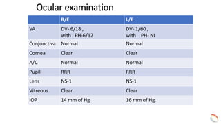
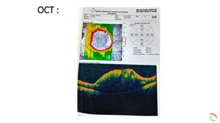
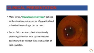
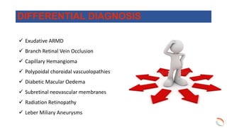
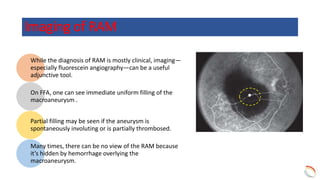
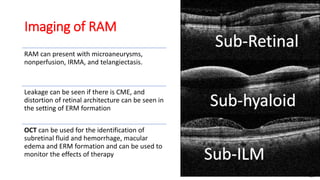
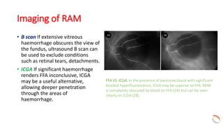
Presenter
Dr. Md. Rezwanul Hasan
Fellow, Vitreo-Retina Department
IIEI&H, Dhaka
Case presentation
Particulars of the Patient
• Name : Mr. Kuddus Dewan
• Age : 60 years
• Sex : Male
• Address : Tongi, Gazipur
Chief complaints
• Gradual dimness of vision in Left eyes
for about 5 days.
Past ocular History
• Use of Presbyopic glass for about 20 years.
Other Medical history
• Diabetic for 5 years.
• Hypertensive for 5 years.
General examination
Appearance Normal
Body build Average
Pulse 80 beats/min
Blood pressure 140/85 mm of Hg. with
medication
Temperature 98⁰F
Anemia Absent
Jaundice Absent
Edema Absent
Ocular examination
R/E L/E
VA DV- 6/18 ,
with PH-6/12
DV- 1/60 ,
with PH- NI
Conjunctiva Normal Normal
Cornea Clear Clear
A/C Normal Normal
Pupil RRR RRR
Lens NS-1 NS-1
Vitreous Clear Clear
IOP 14 mm of Hg 16 mm of Hg.
CFP
Differential Diagnosis
• Retinal Artery Macroaneurysm
• Branch Retinal Vein Occlusion
• Exudative ARMD
• Polypoidal choroidal vascuolopathies (IPCV)
Investigations
Blood Sugar
Fasting - 6.5 mmol/l
2HABF – 8.2 mmol/l
• OCT (L/E)
• FFA (L/E)
OCT :
FFA
OS
Diagnosis :
Ruptured Retinal Artery Macroaneurysm
Treatment :
• Intravitreal Inj. Avastin (L/E)
F/U after 2
months
At Presentation
• Visual Acuity : L/E = 6/60 with
PH - 6/24
After 2 Months
• Visual Acuity : L/E = 1/60 with
PH - NI
• First described in 1973, retinal arterial
macroaneurysm is characterized by the
presence of vascular dilation or outpouching of
a retinal artery or arteriole.
• They are commonly associated with macular
exudation and hemorrhage, which may result in
decreased visual acuity.
• Female preponderance (ratio of 3:1 ), typically
after 60 years of age and with a medical history
of systemic hypertension and/or arteriosclerotic
vascular disease.
Discussion
• Often, RAM is noted on clinical examination but the
patient is asymptomatic.
• The involved artery may be narrowed proximally
and distally to the microaneurysm.
• Severe vision loss can occur from leakage of the
aneurysm with resultant hemorrhage into the
vitreous cavity, subhyaloid space and/or
intraretinally or subretinaly.
CLINICAL PRESENTATION
• Many times, “Hourglass hemorrhage” defined
as the simultaneous presence of preretinal and
subretinal hemorrhage, can be seen.
• Serous fluid can also collect intraretinally.
producing diffuse or focal cystoid macular
oedema with or without the accumulation of
lipid exudates.
CLINICAL PRESENTATION
 Exudative ARMD
 Branch Retinal Vein Occlusion
 Capillary Hemangioma
 Polypoidal choroidal vascuolopathies
 Diabetic Macular Oedema
 Subretinal neovascular membranes
 Radiation Retinopathy
 Leber Miliary Aneurysms
DIFFERENTIAL DIAGNOSIS
• Macroaneurysms more commonly affect the right
eye than the left.
• The supero temporal artery is most commonly
involved.
• However, macroaneurysms also have been
reported in cilioretinal arteries, on the optic nerve
head and associated with congenital retinal
macrovessels.
• Bleeding is a common complication of aneurysm
formation and can occur beneath the retina, the
retinal pigment epithelium (RPE), or the internal
limiting membrane (ILM) or into the vitreous.
VASCULAR SIGNS & SITES OF OCCURRENCE
Distribution of MAs in 34 patients.
Retinal artery macroaneurysms: clinical and fluorescein angiographic features in 34 patients. (2006, November 21). Retinal
artery macroaneurysms: clinical and fluorescein angiographic features in 34 patients. https://doi.org/10.1038/sj.eye.6702068
• The pathogenesis of RAM is thought to be secondary to a
combination of several mechanisms causing blood vessel
wall weakness and subsequent aneurysmal dilatation.
• The main mechanisms thought to underlie RAM
formation include focal ischemia to blood vessel walls,
chronic hypertensive and arteriosclerotic vascular wall
damage, and inherent structural defects in blood vessels.
• Histopathologically, RAMs are found to have arterial
dilatation with variable degrees of artery wall
hyalinization and surrounding retinal exudate or
hemorrhage
Pathogenesis
Fig: Histopathology of a
ruptured RAM with
preretinal, intraretinal and
subretinal hemorrhage
While the diagnosis of RAM is mostly clinical, imaging—
especially fluorescein angiography—can be a useful
adjunctive tool.
On FFA, one can see immediate uniform filling of the
macroaneurysm .
Partial filling may be seen if the aneurysm is
spontaneously involuting or is partially thrombosed.
Many times, there can be no view of the RAM because
it’s hidden by hemorrhage overlying the
macroaneurysm.
Imaging of RAM
Imaging of RAM
RAM can present with microaneurysms,
nonperfusion, IRMA, and telangiectasis.
Leakage can be seen if there is CME, and
distortion of retinal architecture can be seen in
the setting of ERM formation
OCT can be used for the identification of
subretinal fluid and hemorrhage, macular
edema and ERM formation and can be used to
monitor the effects of therapy
Imaging of RAM
• B scan If extensive vitreous
haemorrhage obscures the view of
the fundus, ultrasound B scan can
be used to exclude conditions
such as retinal tears, detachments.
• ICGA If significant haemorrhage
renders FFA inconclusive, ICGA
may be a useful alternative,
allowing deeper penetration
through the areas of
haemorrhage.
FFA VS. ICGA. In the presence of excessive blood with significant
blocked hyperfluorescence, ICGA may be superior to FFA. RAM
is completely obscured by blood on FFA (2A) but can be seen
clearly on ICGA (2B).
Diagnostic challenges Despite several imaging options at our
disposal, diagnosing RAM may prove challenging when
extensive haemorrhage is present.
In such cases, examining the fellow eye closely may help
guide the physician.
Important diagnostic clues for RAM include absence of
drusen and the presence of significant hypertensive
retinopathy in the fellow eye in the setting of an asymmetric
vascular lesion with lipid or blood concentrated around the
bifurcation of an arteriole in the affected eye.
Management
Most macroaneurysms can be observed.
The visual prognosis depends on whether secondary hemorrhage or exudation
involves the central macular region.
In such instances, vision can be reduced to counting fingers or worse.
Spontaneous improvement can occur, particularly when the blood is located
superficially within the retina.
Interventions
• May be required in cases of exudative or
hemorrhagic RAMs. or recurrent/persistent cystoid
macular oedema.
• Anti-vascular endothelial growth factor therapy
has emerged as a useful treatment modality for
RAMs.
• Laser of the artery and surrounding area may
decrease flow and intraluminal pressure, thereby
reducing the macroaneurysm.
• YAG-laser hyaloidotomy incase of dense subhyaloid
haemorrhage has been performed to release the
sequestered blood into the vitreous cavity.
PROGNOSIS
The visual prognosis is excellent for many patients
with macroaneurysm.
The natural history of these lesions suggests that
most close spontaneously, with restoration of near-
normal vision.
TAKE HOME MESSAGE
• RAMs are rare clinical findings occurring
most often in older, hypertensive women.
• While anti-VEGF therapy can be a useful
treatment option to improve vision and
decrease macular oedema, more complex
RAMs may require laser photocoagulation
and / or surgical intervention.
Retinal Artery Macroaneurysm

Retinal Artery Macroaneurysm

  • 1.
    Presenter Dr. Md. RezwanulHasan Fellow, Vitreo-Retina Department IIEI&H, Dhaka Case presentation
  • 2.
    Particulars of thePatient • Name : Mr. Kuddus Dewan • Age : 60 years • Sex : Male • Address : Tongi, Gazipur
  • 3.
    Chief complaints • Gradualdimness of vision in Left eyes for about 5 days.
  • 4.
    Past ocular History •Use of Presbyopic glass for about 20 years.
  • 5.
    Other Medical history •Diabetic for 5 years. • Hypertensive for 5 years.
  • 6.
    General examination Appearance Normal Bodybuild Average Pulse 80 beats/min Blood pressure 140/85 mm of Hg. with medication Temperature 98⁰F Anemia Absent Jaundice Absent Edema Absent
  • 7.
    Ocular examination R/E L/E VADV- 6/18 , with PH-6/12 DV- 1/60 , with PH- NI Conjunctiva Normal Normal Cornea Clear Clear A/C Normal Normal Pupil RRR RRR Lens NS-1 NS-1 Vitreous Clear Clear IOP 14 mm of Hg 16 mm of Hg.
  • 8.
  • 9.
    Differential Diagnosis • RetinalArtery Macroaneurysm • Branch Retinal Vein Occlusion • Exudative ARMD • Polypoidal choroidal vascuolopathies (IPCV)
  • 10.
    Investigations Blood Sugar Fasting -6.5 mmol/l 2HABF – 8.2 mmol/l • OCT (L/E) • FFA (L/E)
  • 11.
  • 12.
  • 13.
    Diagnosis : Ruptured RetinalArtery Macroaneurysm
  • 14.
    Treatment : • IntravitrealInj. Avastin (L/E)
  • 15.
    F/U after 2 months AtPresentation • Visual Acuity : L/E = 6/60 with PH - 6/24 After 2 Months • Visual Acuity : L/E = 1/60 with PH - NI
  • 16.
    • First describedin 1973, retinal arterial macroaneurysm is characterized by the presence of vascular dilation or outpouching of a retinal artery or arteriole. • They are commonly associated with macular exudation and hemorrhage, which may result in decreased visual acuity. • Female preponderance (ratio of 3:1 ), typically after 60 years of age and with a medical history of systemic hypertension and/or arteriosclerotic vascular disease. Discussion
  • 17.
    • Often, RAMis noted on clinical examination but the patient is asymptomatic. • The involved artery may be narrowed proximally and distally to the microaneurysm. • Severe vision loss can occur from leakage of the aneurysm with resultant hemorrhage into the vitreous cavity, subhyaloid space and/or intraretinally or subretinaly. CLINICAL PRESENTATION
  • 18.
    • Many times,“Hourglass hemorrhage” defined as the simultaneous presence of preretinal and subretinal hemorrhage, can be seen. • Serous fluid can also collect intraretinally. producing diffuse or focal cystoid macular oedema with or without the accumulation of lipid exudates. CLINICAL PRESENTATION
  • 19.
     Exudative ARMD Branch Retinal Vein Occlusion  Capillary Hemangioma  Polypoidal choroidal vascuolopathies  Diabetic Macular Oedema  Subretinal neovascular membranes  Radiation Retinopathy  Leber Miliary Aneurysms DIFFERENTIAL DIAGNOSIS
  • 20.
    • Macroaneurysms morecommonly affect the right eye than the left. • The supero temporal artery is most commonly involved. • However, macroaneurysms also have been reported in cilioretinal arteries, on the optic nerve head and associated with congenital retinal macrovessels. • Bleeding is a common complication of aneurysm formation and can occur beneath the retina, the retinal pigment epithelium (RPE), or the internal limiting membrane (ILM) or into the vitreous. VASCULAR SIGNS & SITES OF OCCURRENCE Distribution of MAs in 34 patients. Retinal artery macroaneurysms: clinical and fluorescein angiographic features in 34 patients. (2006, November 21). Retinal artery macroaneurysms: clinical and fluorescein angiographic features in 34 patients. https://doi.org/10.1038/sj.eye.6702068
  • 21.
    • The pathogenesisof RAM is thought to be secondary to a combination of several mechanisms causing blood vessel wall weakness and subsequent aneurysmal dilatation. • The main mechanisms thought to underlie RAM formation include focal ischemia to blood vessel walls, chronic hypertensive and arteriosclerotic vascular wall damage, and inherent structural defects in blood vessels. • Histopathologically, RAMs are found to have arterial dilatation with variable degrees of artery wall hyalinization and surrounding retinal exudate or hemorrhage Pathogenesis Fig: Histopathology of a ruptured RAM with preretinal, intraretinal and subretinal hemorrhage
  • 22.
    While the diagnosisof RAM is mostly clinical, imaging— especially fluorescein angiography—can be a useful adjunctive tool. On FFA, one can see immediate uniform filling of the macroaneurysm . Partial filling may be seen if the aneurysm is spontaneously involuting or is partially thrombosed. Many times, there can be no view of the RAM because it’s hidden by hemorrhage overlying the macroaneurysm. Imaging of RAM
  • 23.
    Imaging of RAM RAMcan present with microaneurysms, nonperfusion, IRMA, and telangiectasis. Leakage can be seen if there is CME, and distortion of retinal architecture can be seen in the setting of ERM formation OCT can be used for the identification of subretinal fluid and hemorrhage, macular edema and ERM formation and can be used to monitor the effects of therapy
  • 24.
    Imaging of RAM •B scan If extensive vitreous haemorrhage obscures the view of the fundus, ultrasound B scan can be used to exclude conditions such as retinal tears, detachments. • ICGA If significant haemorrhage renders FFA inconclusive, ICGA may be a useful alternative, allowing deeper penetration through the areas of haemorrhage. FFA VS. ICGA. In the presence of excessive blood with significant blocked hyperfluorescence, ICGA may be superior to FFA. RAM is completely obscured by blood on FFA (2A) but can be seen clearly on ICGA (2B).
  • 25.
    Diagnostic challenges Despiteseveral imaging options at our disposal, diagnosing RAM may prove challenging when extensive haemorrhage is present. In such cases, examining the fellow eye closely may help guide the physician. Important diagnostic clues for RAM include absence of drusen and the presence of significant hypertensive retinopathy in the fellow eye in the setting of an asymmetric vascular lesion with lipid or blood concentrated around the bifurcation of an arteriole in the affected eye.
  • 26.
    Management Most macroaneurysms canbe observed. The visual prognosis depends on whether secondary hemorrhage or exudation involves the central macular region. In such instances, vision can be reduced to counting fingers or worse. Spontaneous improvement can occur, particularly when the blood is located superficially within the retina.
  • 27.
    Interventions • May berequired in cases of exudative or hemorrhagic RAMs. or recurrent/persistent cystoid macular oedema. • Anti-vascular endothelial growth factor therapy has emerged as a useful treatment modality for RAMs. • Laser of the artery and surrounding area may decrease flow and intraluminal pressure, thereby reducing the macroaneurysm. • YAG-laser hyaloidotomy incase of dense subhyaloid haemorrhage has been performed to release the sequestered blood into the vitreous cavity.
  • 28.
    PROGNOSIS The visual prognosisis excellent for many patients with macroaneurysm. The natural history of these lesions suggests that most close spontaneously, with restoration of near- normal vision.
  • 29.
    TAKE HOME MESSAGE •RAMs are rare clinical findings occurring most often in older, hypertensive women. • While anti-VEGF therapy can be a useful treatment option to improve vision and decrease macular oedema, more complex RAMs may require laser photocoagulation and / or surgical intervention.

Editor's Notes

  • #24 IRMA=intraretinal microvascular abnormalities