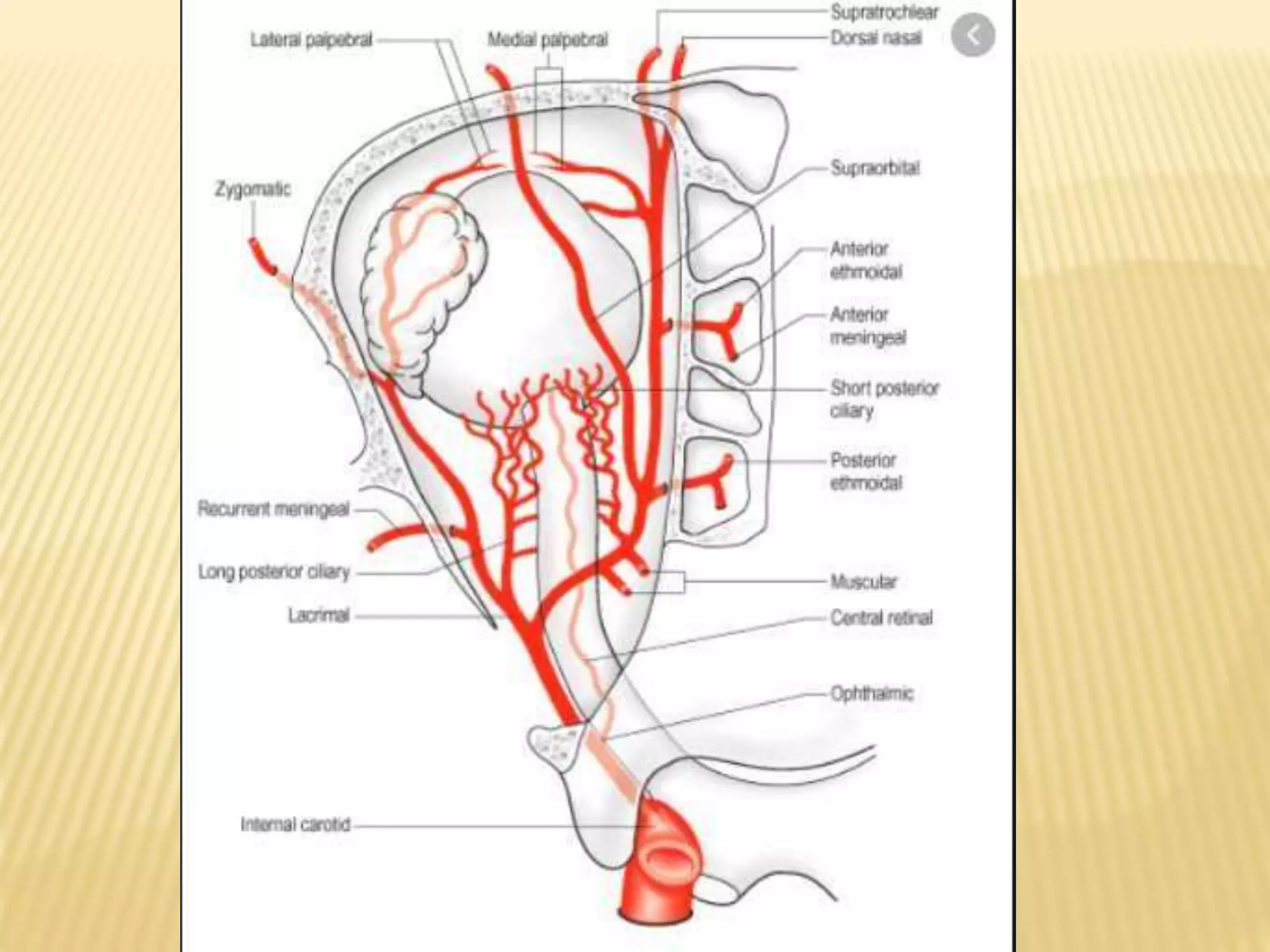
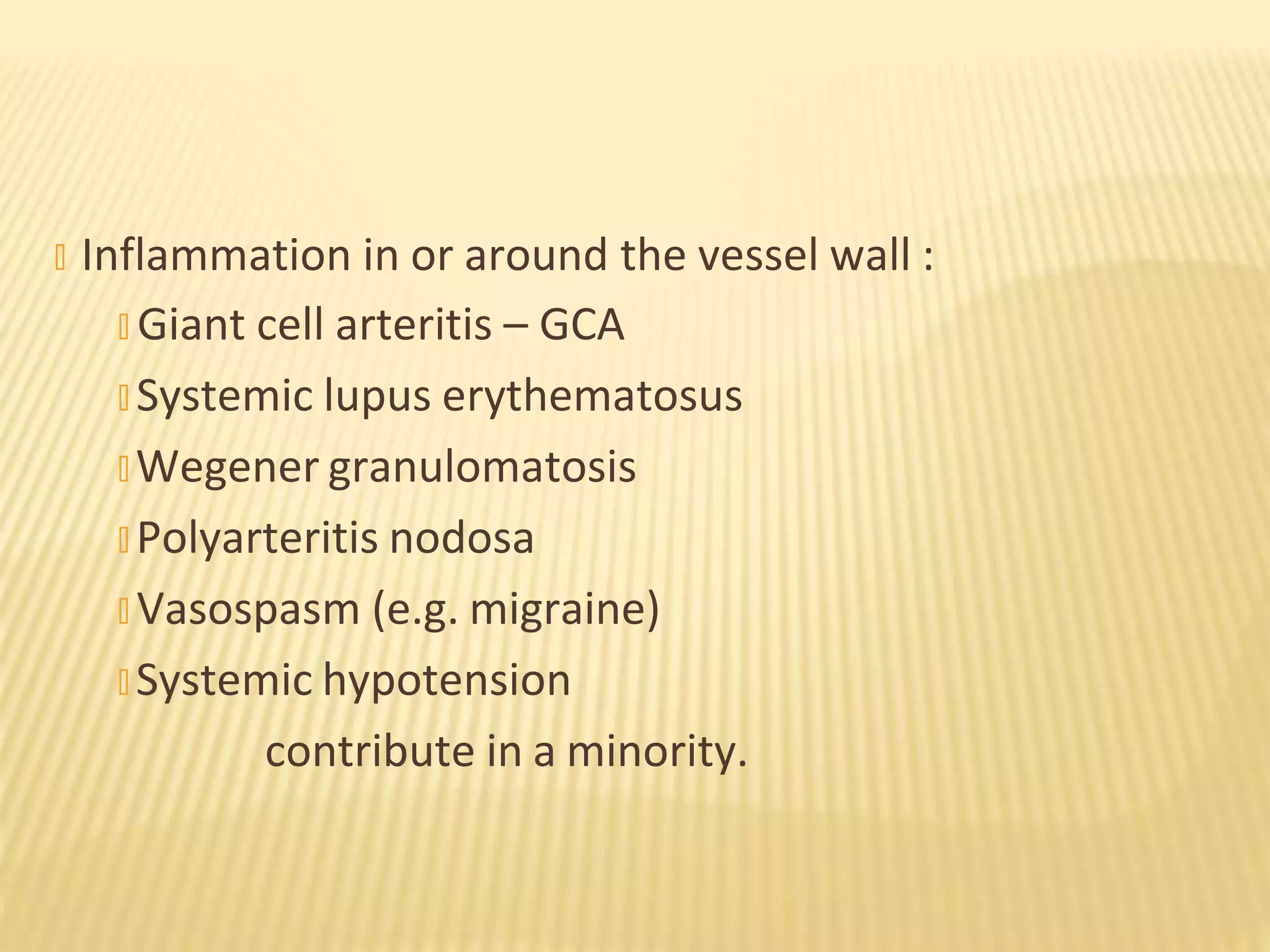
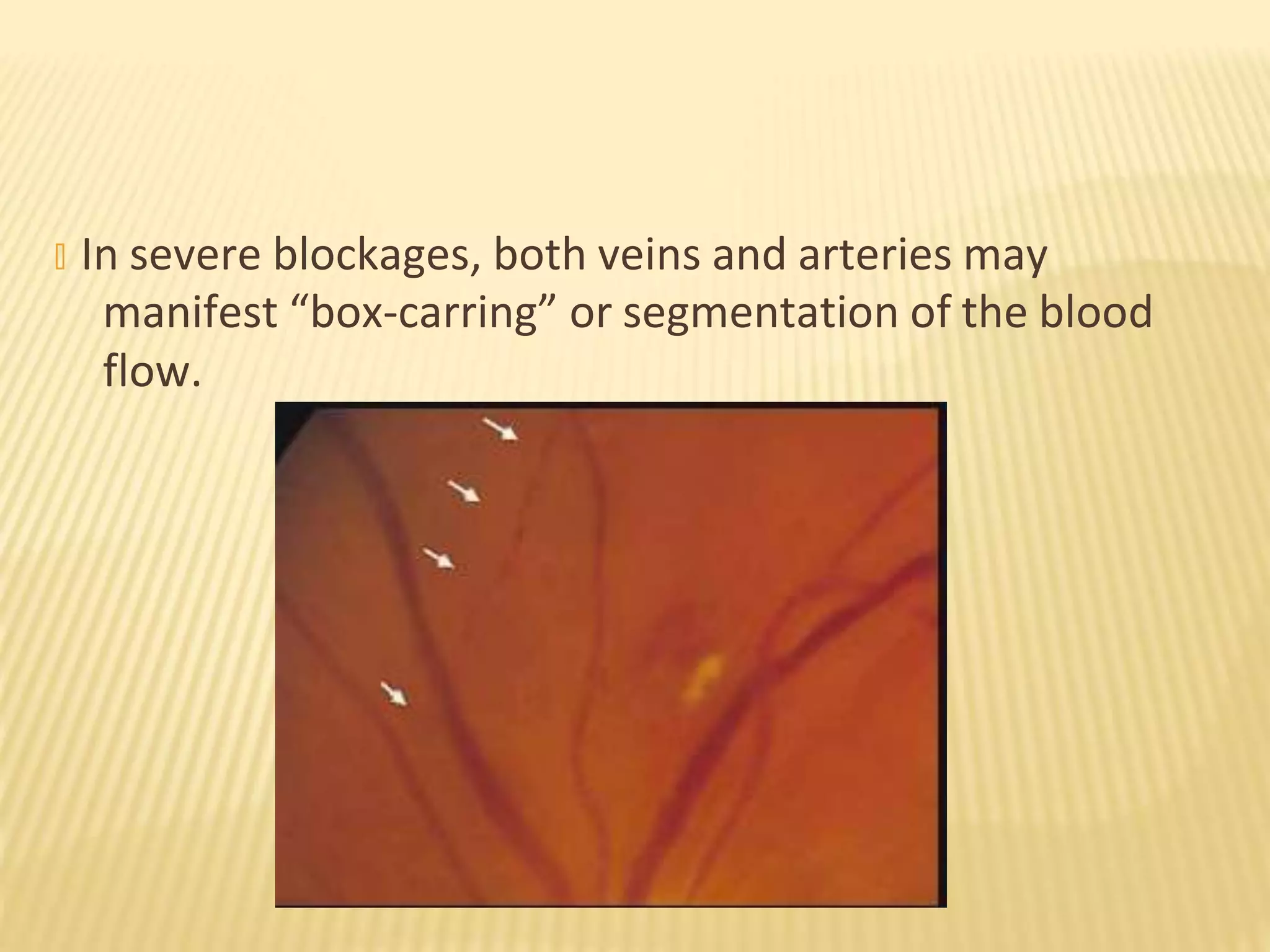
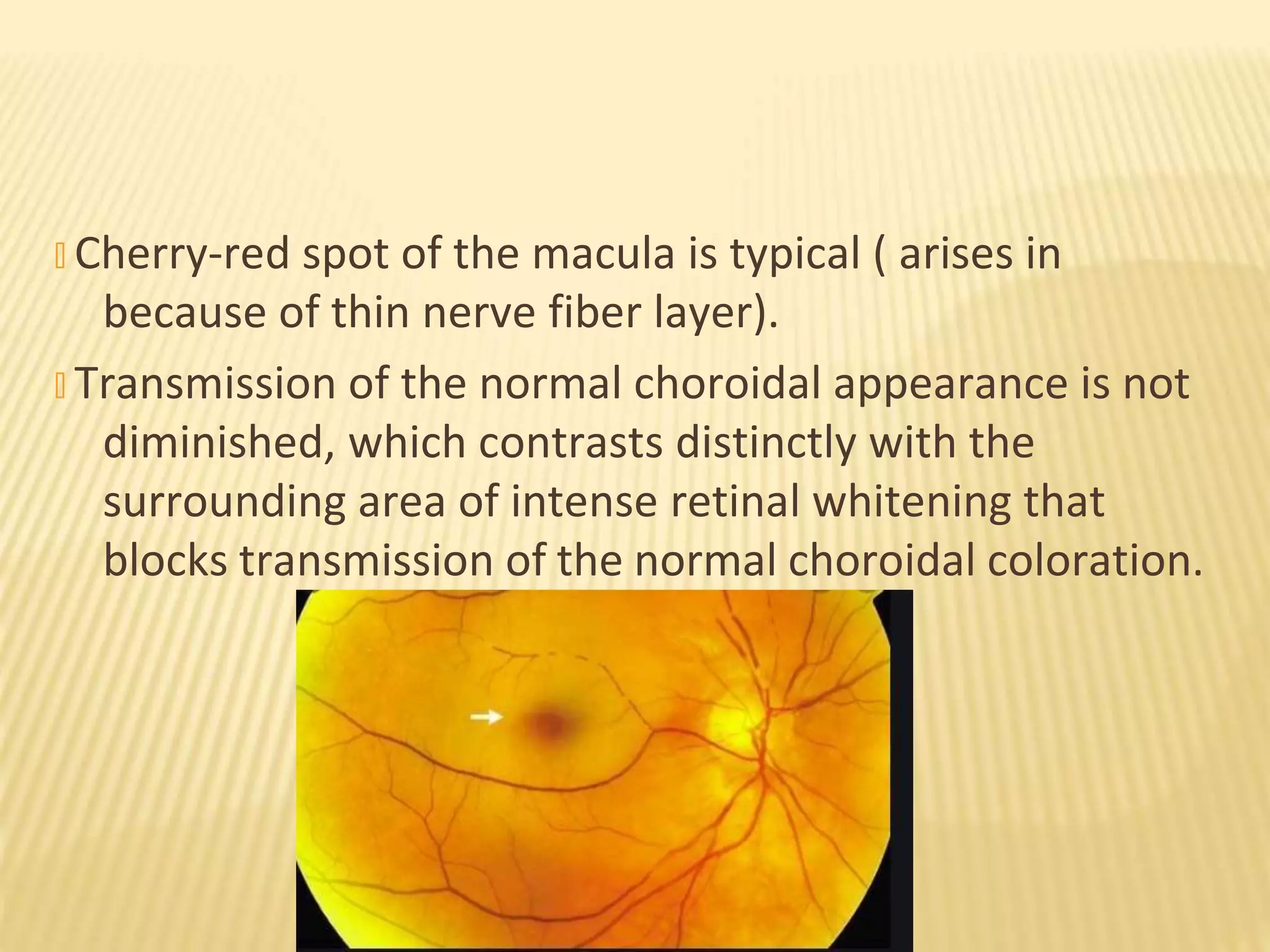
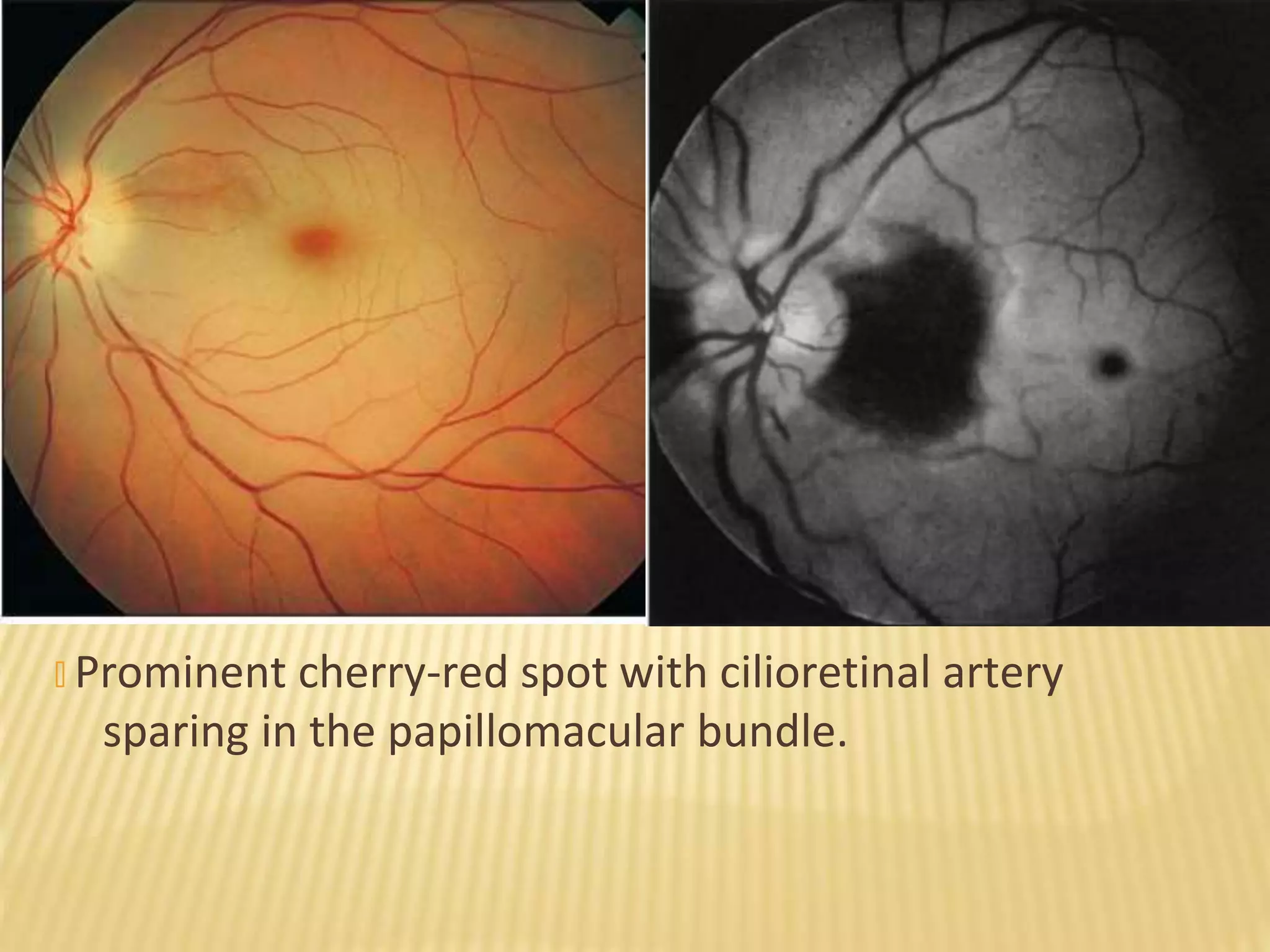
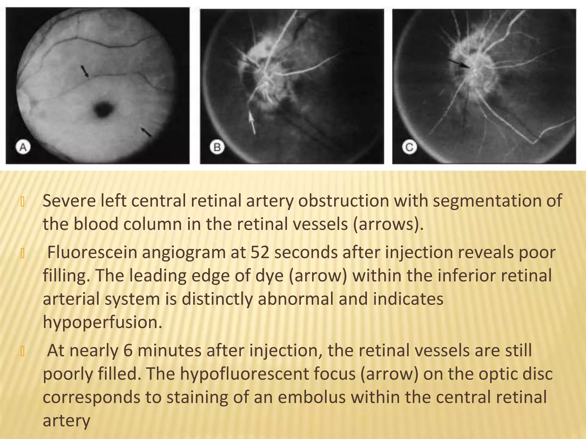
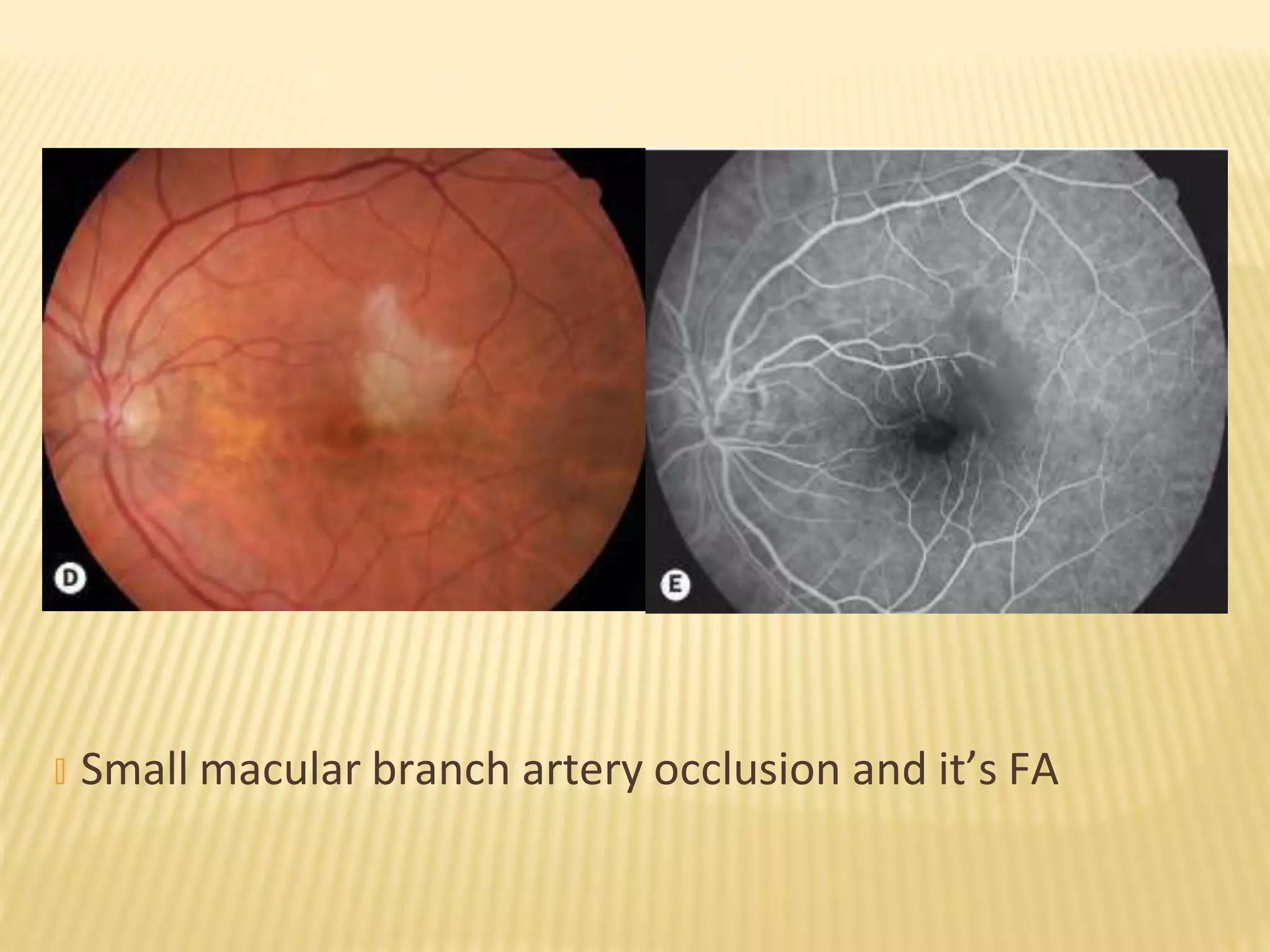
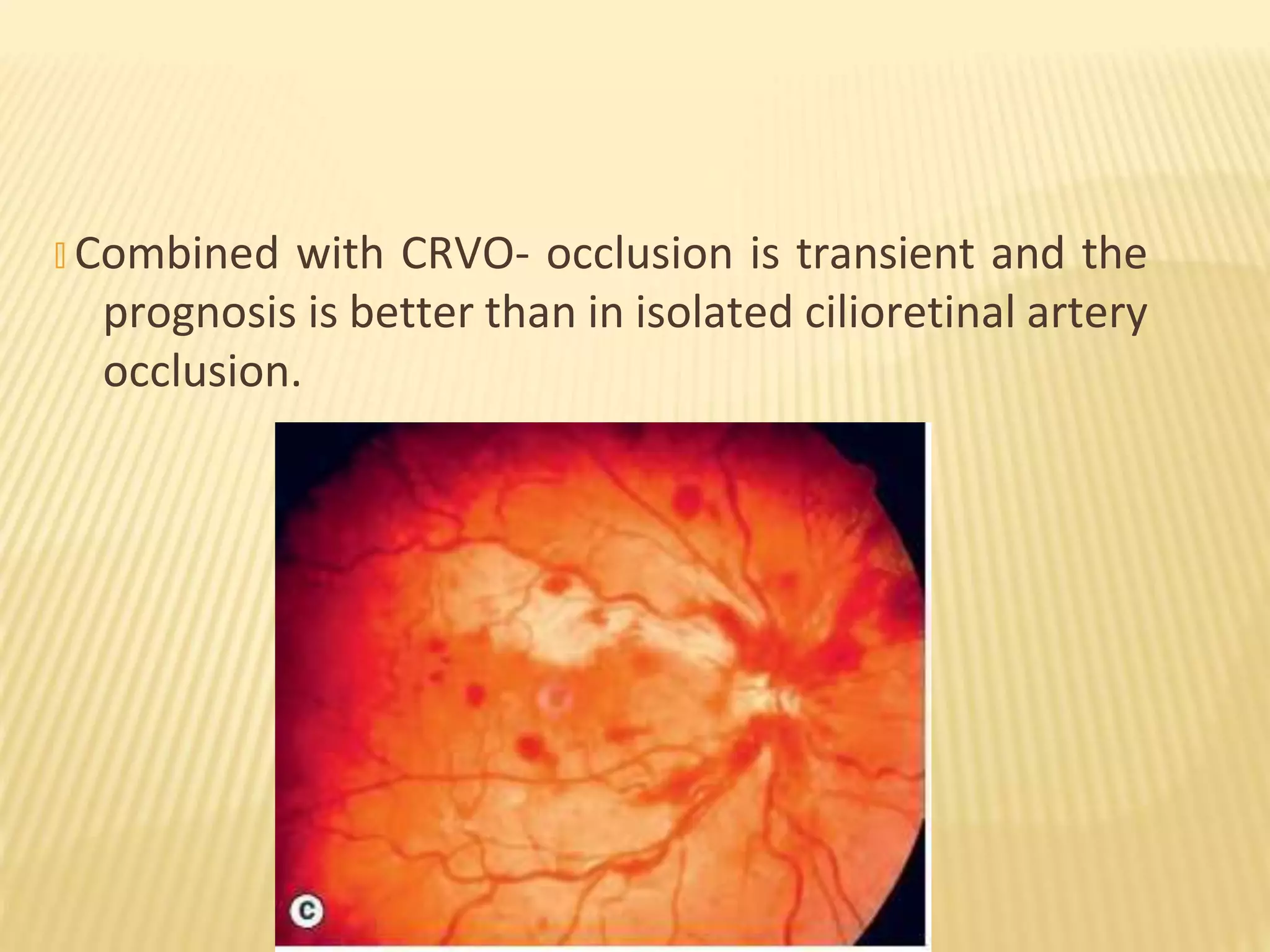
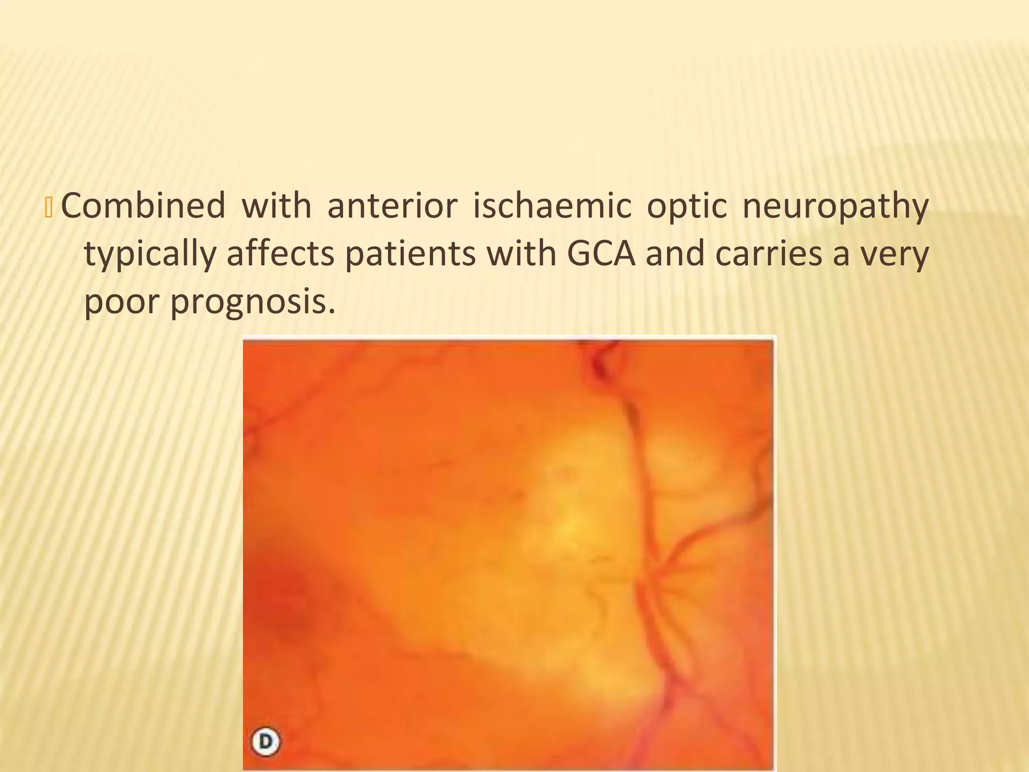
RETINAL ARTERY OCCLUSION
BY
DR ANJALI MAHESHWARI
� Kanski’s – 8th
edition
� Ryan’s textbook of retina
� Yanoff and Duker- 5th
edition
SOURCES
� Introduction
� CRAO
� BRAO
� Cilioretinal artery occlusion
� Ophthalmic artery occlusion
� Combined artery and vein occlusion
� Systemic evaluation
� Treatment
LAYOUT
� Retinal arterial obstructions are divided anatomically
depending on the precise site of obstruction:
� Central - occurs when the blockage is within the optic
nerve substance itself and site of obstruction is not
visible on ophthalmoscopy.
� Branch - occurs when the site of blockage is distal to the
lamina cribrosa of the optic nerve
� More proximal obstructions usually cause a more
chronic form of visual problem − the ocular ischemic
syndrome
INTRODUCTION
� Majority of retinal arterial obstructions are either
thrombotic or embolic in nature.
� Branch retinal artery obstruction is far more likely to be
embolic than is a central retinal artery obstruction
� Retinal artery obstructions selectively affect the inner
retina only
� The outer retina is supplied by the ciliary arteries via the
choriocapillaris and the inner retina by the central
retinal artery (CRA).
� The ophthalmic artery gives rise to both the CRA – its
first branch – and the ciliary arteries.
� Atherosclerosis-related embolism and thrombosis are
thought to be responsible for the majority of cases of
retinal artery occlusion.
BLOOD SUPPLY
� The origin of emboli is most commonly an
atheromatous carotid plaque; the ophthalmic artery is
the first branch of the internal carotid artery, so embolic
material has an easy route to the eye.
� Inflammation in or around the vessel wall :
� Giant cell arteritis – GCA
� Systemic lupus erythematosus
� Wegener granulomatosis
� Polyarteritis nodosa
� Vasospasm (e.g. migraine)
� Systemic hypotension
contribute in a minority.
CENTRAL RETINAL ARTERY OBSTRUCTION.
� Rare event − it has been estimated to account for about
1 in 10000 outpatient visits to the ophthalmologist.
� Men are affected more commonly than women in the
ratio 2:1.
� The mean age at onset is about 60years
� Bilateral involvement occurs in 1–2% of cases
� When both eyes are simultaneously affected by retinal
artery obstruction, the differential diagnosis should
include :
� Cardiac valvular disease
� Giant cell arteritis
� Other vascular inflammations.
� Thrombosis- at level of lamina cribrosa
� Atherosclerosis
� Congenital abnormality of central retinal artery
� Coagulopathies
� Embolism (20-25%) – Origin from atheromatous carotid
plaque.
� Cholesterol (Hollenhorst plaques)
� Platelet-fibrin
� Calcific
ETIOLOGY
� Inflammatory
� Vasculitis( Varicella, SLE, Wegner’s, PAN)
� Optic neuritis
� Orbital disorder (Mucormycosis)
� Local trauma
� Arterial spasms (dissecting aneurysm)
� Thrombophilic disorders (In one-third of young patients)
� Hyperhomocystinemia
� Anti-phospholipid antibody syndrome
� Sickling hemoglobinopathies
� Susac syndrome ( Retino-cochleo-cerebral syndrome)
� Protein C& S deficiency
� Anti-thrombin III deficiency
� Rare causes
� Radiation retinopathy
� Emboli secondary to depot medications around eyes
� Optic nerve drusens
� Peripapillary arterial loops
� Medical examination and manipulation Eg: Carotid
angiography, angioplasty , chiropractic neck
manipulation
� Transient monocular loss of vision
� Amaurosis fugax study group divides cause into five
categories:
� Embolic
� Ocular
� Neurologic
� Hemodynamic
� Idiopathic
Recovery is in the same pattern as lost.
AMAUROSIS FUGAX
� The hallmark symptom of acute central retinal artery
obstruction is abrupt, painless loss of vision.
� Pain is unusual and suggests associated ocular ischemic
syndrome (OIS).
� Amaurosis fugax precedes visual loss in about 10% of
patients.
� In cases associated with arterial spasm, a relapsing and
remitting course of visual loss precedes central retinal
artery obstruction.
CLINICAL FEATURES
� Visual acuity of 20/800 (6/240) or worse.
� No light perception vision is uncommon except in case
of ophthalmic artery obstruction or temporal arteritis.
� Patent cilioretinal artery - perfuses the fovea, normal
central acuity may be present.
� Afferent pupillary defect on the affected side.
� Anterior segment examination is normal
(Except in concurrent OIS with neovascularization of the
iris)
obstruction, the fundus may appear relatively normal.
� Decreased blood flow results in ischemic whitening of
the retina in the territory of the obstructed artery.
� Most pronounced in the posterior pole (where the
nerve fiber layer of the retina is thickest).
� Acutely arteries appear thin and attenuated.
FUNDUS
� Within the first few minutes to hours after the
� Retinal whitening is very subtle and the retinal vessels
appear normal.
� Fluorescein angiography reveals abnormal arterial filling
with a leading edge of dye that confirms central retinal
artery obstruction.
� The same eye 24 hours later- intense retinal whitening
with a cherry-red spot is present.
� In severe blockages, both veins and arteries may
manifest “box-carring” or segmentation of the blood
flow.
� Cherry-red spot of the macula is typical ( arises in
because of thin nerve fiber layer).
� Transmission of the normal choroidal appearance is not
diminished, which contrasts distinctly with the
surrounding area of intense retinal whitening that
blocks transmission of the normal choroidal coloration.
� It also depends on the size of fovea.
� Tay–Sachs disease
� Farber’s disease
� Sandhoff’s disease
� Niemann–Pick disease
� Goldberg’s syndrome
� Gaucher’s disease
� Ganglioside GMI, type 2
� Hurler’s syndrome (mucopolysaccharidosis 1 H)
� β-Galactosidase deficiency (mucopolysaccharidosis VII)
� Hallevorden–Spatz disease
OTHER CAUSES OF CHERRY RED SPOT
� Splinter retinal hemorrhages on the disc are common.
� If pallid swelling is present, temporal arteritis must be
ruled out.
� A patent cilioretinal artery results in a small area of
retina that appears normal .
� Prominent cherry-red spot with cilioretinal artery
sparing in the papillomacular bundle.
� By 6 weeks after the acute event:
� Retinal whitening resolves
� Optic disc develops pallor
� Arterial collaterals forms on the optic disc
� No foveolar light reflex apparent
� Fine changes in the retinal pigment epithelium may
be visible.
� Secondary ocular neovascularization occuar 8 weeks
after the obstruction
� Iris neovascularization occurs in about 18% of patients
with many developing to neovascular glaucoma.
� Neovascularization of the optic disc occurs after about
2% of central retinal artery obstruction
� Vitreous hemorrhage may also occur.
� Central retinal artery
obstruction caused by a
platelet-fibrin embolus.
� After 3 months,
extensive
neovascularization of
the disc is present.
DIAGNOSIS AND ANCILLARY TESTING
� Diffuse ischemic retinal whitening is present in the
setting of abrupt, painless visual loss.
� Fluorescein angiography
� Optical coherence tomography
� Electroretinogram
� Visual field
FLUORESCEIN ANGIOGRAPHY
� Delayed arm-to-retina time with a leading edge of dye
visible in the retinal arteries is typical
� Arteriovenous transit is delayed :time elapsed from the
appearance of dye within the arteries of the temporal
vascular arcade until the corresponding veins are
completely filled; normal is less than or equal to 11
seconds
� Late staining of the disc is common.
� Severe left central retinal artery obstruction with segmentation of
the blood column in the retinal vessels (arrows).
� Fluorescein angiogram at 52 seconds after injection reveals poor
filling. The leading edge of dye (arrow) within the inferior retinal
arterial system is distinctly abnormal and indicates
hypoperfusion.
� At nearly 6 minutes after injection, the retinal vessels are still
poorly filled. The hypofluorescent focus (arrow) on the optic disc
corresponds to staining of an embolus within the central retinal
artery
� Complete lack of filling of the retinal arteries is unusual
and occurs in less than 2% of cases.
� The choroidal vascular bed in eyes usually fills normally,
delays of five seconds or greater seen in about 10% of
cases
� A marked prolongation of choroidal filling in the
presence of a cherry-red spot should arouse suspicion
of an ophthalmic artery obstruction or a concomitant
carotid artery obstruction
� The retinal circulation has propensity to re-establish the
circulation following an acute central retinal artery
obstruction.
� Arterial narrowing and visual loss may persist, but the
fluorescein angiogram can revert to normal at varying
times after the insult.
OPTICAL COHERENCE TOMOGRAPHY
� Macular optical coherence tomography (OCT) in the
acute phase shows inner retinal thickening with
shadowing of the outer retina that can be mistaken for
subretinal fluid.
� When the retinal whitening resolves, OCT reveals
severe inner retinal thinning.
� OCT angiography of prior central retinal artery
obstruction demonstrates decreased retinal vascularity
and attenuation of vessels in the macula
� Optical Coherence
Tomography
Angiography and
Corresponding Optical
Coherence
Tomography Structural
B-Scan From an Eye
With a Prior Central
Retinal Artery
Obstruction.
� Paucity of small retinal
vessels in the macula
and the attenuation of
the large retinal
vessels. Structural B-
scan shows loss of
inner retinal tissue.
� Diminution in the amplitude of the b-wave
(corresponding to the function of the Muller and/or
bipolar cells) secondary to inner layer retinal ischemia.
� The a-wave, which corresponds to photoreceptor
function, is generally unaffected.
� In some eyes the study is normal in the presence of
decreased vision, possibly because of the re-
establishment of retinal blood flow
ELECTRORETINOGRAPHY
� Electroretinograms of a normal right eye (upper
tracing) and a left eye (lower tracing) affected by a
CRAO.
� The “b” wave is diminished in the lower tracing of the
CRAO eye, but the “a” wave is normal.
� Demonstrate a remaining temporal island of vision,
presumably because the choroid nourished the
corresponding nasal retina.
� In the presence of a patent cilioretinal artery, small
areas of central vision are preserved.
� Depending upon the degree and the extent of the
obstruction, varied portions of the peripheral field may
remain.
VISUAL FIELD
� About 50%–60% of patients have concurrent systemic
arterial hypertension, and diabetes is present in 25%.
� Systemic evaluation reveals no definite cause for the
obstruction in over 50% of affected patients.
� The Retinal Emboli of Cardiac Origin Study Group, a
multicenter study, reported on the cardiac findings
associated with acute retinal arterial occlusion
SYSTEMIC ASSOCIATIONS
ASSOCIATED SYSTEMIC CONDITIONS
� Single or multiple branch retinal artery obstruction
� Cilioretinal artery obstruction
� Severe commotio retinas
� Necrotizing herpetic retinitis
DIFFRENTIAL DIAGNOSIS
� Most central retinal artery obstructions result in severe,
permanent loss of vision.
� About one-third of patients experience some
improvement in final vision in terms of presentation
acuity either with or without conventional treatment.
� Three or more Snellen lines of improved visual acuity
occur in only about 10% of untreated patients.
� Spontaneous recovery may be more common in young
children.
COURSE AND OUTCOME
� Rare event, less common than central retinal artery
obstruction overall.
� Exception - comparative incidence with young patients,
in whom branch retinal artery obstruction is the more
common type of retinal artery obstruction.
� Men are more affected than women by a 2 : 1 ratio,
reflects the higher incidence of vasculopathic disease in
men.
� Young patients (less than 50 years of age), women and
men are affected equally.
BRANCHED RETINAL ARTERY OCCLUSION
� Mean age of affected patients is 60 years (Range -
second decade to tenth).
� The right eye (60%) is affected more commonly than the
left (40%), which reflects the greater possibility of
cardiac or aortic emboli traveling to the right carotid
artery.
� Branch retinal artery obstruction strikes the temporal
retinal circulation far more frequently than the nasal.
� Over two-thirds of branch retinal artery obstructions are
secondary to emboli to the retinal circulation.
� In most cases, the emboli are clearly visible in the
arterial tree.
� Emboli to the retinal circulation may originate at any
point in the proximal circulation from the heart to the
ophthalmic artery.
� Risk factors reflect the vasculopathic mechanisms that
produce disease within the cardiovascular system:
� Predisposing family history
� Hypertension
� Elevated lipid levels
� Cigarette smoking
� Diabetes mellitus
� Three main types of retinal emboli have been identified:
• Cholesterol (Hollenhorst plaque)
• Platelet-fibrin
• Calcific
� Cholesterol emboli typically emanate from
atheromatous plaques of the ipsilateral carotid artery
system, aorta or heart valves may also be a source.
� Yellow–orange in color, refractile, and globular or
rectangular in shape.
� They are small and can be seen intravascularly without
blockage of blood flow.
� Cholesterol embolus (arrow) in the fundus of an
asymptomatic woman.
� Embolus is present at the bifurcation, since it is trapped
as the lumen of the artery narrows
� Platelet-fibrin emboli are long, smooth, white-colored,
intra-arterial plugs that may be mobile or break up over
time.
� Usually they are associated with carotid or cardiac
thromboses.
� Calcific emboli are solid, white, nonrefractile plugs
associated with calcification of heart valves or the aorta.
� Less commonly seen embolic types :
� Tumor cells from atrial myxoma or a systemic
metastasis
� Septic emboli associated with septicemia or
endocarditis
� Fat emboli associated with large bone fractures
� Emboli dislodged during angioplasty or angiography
� Depot drug preparations from intra-arterial
injections around the eye or face
� Rarely, local ocular conditions produce branch retinal
artery obstruction including:
� Inflammatory diseases such as toxoplasmosis
� Acute retinal necrosis
� Mechanical compression from anterior ischemic optic
neuropathy
� Structural entities such as optic disc drusen or
prepapillary arterial loops
� Systemic hematological or clotting problems -
isolated branch retinal artery obstruction or multiple
recurrent branch retinal artery obstruction
� Systemic vasculitides, such as polyarteritis nodosa
� Local vasculitis associated with varicella infection
� Oral contraceptive use and cigarette smoking - in
young, otherwise healthy women
� Symptoms.
� Sudden and profound painless altitudinal or sectoral
visual field loss.
� Can sometimes go unnoticed, particularly if central
vision is spared.
� VA
� Variable
� In patients where central vision is severely
compromised, the prognosis is commonly poor
unless the obstruction is relieved within a few hours .
CLINICAL FEATURES
� RAPD is often present.
� Amaurosis fugax occurs in about one-fourth of patients
prior to frank obstruction, especially case of carotid
disease.
� Bilateral simultaneous branch retinal artery obstruction,
which can mimic homonymous field defects can occur
rarely.
� Fundus signs may be subtle :
� Attenuation of arteries and veins with sludging and
segmentation of the blood column (‘cattle trucking/
boxcarring’).
� Cloudy white oedematous (ground glass) retina
corresponding to the area of ischaemia.
� One or more occluding emboli may be seen,
especially at bifurcation points.
� The affected artery is likely to remain attenuated.
� Recanalization may leave absent ophthalmoscopic
signs.
� A syndrome of multiple, recurrent, bilateral branch
retinal artery obstruction in young, otherwise healthy
patients has been reported.
� A few of the patients also manifest vestibuloauditory
� Susac’s syndrome, a rare disorder that manifests as a
microangiopathy of the central nervous system.
� Inferotemporal occlusion – embolus visible over the
disc
� Superior branch retinal artery occlusion due to an
embolus at the disc
� FA shows lack of arterial filling of the involved artery and
hypofluorescence of the involved segment due to
blockage of background fluorescence by retinal swelling
� Small macular branch artery occlusion and it’s FA
� In the chronic phase, when the retinal whitening has
diminished, a loss of the nerve fiber layer in the affected
area is apparent.
� Arteriolar collaterals on the optic disc or at the site of
obstruction may develop
� Visual field testing confirms the defect, which rarely
recovers.
� FA shows delay in arterial filling and hypofluorescence of
the involved segment due to blockage of background
fluorescence by retinal swelling
� OCTA demonstrates vascular features that correlate with
FA and provide improved visualization of the
microvasculature compared to FA.
ANCILLARY TESTING
� OCT initially reveals thickening and hyperreflectivity
consistent with intracellular edema of the inner retina in
the territory of the obstructed artery.
� Over time, corresponding inner retina will be severely
thinned.
� Review in 3 months is warranted to review the
appearance of the fundus and visual fields
� Provide advice on prognosis and confirm that systemic
management has been carried out appropriately.
� Cotton–wool spot(s)
� Central retinal artery obstruction
� Cilioretinal artery obstruction
� Retinal astrocytoma
� Inflammatory or infectious retinitis
DIFFRENTIAL DIAGNOSIS
CILIORETINAL ARTERY OCLUSION
� A cilioretinal artery is present in 15–50% of eyes
� Its main importance is that when present it may
facilitate preservation of central vision following central
retinal artery occlusion, provided the fovea is supplied.
shows hypofluorescence in the affected area due to
reduced filling and masking by retinal oedema
� Isolated- rare; occur in young patients with an
associated systemic vasculitis.
�
� Isolated; (B) FA of the eye in (A) shows
hypofluorescence in the affected area due to
reduced filling and masking by retinal oedema;
Isolated cilioretinal artery occlusion and FA of the eye
� Isolated cilioretinal artery obstruction
� Fluorescein angiogram demonstrates poor filling of the
obstructed cilioretinal artery and retinal capillary
nonperfusion within the area of distribution of the vessel.
� Combined with CRVO- occlusion is transient and the
prognosis is better than in isolated cilioretinal artery
occlusion.
� Combined with anterior ischaemic optic neuropathy
typically affects patients with GCA and carries a very
poor prognosis.
� Acute simultaneous obstruction of both the retinal and
choroidal circulations is referred to as an ophthalmic
artery obstruction.
� Cases of ophthalmic artery obstruction usually have
associated local orbital or systemic diseases including:
� Orbital mucormycosis
� Orbital trauma
� Retrobulbar anesthesia
� Depot corticosteroid injection
� Atrial myxoma
� Carotid artery disease.
OPHTHALMIC ARTERY OCCLUSION
� Ophthalmic artery obstructions can be differentiated
clinically from central retinal artery obstruction by the
following features:
• Severe visual loss—bare or no light perception.
• Intense ischemic retinal whitening that extends
beyond the macular area.
• Little to no cherry-red spot.
• Marked choroidal perfusion defects on fluorescein
angiography.
• Nonrecordable electroretinogram.
• Late retinal pigment epithelium alterations
COMBINED ARTERY AND VEIN OBSTRUCTIONS
� Central retinal artery obstruction combined with
simultaneous central retinal vein obstruction rarely
occurs.
� Patients typically present with acute, severe loss of
vision.
� Examination shows a cherry-red spot combined with
features of a central retinal vein obstruction, which
include dilated, tortuous veins that have retinal
hemorrhages in all four quadrants
� Associated systemic or local disease is the rule—
� Collagen vascular disorders
� Leukemia
� Orbital trauma
� Retrobulbar injections
� Mucormycosis
� The visual prognosis is generally poor and the risk of
neovascularization of the iris is about 75%.
� Urgent specialist vascular evaluation, typically within 24
hours, is the standard of care following a retinal arterial
event, including amaurosis fugax
� Risk of a stroke is relatively high in the first few days
after a transient ischaemic attack (TIA).
� The detection of atrial fibrillation is of particular
importance as admission for anticoagulation may be
indicated.
SYSTEMIC EVALUATION
� Smoking
� Symptoms of GCA (1–2% of central retinal artery
occlusion – CRAO) :
� Headache, jaw claudication, scalp tenderness, limb
girdle pain, weight loss and existing polymyalgia
rheumatica
� Extremely unlikely under 55–60 years.
� Constitutes an ophthalmic emergency
ALL PATIENTS
� Pulse - detect arrhythmia, particularly atrial fibrillation.
� Blood pressure.
� Cardiac auscultation for a murmur.
� Carotid auscultation is of limited value as the absence
of a bruit does not exclude significant stenosis.
� ECG to detect arrhythmia and other cardiac disease.
� ESR and CRP to identify possible GCA.
� Other blood tests include :
� CBC (platelets may be raised in GCA)
� Glucose
� Lipids
� Urea and electrolytes
� Carotid duplex scanning is a non-invasive screening test
involving a combination of high-resolution real-time
ultrasonography with Doppler flow analysis. If significant
stenosis is present, surgical management may be
considered
� Considered on a targeted basis in some patients,
particularly if younger and with no known
cardiovascular risk factors, or there is an atypical clinical
picture.
SELECTED PATIENTS
� Further carotid imaging
� Cranial magnetic resonance imaging (MRI) or computed
tomography (CT) may be indicated to rule out
intracranial or orbital pathology.
� Echocardiography- Usually performed in young patients
or if there is a specific indication such as a history of
rheumatic fever, known cardiac valvular disease, or
intravenous drug use.
� Chest X-ray. Sarcoidosis, tuberculosis, left ventricular
hypertrophy in hypertension.
� 24-hour ECG to exclude intermittent arrhythmia.
� Additional blood tests :
� Fasting plasma homocysteine level to exclude
hyperhomocysteinaemia.
� ‘Thrombophilia screen’ - refers to heritable
thrombophilias, which have predominantly been
implicated in venous rather than arterial thromboses.
� Plasma protein electrophoresis to detect
dysproteinaemias such as multiple myeloma.
� Thyroid function tests, especially if atrial fibrillation is
present; may be associated with dyslipidaemia.
� Autoantibodies: Rheumatoid factor, anticardiolipin
antibody, antinuclear antibody, anti-double stranded
DNA antibodies, principally looking for vasculitis in
younger patients.
� Syphilis serology.
� Blood cultures.
� Retinal artery occlusion is an emergency because it
causes irreversible visual loss unless the retinal
circulation is re-established prior to the development of
retinal infarction.
� Theoretically, timely dislodgement of thrombus or
emboli may ameliorate subsequent visual loss
� Treatments may be tried in patients with occlusions of
less than 24–48 hours’ duration at presentation, though
evidence of benefit is limited.
TREATMENT
� The number of measures tried and the intensity of
treatment should be tailored to the individual
� More aggressive if lower duration of occlusion, good
general health, monocularity
� More aggressive systemic treatment may be avoided
in the frail elderly
� Lack of evidence for clear benefit and the risks, should
be discussed before use
� Adoption of a supine posture:
� Might improve ocular perfusion and should always be
implemented
� Ocular massage :
� Using a three-mirror contact lens (allows direct artery
visualization).
� Aim is to mechanically collapse the arterial lumen
and cause prompt changes in arterial flow, improving
perfusion and potentially dislodging an embolus or
thrombus.
� One described method consists of positive pressure
for 10–15 seconds followed by release, continued for
3–5 minutes.
� Self-massage through closed eyelids can be
continued by the patient.
� Breathing a high oxygen (95%) and carbon dioxide (5%)
mixture, ‘carbogen’, has been advocated for a possible
dual effect of retarding ischaemia and vasodilatation.
� ‘Rebreathing’ into a paper bag in order to elevate blood
carbon dioxide and respiratory acidosis has been
advocated, as this may promote vasodilatation
� Anterior chamber paracentesis
� Using 27-gauge needle to withdraw 0.1–0.2 ml of
aqueous is controversial but has been advocated by
some authorities.
� Povidone-iodine 5% and topical antibiotic are instilled
a few minutes prior to the procedure, with a short
course of antibiotic afterwards.
� It may be prudent for ocular massage to be avoided
following paracentesis.
� Transluminal Nd:YAG laser embolysis or embolectomy :
� BRAO or CRAO in which an occluding embolus is
visible
� Shots of 0.5–1.0 mJ or higher are applied directly to
the embolus using a fundus contact lens.
� Embolectomy has been said to occur if the embolus is
ejected into the vitreous via a hole in the arteriole.
� The number of shots described in reports is
extremely variable.
� The main complication is vitreous haemorrhage
� Topical apraclonidine 1%, timolol 0.5% and intravenous
acetazolamide 500 mg to achieve a more sustained
lowering of intraocular pressure.
� Sublingual isosorbide dinitrate to induce vasodilatation
� Hyperosmotic agents
� Mannitol or glycerol have been used for their
possibly more rapid IOP-lowering effect as well as
increased intravascular volume
� Thrombolysis:
� Extrapolating from successful treatment of stroke
and myocardial infarction, various strategies have
been used to deliver thrombolytic agents to the
ophthalmic artery, including local arterial (internal
carotid and ophthalmic) and intravenous infusion.
� A recent large trial of local intra-arterial fibrinolysis
with recombinant tissue plasminogen activator (rtPA)
showed no benefit over conservative treatment that
included isovolaemic haemodilution
� Nearly 40% adverse reaction rate in the rtPA group.
� Aim should be to decrease IOP as it cause :
� Dislodgement of embolus
� Produce retinal dilatation and increase perfusion
OPD MANGEMENT
� Ocular massage is of utmost important especially with
goldmann contact lens – done till re-establishment of
continuous laminar flow and increase in width of blood
column and disappearance of fragmented flow.
� IOP lowering agents
� Carbogen inhalation
� AC paracentesis
THANK YOU

retinalarteryocclusions-200830094234.docx

  • 1.
  • 2.
    � Kanski’s –8th edition � Ryan’s textbook of retina � Yanoff and Duker- 5th edition SOURCES
  • 3.
    � Introduction � CRAO �BRAO � Cilioretinal artery occlusion � Ophthalmic artery occlusion � Combined artery and vein occlusion � Systemic evaluation � Treatment LAYOUT
  • 4.
    � Retinal arterialobstructions are divided anatomically depending on the precise site of obstruction: � Central - occurs when the blockage is within the optic nerve substance itself and site of obstruction is not visible on ophthalmoscopy. � Branch - occurs when the site of blockage is distal to the lamina cribrosa of the optic nerve � More proximal obstructions usually cause a more chronic form of visual problem − the ocular ischemic syndrome INTRODUCTION
  • 5.
    � Majority ofretinal arterial obstructions are either thrombotic or embolic in nature. � Branch retinal artery obstruction is far more likely to be embolic than is a central retinal artery obstruction � Retinal artery obstructions selectively affect the inner retina only
  • 6.
    � The outerretina is supplied by the ciliary arteries via the choriocapillaris and the inner retina by the central retinal artery (CRA). � The ophthalmic artery gives rise to both the CRA – its first branch – and the ciliary arteries. � Atherosclerosis-related embolism and thrombosis are thought to be responsible for the majority of cases of retinal artery occlusion. BLOOD SUPPLY
  • 8.
    � The originof emboli is most commonly an atheromatous carotid plaque; the ophthalmic artery is the first branch of the internal carotid artery, so embolic material has an easy route to the eye.
  • 9.
    � Inflammation inor around the vessel wall : � Giant cell arteritis – GCA � Systemic lupus erythematosus � Wegener granulomatosis � Polyarteritis nodosa � Vasospasm (e.g. migraine) � Systemic hypotension contribute in a minority.
  • 10.
    CENTRAL RETINAL ARTERYOBSTRUCTION. � Rare event − it has been estimated to account for about 1 in 10000 outpatient visits to the ophthalmologist. � Men are affected more commonly than women in the ratio 2:1. � The mean age at onset is about 60years � Bilateral involvement occurs in 1–2% of cases
  • 11.
    � When botheyes are simultaneously affected by retinal artery obstruction, the differential diagnosis should include : � Cardiac valvular disease � Giant cell arteritis � Other vascular inflammations.
  • 12.
    � Thrombosis- atlevel of lamina cribrosa � Atherosclerosis � Congenital abnormality of central retinal artery � Coagulopathies � Embolism (20-25%) – Origin from atheromatous carotid plaque. � Cholesterol (Hollenhorst plaques) � Platelet-fibrin � Calcific ETIOLOGY
  • 13.
    � Inflammatory � Vasculitis(Varicella, SLE, Wegner’s, PAN) � Optic neuritis � Orbital disorder (Mucormycosis) � Local trauma � Arterial spasms (dissecting aneurysm)
  • 14.
    � Thrombophilic disorders(In one-third of young patients) � Hyperhomocystinemia � Anti-phospholipid antibody syndrome � Sickling hemoglobinopathies � Susac syndrome ( Retino-cochleo-cerebral syndrome) � Protein C& S deficiency � Anti-thrombin III deficiency
  • 15.
    � Rare causes �Radiation retinopathy � Emboli secondary to depot medications around eyes � Optic nerve drusens � Peripapillary arterial loops � Medical examination and manipulation Eg: Carotid angiography, angioplasty , chiropractic neck manipulation
  • 16.
    � Transient monocularloss of vision � Amaurosis fugax study group divides cause into five categories: � Embolic � Ocular � Neurologic � Hemodynamic � Idiopathic Recovery is in the same pattern as lost. AMAUROSIS FUGAX
  • 17.
    � The hallmarksymptom of acute central retinal artery obstruction is abrupt, painless loss of vision. � Pain is unusual and suggests associated ocular ischemic syndrome (OIS). � Amaurosis fugax precedes visual loss in about 10% of patients. � In cases associated with arterial spasm, a relapsing and remitting course of visual loss precedes central retinal artery obstruction. CLINICAL FEATURES
  • 18.
    � Visual acuityof 20/800 (6/240) or worse. � No light perception vision is uncommon except in case of ophthalmic artery obstruction or temporal arteritis. � Patent cilioretinal artery - perfuses the fovea, normal central acuity may be present. � Afferent pupillary defect on the affected side.
  • 19.
    � Anterior segmentexamination is normal (Except in concurrent OIS with neovascularization of the iris)
  • 20.
    obstruction, the fundusmay appear relatively normal. � Decreased blood flow results in ischemic whitening of the retina in the territory of the obstructed artery. � Most pronounced in the posterior pole (where the nerve fiber layer of the retina is thickest). � Acutely arteries appear thin and attenuated. FUNDUS � Within the first few minutes to hours after the
  • 21.
    � Retinal whiteningis very subtle and the retinal vessels appear normal. � Fluorescein angiography reveals abnormal arterial filling with a leading edge of dye that confirms central retinal artery obstruction. � The same eye 24 hours later- intense retinal whitening with a cherry-red spot is present.
  • 22.
    � In severeblockages, both veins and arteries may manifest “box-carring” or segmentation of the blood flow.
  • 23.
    � Cherry-red spotof the macula is typical ( arises in because of thin nerve fiber layer). � Transmission of the normal choroidal appearance is not diminished, which contrasts distinctly with the surrounding area of intense retinal whitening that blocks transmission of the normal choroidal coloration.
  • 24.
    � It alsodepends on the size of fovea.
  • 25.
    � Tay–Sachs disease �Farber’s disease � Sandhoff’s disease � Niemann–Pick disease � Goldberg’s syndrome � Gaucher’s disease � Ganglioside GMI, type 2 � Hurler’s syndrome (mucopolysaccharidosis 1 H) � β-Galactosidase deficiency (mucopolysaccharidosis VII) � Hallevorden–Spatz disease OTHER CAUSES OF CHERRY RED SPOT
  • 26.
    � Splinter retinalhemorrhages on the disc are common. � If pallid swelling is present, temporal arteritis must be ruled out. � A patent cilioretinal artery results in a small area of retina that appears normal .
  • 27.
    � Prominent cherry-redspot with cilioretinal artery sparing in the papillomacular bundle.
  • 28.
    � By 6weeks after the acute event: � Retinal whitening resolves � Optic disc develops pallor � Arterial collaterals forms on the optic disc � No foveolar light reflex apparent � Fine changes in the retinal pigment epithelium may be visible.
  • 29.
    � Secondary ocularneovascularization occuar 8 weeks after the obstruction � Iris neovascularization occurs in about 18% of patients with many developing to neovascular glaucoma. � Neovascularization of the optic disc occurs after about 2% of central retinal artery obstruction � Vitreous hemorrhage may also occur.
  • 30.
    � Central retinalartery obstruction caused by a platelet-fibrin embolus. � After 3 months, extensive neovascularization of the disc is present.
  • 31.
    DIAGNOSIS AND ANCILLARYTESTING � Diffuse ischemic retinal whitening is present in the setting of abrupt, painless visual loss. � Fluorescein angiography � Optical coherence tomography � Electroretinogram � Visual field
  • 32.
    FLUORESCEIN ANGIOGRAPHY � Delayedarm-to-retina time with a leading edge of dye visible in the retinal arteries is typical � Arteriovenous transit is delayed :time elapsed from the appearance of dye within the arteries of the temporal vascular arcade until the corresponding veins are completely filled; normal is less than or equal to 11 seconds � Late staining of the disc is common.
  • 33.
    � Severe leftcentral retinal artery obstruction with segmentation of the blood column in the retinal vessels (arrows). � Fluorescein angiogram at 52 seconds after injection reveals poor filling. The leading edge of dye (arrow) within the inferior retinal arterial system is distinctly abnormal and indicates hypoperfusion. � At nearly 6 minutes after injection, the retinal vessels are still poorly filled. The hypofluorescent focus (arrow) on the optic disc corresponds to staining of an embolus within the central retinal artery
  • 34.
    � Complete lackof filling of the retinal arteries is unusual and occurs in less than 2% of cases. � The choroidal vascular bed in eyes usually fills normally, delays of five seconds or greater seen in about 10% of cases � A marked prolongation of choroidal filling in the presence of a cherry-red spot should arouse suspicion of an ophthalmic artery obstruction or a concomitant carotid artery obstruction
  • 35.
    � The retinalcirculation has propensity to re-establish the circulation following an acute central retinal artery obstruction. � Arterial narrowing and visual loss may persist, but the fluorescein angiogram can revert to normal at varying times after the insult.
  • 36.
    OPTICAL COHERENCE TOMOGRAPHY �Macular optical coherence tomography (OCT) in the acute phase shows inner retinal thickening with shadowing of the outer retina that can be mistaken for subretinal fluid. � When the retinal whitening resolves, OCT reveals severe inner retinal thinning. � OCT angiography of prior central retinal artery obstruction demonstrates decreased retinal vascularity and attenuation of vessels in the macula
  • 37.
    � Optical Coherence Tomography Angiographyand Corresponding Optical Coherence Tomography Structural B-Scan From an Eye With a Prior Central Retinal Artery Obstruction. � Paucity of small retinal vessels in the macula and the attenuation of the large retinal vessels. Structural B- scan shows loss of inner retinal tissue.
  • 38.
    � Diminution inthe amplitude of the b-wave (corresponding to the function of the Muller and/or bipolar cells) secondary to inner layer retinal ischemia. � The a-wave, which corresponds to photoreceptor function, is generally unaffected. � In some eyes the study is normal in the presence of decreased vision, possibly because of the re- establishment of retinal blood flow ELECTRORETINOGRAPHY
  • 39.
    � Electroretinograms ofa normal right eye (upper tracing) and a left eye (lower tracing) affected by a CRAO. � The “b” wave is diminished in the lower tracing of the CRAO eye, but the “a” wave is normal.
  • 40.
    � Demonstrate aremaining temporal island of vision, presumably because the choroid nourished the corresponding nasal retina. � In the presence of a patent cilioretinal artery, small areas of central vision are preserved. � Depending upon the degree and the extent of the obstruction, varied portions of the peripheral field may remain. VISUAL FIELD
  • 41.
    � About 50%–60%of patients have concurrent systemic arterial hypertension, and diabetes is present in 25%. � Systemic evaluation reveals no definite cause for the obstruction in over 50% of affected patients. � The Retinal Emboli of Cardiac Origin Study Group, a multicenter study, reported on the cardiac findings associated with acute retinal arterial occlusion SYSTEMIC ASSOCIATIONS
  • 42.
  • 45.
    � Single ormultiple branch retinal artery obstruction � Cilioretinal artery obstruction � Severe commotio retinas � Necrotizing herpetic retinitis DIFFRENTIAL DIAGNOSIS
  • 46.
    � Most centralretinal artery obstructions result in severe, permanent loss of vision. � About one-third of patients experience some improvement in final vision in terms of presentation acuity either with or without conventional treatment. � Three or more Snellen lines of improved visual acuity occur in only about 10% of untreated patients. � Spontaneous recovery may be more common in young children. COURSE AND OUTCOME
  • 47.
    � Rare event,less common than central retinal artery obstruction overall. � Exception - comparative incidence with young patients, in whom branch retinal artery obstruction is the more common type of retinal artery obstruction. � Men are more affected than women by a 2 : 1 ratio, reflects the higher incidence of vasculopathic disease in men. � Young patients (less than 50 years of age), women and men are affected equally. BRANCHED RETINAL ARTERY OCCLUSION
  • 48.
    � Mean ageof affected patients is 60 years (Range - second decade to tenth). � The right eye (60%) is affected more commonly than the left (40%), which reflects the greater possibility of cardiac or aortic emboli traveling to the right carotid artery. � Branch retinal artery obstruction strikes the temporal retinal circulation far more frequently than the nasal.
  • 49.
    � Over two-thirdsof branch retinal artery obstructions are secondary to emboli to the retinal circulation. � In most cases, the emboli are clearly visible in the arterial tree. � Emboli to the retinal circulation may originate at any point in the proximal circulation from the heart to the ophthalmic artery.
  • 50.
    � Risk factorsreflect the vasculopathic mechanisms that produce disease within the cardiovascular system: � Predisposing family history � Hypertension � Elevated lipid levels � Cigarette smoking � Diabetes mellitus
  • 51.
    � Three maintypes of retinal emboli have been identified: • Cholesterol (Hollenhorst plaque) • Platelet-fibrin • Calcific
  • 52.
    � Cholesterol embolitypically emanate from atheromatous plaques of the ipsilateral carotid artery system, aorta or heart valves may also be a source. � Yellow–orange in color, refractile, and globular or rectangular in shape. � They are small and can be seen intravascularly without blockage of blood flow.
  • 53.
    � Cholesterol embolus(arrow) in the fundus of an asymptomatic woman. � Embolus is present at the bifurcation, since it is trapped as the lumen of the artery narrows
  • 54.
    � Platelet-fibrin emboliare long, smooth, white-colored, intra-arterial plugs that may be mobile or break up over time. � Usually they are associated with carotid or cardiac thromboses.
  • 55.
    � Calcific emboliare solid, white, nonrefractile plugs associated with calcification of heart valves or the aorta.
  • 56.
    � Less commonlyseen embolic types : � Tumor cells from atrial myxoma or a systemic metastasis � Septic emboli associated with septicemia or endocarditis � Fat emboli associated with large bone fractures � Emboli dislodged during angioplasty or angiography � Depot drug preparations from intra-arterial injections around the eye or face
  • 57.
    � Rarely, localocular conditions produce branch retinal artery obstruction including: � Inflammatory diseases such as toxoplasmosis � Acute retinal necrosis � Mechanical compression from anterior ischemic optic neuropathy � Structural entities such as optic disc drusen or prepapillary arterial loops
  • 58.
    � Systemic hematologicalor clotting problems - isolated branch retinal artery obstruction or multiple recurrent branch retinal artery obstruction � Systemic vasculitides, such as polyarteritis nodosa � Local vasculitis associated with varicella infection � Oral contraceptive use and cigarette smoking - in young, otherwise healthy women
  • 59.
    � Symptoms. � Suddenand profound painless altitudinal or sectoral visual field loss. � Can sometimes go unnoticed, particularly if central vision is spared. � VA � Variable � In patients where central vision is severely compromised, the prognosis is commonly poor unless the obstruction is relieved within a few hours . CLINICAL FEATURES
  • 60.
    � RAPD isoften present. � Amaurosis fugax occurs in about one-fourth of patients prior to frank obstruction, especially case of carotid disease. � Bilateral simultaneous branch retinal artery obstruction, which can mimic homonymous field defects can occur rarely.
  • 61.
    � Fundus signsmay be subtle : � Attenuation of arteries and veins with sludging and segmentation of the blood column (‘cattle trucking/ boxcarring’). � Cloudy white oedematous (ground glass) retina corresponding to the area of ischaemia. � One or more occluding emboli may be seen, especially at bifurcation points. � The affected artery is likely to remain attenuated. � Recanalization may leave absent ophthalmoscopic signs.
  • 62.
    � A syndromeof multiple, recurrent, bilateral branch retinal artery obstruction in young, otherwise healthy patients has been reported. � A few of the patients also manifest vestibuloauditory � Susac’s syndrome, a rare disorder that manifests as a microangiopathy of the central nervous system.
  • 63.
    � Inferotemporal occlusion– embolus visible over the disc � Superior branch retinal artery occlusion due to an embolus at the disc
  • 64.
    � FA showslack of arterial filling of the involved artery and hypofluorescence of the involved segment due to blockage of background fluorescence by retinal swelling
  • 65.
    � Small macularbranch artery occlusion and it’s FA
  • 66.
    � In thechronic phase, when the retinal whitening has diminished, a loss of the nerve fiber layer in the affected area is apparent. � Arteriolar collaterals on the optic disc or at the site of obstruction may develop
  • 67.
    � Visual fieldtesting confirms the defect, which rarely recovers. � FA shows delay in arterial filling and hypofluorescence of the involved segment due to blockage of background fluorescence by retinal swelling � OCTA demonstrates vascular features that correlate with FA and provide improved visualization of the microvasculature compared to FA. ANCILLARY TESTING
  • 68.
    � OCT initiallyreveals thickening and hyperreflectivity consistent with intracellular edema of the inner retina in the territory of the obstructed artery. � Over time, corresponding inner retina will be severely thinned.
  • 69.
    � Review in3 months is warranted to review the appearance of the fundus and visual fields � Provide advice on prognosis and confirm that systemic management has been carried out appropriately.
  • 70.
    � Cotton–wool spot(s) �Central retinal artery obstruction � Cilioretinal artery obstruction � Retinal astrocytoma � Inflammatory or infectious retinitis DIFFRENTIAL DIAGNOSIS
  • 71.
    CILIORETINAL ARTERY OCLUSION �A cilioretinal artery is present in 15–50% of eyes � Its main importance is that when present it may facilitate preservation of central vision following central retinal artery occlusion, provided the fovea is supplied.
  • 72.
    shows hypofluorescence inthe affected area due to reduced filling and masking by retinal oedema � Isolated- rare; occur in young patients with an associated systemic vasculitis. � � Isolated; (B) FA of the eye in (A) shows hypofluorescence in the affected area due to reduced filling and masking by retinal oedema; Isolated cilioretinal artery occlusion and FA of the eye
  • 73.
    � Isolated cilioretinalartery obstruction � Fluorescein angiogram demonstrates poor filling of the obstructed cilioretinal artery and retinal capillary nonperfusion within the area of distribution of the vessel.
  • 74.
    � Combined withCRVO- occlusion is transient and the prognosis is better than in isolated cilioretinal artery occlusion.
  • 75.
    � Combined withanterior ischaemic optic neuropathy typically affects patients with GCA and carries a very poor prognosis.
  • 76.
    � Acute simultaneousobstruction of both the retinal and choroidal circulations is referred to as an ophthalmic artery obstruction. � Cases of ophthalmic artery obstruction usually have associated local orbital or systemic diseases including: � Orbital mucormycosis � Orbital trauma � Retrobulbar anesthesia � Depot corticosteroid injection � Atrial myxoma � Carotid artery disease. OPHTHALMIC ARTERY OCCLUSION
  • 77.
    � Ophthalmic arteryobstructions can be differentiated clinically from central retinal artery obstruction by the following features: • Severe visual loss—bare or no light perception. • Intense ischemic retinal whitening that extends beyond the macular area. • Little to no cherry-red spot. • Marked choroidal perfusion defects on fluorescein angiography. • Nonrecordable electroretinogram. • Late retinal pigment epithelium alterations
  • 78.
    COMBINED ARTERY ANDVEIN OBSTRUCTIONS � Central retinal artery obstruction combined with simultaneous central retinal vein obstruction rarely occurs. � Patients typically present with acute, severe loss of vision. � Examination shows a cherry-red spot combined with features of a central retinal vein obstruction, which include dilated, tortuous veins that have retinal hemorrhages in all four quadrants
  • 80.
    � Associated systemicor local disease is the rule— � Collagen vascular disorders � Leukemia � Orbital trauma � Retrobulbar injections � Mucormycosis � The visual prognosis is generally poor and the risk of neovascularization of the iris is about 75%.
  • 81.
    � Urgent specialistvascular evaluation, typically within 24 hours, is the standard of care following a retinal arterial event, including amaurosis fugax � Risk of a stroke is relatively high in the first few days after a transient ischaemic attack (TIA). � The detection of atrial fibrillation is of particular importance as admission for anticoagulation may be indicated. SYSTEMIC EVALUATION
  • 82.
    � Smoking � Symptomsof GCA (1–2% of central retinal artery occlusion – CRAO) : � Headache, jaw claudication, scalp tenderness, limb girdle pain, weight loss and existing polymyalgia rheumatica � Extremely unlikely under 55–60 years. � Constitutes an ophthalmic emergency ALL PATIENTS
  • 83.
    � Pulse -detect arrhythmia, particularly atrial fibrillation. � Blood pressure. � Cardiac auscultation for a murmur. � Carotid auscultation is of limited value as the absence of a bruit does not exclude significant stenosis. � ECG to detect arrhythmia and other cardiac disease. � ESR and CRP to identify possible GCA.
  • 84.
    � Other bloodtests include : � CBC (platelets may be raised in GCA) � Glucose � Lipids � Urea and electrolytes
  • 85.
    � Carotid duplexscanning is a non-invasive screening test involving a combination of high-resolution real-time ultrasonography with Doppler flow analysis. If significant stenosis is present, surgical management may be considered
  • 86.
    � Considered ona targeted basis in some patients, particularly if younger and with no known cardiovascular risk factors, or there is an atypical clinical picture. SELECTED PATIENTS
  • 87.
    � Further carotidimaging � Cranial magnetic resonance imaging (MRI) or computed tomography (CT) may be indicated to rule out intracranial or orbital pathology. � Echocardiography- Usually performed in young patients or if there is a specific indication such as a history of rheumatic fever, known cardiac valvular disease, or intravenous drug use. � Chest X-ray. Sarcoidosis, tuberculosis, left ventricular hypertrophy in hypertension. � 24-hour ECG to exclude intermittent arrhythmia.
  • 88.
    � Additional bloodtests : � Fasting plasma homocysteine level to exclude hyperhomocysteinaemia. � ‘Thrombophilia screen’ - refers to heritable thrombophilias, which have predominantly been implicated in venous rather than arterial thromboses. � Plasma protein electrophoresis to detect dysproteinaemias such as multiple myeloma. � Thyroid function tests, especially if atrial fibrillation is present; may be associated with dyslipidaemia.
  • 89.
    � Autoantibodies: Rheumatoidfactor, anticardiolipin antibody, antinuclear antibody, anti-double stranded DNA antibodies, principally looking for vasculitis in younger patients. � Syphilis serology. � Blood cultures.
  • 91.
    � Retinal arteryocclusion is an emergency because it causes irreversible visual loss unless the retinal circulation is re-established prior to the development of retinal infarction. � Theoretically, timely dislodgement of thrombus or emboli may ameliorate subsequent visual loss � Treatments may be tried in patients with occlusions of less than 24–48 hours’ duration at presentation, though evidence of benefit is limited. TREATMENT
  • 92.
    � The numberof measures tried and the intensity of treatment should be tailored to the individual � More aggressive if lower duration of occlusion, good general health, monocularity � More aggressive systemic treatment may be avoided in the frail elderly � Lack of evidence for clear benefit and the risks, should be discussed before use
  • 93.
    � Adoption ofa supine posture: � Might improve ocular perfusion and should always be implemented
  • 94.
    � Ocular massage: � Using a three-mirror contact lens (allows direct artery visualization). � Aim is to mechanically collapse the arterial lumen and cause prompt changes in arterial flow, improving perfusion and potentially dislodging an embolus or thrombus. � One described method consists of positive pressure for 10–15 seconds followed by release, continued for 3–5 minutes. � Self-massage through closed eyelids can be continued by the patient.
  • 95.
    � Breathing ahigh oxygen (95%) and carbon dioxide (5%) mixture, ‘carbogen’, has been advocated for a possible dual effect of retarding ischaemia and vasodilatation. � ‘Rebreathing’ into a paper bag in order to elevate blood carbon dioxide and respiratory acidosis has been advocated, as this may promote vasodilatation
  • 96.
    � Anterior chamberparacentesis � Using 27-gauge needle to withdraw 0.1–0.2 ml of aqueous is controversial but has been advocated by some authorities. � Povidone-iodine 5% and topical antibiotic are instilled a few minutes prior to the procedure, with a short course of antibiotic afterwards. � It may be prudent for ocular massage to be avoided following paracentesis.
  • 97.
    � Transluminal Nd:YAGlaser embolysis or embolectomy : � BRAO or CRAO in which an occluding embolus is visible � Shots of 0.5–1.0 mJ or higher are applied directly to the embolus using a fundus contact lens. � Embolectomy has been said to occur if the embolus is ejected into the vitreous via a hole in the arteriole. � The number of shots described in reports is extremely variable. � The main complication is vitreous haemorrhage
  • 98.
    � Topical apraclonidine1%, timolol 0.5% and intravenous acetazolamide 500 mg to achieve a more sustained lowering of intraocular pressure. � Sublingual isosorbide dinitrate to induce vasodilatation � Hyperosmotic agents � Mannitol or glycerol have been used for their possibly more rapid IOP-lowering effect as well as increased intravascular volume
  • 99.
    � Thrombolysis: � Extrapolatingfrom successful treatment of stroke and myocardial infarction, various strategies have been used to deliver thrombolytic agents to the ophthalmic artery, including local arterial (internal carotid and ophthalmic) and intravenous infusion. � A recent large trial of local intra-arterial fibrinolysis with recombinant tissue plasminogen activator (rtPA) showed no benefit over conservative treatment that included isovolaemic haemodilution � Nearly 40% adverse reaction rate in the rtPA group.
  • 100.
    � Aim shouldbe to decrease IOP as it cause : � Dislodgement of embolus � Produce retinal dilatation and increase perfusion OPD MANGEMENT
  • 101.
    � Ocular massageis of utmost important especially with goldmann contact lens – done till re-establishment of continuous laminar flow and increase in width of blood column and disappearance of fragmented flow. � IOP lowering agents � Carbogen inhalation � AC paracentesis
  • 102.