SlideShare a Scribd company logo
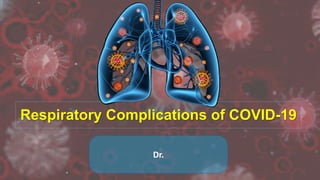
Respiratory Complications of COVID-19
Dr.
• At the end of 2019
• SARS-CoV-2: a positive-stranded RNA virus
• The virus was initially isolated from the Broncho alveolar lavage
• Viable virus is detected in aerosols for up to 3 hours, on plastic and
stainless steel surfaces up to 72 hours
Transmission
There are three modes of
transmission of SARS-CoV-2
which include:
1. Droplets transmission
2. Contact transmission
3. Aerosol transmission
 China-CDC reports the incubation
period to be 3-7 days.
 The mean incubation period was
reported to be 5.2 days, and
 The 95% percentile of the
distribution was up to 12.5 days.
What type of damage can coronavirus cause in
the lungs?
COVID-19, can cause lasting lung damage. Some people (5%) will have
complications, which may be life-threatening. It can cause-
 Pneumonia
 Acute Respiratory Failure
 Acute respiratory distress syndrome (ARDS)
 Sepsis can also cause lasting harm to the lungs
 Superinfection / Secondary infection
 Blood clots
Many of these complications may be caused by
Cytokine release syndrome or cytokine storm
 In the majority of the cases i.e.
80% will exhibit mild
symptoms
 14% will have pneumonia
 5% will suffer from septic
shock and organ failure
(mostly respiratory failure)
 2% cases it will be fatal.
 The pneumonia tends to take hold in both lungs.
Predominantly basal, peripheral and sub-
pleural region.
 Due to the novelty of the Covid 19 strain, there is no
specific treatment and mostly given supportive
care.
 A spike in pneumonia cases was the first sign of the new coronavirus in
China
 Air sacs become filled with fluid, pus, and cell debris, limiting their ability
to take in oxygen and causing shortness of breath, cough and other
symptoms
(a) in a 61-year-old man shows bilateral
patchy, somewhat nodular opacities in
the mid to lower lungs.
(b) in a 33-year-old woman, CT Images
show multiple ground glass opacities
in the periphery of the bilateral lungs. The
bilateral, peripheral patterns of
opacities without subpleural sparing
are common and characteristic CT
findings of the 2019 novel coronavirus
pneumonia.
(c) CT image of a 71-year-old male
shows consolidation in the peripheral
right upper lobe and a patchy area of
ground glass opacity with some
associated consolidation intra- and
interlobular septal thickening within the
left upper lobe.
Chest radiograph
(a) Chest CT in a 75-year-old male show
multiple patchy areas of pure ground glass
opacity (GGO) and GGO with reticular and/or
interlobular septal thickening.
(b) Chest CT image of a 38-year-old male
shows multiple patches, grid-like lobule, and
thickening of interlobular septa, typical “paving
stone-like” signs.
(c) An axial CT image obtained in 65-year-old
female shows bilateral ground glass and
consolidative opacities with a striking
peripheral distribution.
(d) CT image of a 65-year-old male shows
large consolidation in the right middle lobe,
patchy consolidation in the posterior and basal
segment of right lower lobe, with air
bronchogram inside.
Typical CT findings of COVID-19
CT manifestations of different stages of COVID-19
 One of the leading cause of death of COVID-19 is acute hypoxaemic
respiratory failure.
 Diffuse alveolar damage with interstitial thickening leading to compromised
gas exchange.
 The compensatory ventilatory response to hypoxaemia, increased minute
ventilation, may lead to extreme hypocapnia.
 Hypocapnic hypoxia is not usually accompanied by air hunger; instead, a
paradoxical feeling of calm and well-being, impassive, cooperative, and
hemodynamically stable.
 This phenomenon has been coined ‘Silent hypoxia’. Apart from a rapid
respiratory rate clinical presentation can be misleading.
 These patients are evaluated by GPs, often by telephone in pre-hospital
context.
 Pulse oximetry readings should be interpreted.
 In COVID-19 patients, a low end-tidal CO2 values (1.4–2.0 kPa) in COVID-
19 patients should alert the physician that respiratory failure is evolving.
 Recent guidance recommends a target oxygen saturation of 92-96% .
 Oxygen delivered through high flow nasal cannulas is beneficial which
provide up to 60 L/min of nearly 100% oxygen.
Acute Respiratory Distress Syndrome
(ARDS)
 As COVID-19 pneumonia progresses, more of the air sacs
become filled with fluid which can lead to ARDS.
 With ARDS, the body has trouble getting oxygen into the
bloodstream.
 Patients with ARDS may require ventilator support.
Effect of ARDS on lungs
 The virus works by damaging the wall and the lining of the
alveolus and capillaries.
 The debris from the damage, accumulates on the alveolus wall and
thickens the lining.
 More difficult to transfer oxygen to the red blood cells, which
causes difficulty in breathing.
 The lack of oxygen to the internal organs impairs the functioning of
the organs. Then the body fights to increase oxygen intake.
Severity
ARDS Severity PaO2/FiO2* Mortality**
Mild 200 – 300 27%
Moderate 100 – 200 32%
Severe < 100 45%
*on PEEP 5+; This value excludes hypoxemia caused by atelectasis.
Differences from ARDS caused by other factors
The reported onset of COVID-19-related ARDS was similar in different studies
 Huang et al. first reported 41 cases of COVID-19 in which the median time from
onset of symptoms to ARDS was 9.0 days (8.0–14.0).
 Subsequently, Wang et al. reported 138 cases of COVID-19 in which the median
time from the first symptom to ARDS was 8.0 days (6.0–12.0).
 Zhou et al. reported the median time from illness onset to ARDS was 12.0 days
(8.0–15.0).
 As the onset time of COVID-19-related ARDS was 8–12 days, it suggested that
the 1-week onset limit defined by ARDS Berlin criteria did not apply to COVID-19-
related ARDS.
 ARDS resulting in reduced lung compliance and severe hypoxemia.
 Lung compliance might be relatively normal in some COVID-19-related ARDS
patients. This was obviously inconsistent with ARDS caused by other factors.
 CT findings of COVID-19 showed consolidation and exudation, it was not a
“typical” ARDS image.
Differences from ARDS caused by other factors
Figure 1: Early and late-stage X-ray findings in patients with COVID-19
How do doctors’ ascertain the onset of ARDS
in Covid 19 infected individuals?
Key indicators to judge the onset and severity of
ARDS in infected individuals:
• Hypoxia – due to damage to the alveolus
• Breathing difficulties and shortness of breath
• Chest X-rays- exhibit an opaque and glassy look
against the black background
• Worsening symptoms over the course of time
Sepsis
 Another complication of a severe
case of COVID-19.
 It occurs when an infection reaches
and spreads through the
bloodstream, causing tissue damage.
 Sepsis, even when survived, can
leave a patient with lasting damage
to the lungs and other organs.
Superinfection / Secondary infection
A review of several studies found that secondary infection is a
possible but not common complication.
Strep and Staph are common culprits.
This can be serious enough to raise the risk of death.
Blood Clots
• Disseminated intravascular coagulation (DIC) causes Unusual
clots form, which can lead to internal bleeding or organ failure.
• Abnormally aggressive coagulation has been noted with COVID-
19, which is called COVID-19-associated coagulopathy (CAC).
• D-dimer level >1 μg/mL has been identified as a risk factor for poor
outcome.
• D-dimer over 2,660 µg/L had 100% sensitivity and 67%
specificity for PE on CT angiography
• Contrast-enhanced CT or CTPA should be performed to rule out
PE if supplementary oxygen is needed.
• prophylactic anticoagulation (enoxaparin 40 mg once daily in
ward patients, or twice daily in obese and ICU patients).
Blood Clots
COVID-19: Anticoagulation Recommended
Even After Discharge
 ISTH had recommended prophylactic-dose low molecular weight heparin
(LMWH) for all hospitalized COVID-19 patients, unless they have
contraindications such as active bleeding or low platelet count (<25×109/L).
 Up to 45 days of prophylaxis could be considered for low-bleeding-risk
patients but elevated VTE risk due to reduced mobility or comorbidities.
 Elevated D-dimer more than twice the upper normal limit was also
suggested as a high-risk group to get post-discharge extended prophylaxis
 UNC's algorithm calls for 30 days of direct oral anticoagulants (DOAC)
use after discharge with COVID-19.
Formation of blood clots in different organs raises the
chance of fatality
(a,b) Axial CT images (lung windows)
show peripheral ground-glass opacities
(arrow) associated with areas of
consolidation in dependent portions of
the lung (arrowheads). Interlobular
reticulations, bronchiectasis (black
arrow) and lung architectural distortion
are present. Involvement of the lung
volume was estimated to be between 25%
and 50%.
(c,d) Coronal CT reformations
(mediastinum windows) show bilateral
lobar and segmental pulmonary
embolism (black arrows).
Figure: Pulmonary CT angiography of a 68 year old male. The CT scan was obtained 10 days after the onset
of COVID-19 symptoms and on the day the patient was transferred to the intensive care unit.
Three Factors in Coronavirus Lung Damage
• Disease severity - Milder cases are less likely to
cause lasting scars in the lung tissue.
• Health conditions- The weaker immunity is
unable to stop the virus and aggravates the crisis.
Existing health problems- such as DM, COPD,
Heart disease and on immune-suppressing
medications can raise the risk for severe disease.
• Age- Older people are also more vulnerable for a
severe case of COVID-19.
Can coronavirus patients lessen the chance of lung damage?
People living with Diabetes, Heart disease and Respiratory issues
(COPD/Asthma) should be especially careful to manage those
conditions with monitoring and taking their medications as directed.
Proper nutrition and hydration
can also help patients avoid complications of COVID-19.
is COVID-19 lung damage reversible?
 After a serious case of COVID-19, recovery from lung damage
takes time.
 Over time, the tissue heals, but it can take three months to a year
or more for a person’s lung function to return to pre-COVID-19
levels.
 So, doctors and patients alike should be prepared for continuing
treatment and therapy.
Conclusion
 While a vaccine for Covid 19 is still under work and might take a while before it is
tested and certified for usage, our best bet right now is to stay safe, stay indoors
and avoid crowds.
 A few simple precautions like maintaining hygiene and sanitizing our
environment will go a long way in countering the spread of Covid 19 infection.
Thank You

More Related Content

What's hot

Covid-19 Diagnosis and Mx
Covid-19 Diagnosis and MxCovid-19 Diagnosis and Mx
Covid-19 Diagnosis and Mx
Mustafa Diaa
 
Corona Virus (SARS-Cov-2) and covid 19 disease
Corona Virus (SARS-Cov-2) and covid 19 diseaseCorona Virus (SARS-Cov-2) and covid 19 disease
Corona Virus (SARS-Cov-2) and covid 19 disease
OKELLO ISAAC OPIO
 
COVID-19
COVID-19 COVID-19
COVID-19
Bigyan Chhetri
 
Coronavirus
CoronavirusCoronavirus
Coronavirus
Sushil Patel
 
Possible Neurological Complications following COVID-19 Vaccines
Possible Neurological Complications following COVID-19 VaccinesPossible Neurological Complications following COVID-19 Vaccines
Possible Neurological Complications following COVID-19 Vaccines
Institute for Clinical Research (ICR)
 
Overview about Covid19 Pandemic
Overview about Covid19 Pandemic Overview about Covid19 Pandemic
Overview about Covid19 Pandemic
786
 
Hypoxemia for residents, 2 19-15
Hypoxemia for residents, 2 19-15Hypoxemia for residents, 2 19-15
Hypoxemia for residents, 2 19-15
katejohnpunag
 
Corona virus
Corona virusCorona virus
Corona virus
Xayneb Zia
 
Covid 19
Covid 19Covid 19
Neurological manifestations of sars cov 2
Neurological manifestations of sars cov 2Neurological manifestations of sars cov 2
Neurological manifestations of sars cov 2
Serge Eddy Mba
 
COVID-19
COVID-19COVID-19
COVID-19
Muhammad Zaid
 
Covid-19 :Cardiovascular considerations
Covid-19 :Cardiovascular considerationsCovid-19 :Cardiovascular considerations
Covid-19 :Cardiovascular considerations
Dr.Mahmoud Abbas
 
PEDIATRIC PULMONARY HYPERTENSION- AHA & ATS GUIDELINES
PEDIATRIC PULMONARY HYPERTENSION- AHA & ATS GUIDELINESPEDIATRIC PULMONARY HYPERTENSION- AHA & ATS GUIDELINES
PEDIATRIC PULMONARY HYPERTENSION- AHA & ATS GUIDELINES
Dr. Murtaza Kamal MD,DNB,DrNB Ped Cardiology
 
COVID-19 reviews- By Sabnam Khatun
COVID-19 reviews- By Sabnam KhatunCOVID-19 reviews- By Sabnam Khatun
COVID-19 reviews- By Sabnam Khatun
MD SHORAB ALAM
 
Infective endocarditis updated
Infective endocarditis updatedInfective endocarditis updated
Infective endocarditis updated
Tasneem Bashir • تسنيم بشير
 
Covid 19 rajni sharma
Covid 19 rajni sharmaCovid 19 rajni sharma
Covid 19 rajni sharma
DR.RAJNI SHARMA
 
HIV and respiratory infections
HIV and respiratory infectionsHIV and respiratory infections
HIV and respiratory infections
Arwa H. Al-Onayzan
 
08.07.20 | Neurologic Complications of COVID-19
08.07.20 | Neurologic Complications of COVID-1908.07.20 | Neurologic Complications of COVID-19
08.07.20 | Neurologic Complications of COVID-19
UC San Diego AntiViral Research Center
 

What's hot (20)

Covid-19 Diagnosis and Mx
Covid-19 Diagnosis and MxCovid-19 Diagnosis and Mx
Covid-19 Diagnosis and Mx
 
Corona Virus (SARS-Cov-2) and covid 19 disease
Corona Virus (SARS-Cov-2) and covid 19 diseaseCorona Virus (SARS-Cov-2) and covid 19 disease
Corona Virus (SARS-Cov-2) and covid 19 disease
 
COVID-19
COVID-19 COVID-19
COVID-19
 
Coronavirus
CoronavirusCoronavirus
Coronavirus
 
Possible Neurological Complications following COVID-19 Vaccines
Possible Neurological Complications following COVID-19 VaccinesPossible Neurological Complications following COVID-19 Vaccines
Possible Neurological Complications following COVID-19 Vaccines
 
Overview about Covid19 Pandemic
Overview about Covid19 Pandemic Overview about Covid19 Pandemic
Overview about Covid19 Pandemic
 
Hypoxemia for residents, 2 19-15
Hypoxemia for residents, 2 19-15Hypoxemia for residents, 2 19-15
Hypoxemia for residents, 2 19-15
 
Corona virus
Corona virusCorona virus
Corona virus
 
Tetralogy Of Fallot
Tetralogy Of FallotTetralogy Of Fallot
Tetralogy Of Fallot
 
Covid 19
Covid 19Covid 19
Covid 19
 
Neurological manifestations of sars cov 2
Neurological manifestations of sars cov 2Neurological manifestations of sars cov 2
Neurological manifestations of sars cov 2
 
COVID-19
COVID-19COVID-19
COVID-19
 
Covid-19 :Cardiovascular considerations
Covid-19 :Cardiovascular considerationsCovid-19 :Cardiovascular considerations
Covid-19 :Cardiovascular considerations
 
PEDIATRIC PULMONARY HYPERTENSION- AHA & ATS GUIDELINES
PEDIATRIC PULMONARY HYPERTENSION- AHA & ATS GUIDELINESPEDIATRIC PULMONARY HYPERTENSION- AHA & ATS GUIDELINES
PEDIATRIC PULMONARY HYPERTENSION- AHA & ATS GUIDELINES
 
COVID-19 reviews- By Sabnam Khatun
COVID-19 reviews- By Sabnam KhatunCOVID-19 reviews- By Sabnam Khatun
COVID-19 reviews- By Sabnam Khatun
 
Infective endocarditis updated
Infective endocarditis updatedInfective endocarditis updated
Infective endocarditis updated
 
Covid 19 rajni sharma
Covid 19 rajni sharmaCovid 19 rajni sharma
Covid 19 rajni sharma
 
HIV and respiratory infections
HIV and respiratory infectionsHIV and respiratory infections
HIV and respiratory infections
 
Ards
ArdsArds
Ards
 
08.07.20 | Neurologic Complications of COVID-19
08.07.20 | Neurologic Complications of COVID-1908.07.20 | Neurologic Complications of COVID-19
08.07.20 | Neurologic Complications of COVID-19
 

Similar to Respiratory Complications of COVID-19

The Pathology of Acute Lung Injury
The Pathology of Acute Lung InjuryThe Pathology of Acute Lung Injury
The Pathology of Acute Lung Injury
Dr. Anunoy Samanta
 
Radiological Presentation of COVID 19
Radiological Presentation of COVID 19Radiological Presentation of COVID 19
Radiological Presentation of COVID 19
Gamal Agmy
 
A-CASE-REPORT-OF-ARTERIAL-AND-VENOUS-THROMBOEMBOLIC-ILLNESS-IN-A-COVID-19-PAT...
A-CASE-REPORT-OF-ARTERIAL-AND-VENOUS-THROMBOEMBOLIC-ILLNESS-IN-A-COVID-19-PAT...A-CASE-REPORT-OF-ARTERIAL-AND-VENOUS-THROMBOEMBOLIC-ILLNESS-IN-A-COVID-19-PAT...
A-CASE-REPORT-OF-ARTERIAL-AND-VENOUS-THROMBOEMBOLIC-ILLNESS-IN-A-COVID-19-PAT...
Pubrica
 
Ild seminar ppt
Ild seminar pptIld seminar ppt
Ild seminar ppt
mikru taye
 
Overview of interstitial lung diseases
Overview of interstitial lung diseasesOverview of interstitial lung diseases
Overview of interstitial lung diseases
Beka Aberra
 
Imaging in covid 19
Imaging in covid 19Imaging in covid 19
Imaging in covid 19
LALIT KARKI
 
Covid 19
Covid 19Covid 19
PATHOLOGY OF COVID-19
PATHOLOGY OF COVID-19PATHOLOGY OF COVID-19
PATHOLOGY OF COVID-19
DrRashmiBudha
 
Management of co vid 19
Management of co vid 19Management of co vid 19
Management of co vid 19
Rajesh Mandal
 
Chronic obstructive pulmonary disease (copd)
Chronic obstructive pulmonary disease (copd)Chronic obstructive pulmonary disease (copd)
Chronic obstructive pulmonary disease (copd)
Rahil Dalal
 
Radiological manifestation covid19
Radiological manifestation covid19 Radiological manifestation covid19
Radiological manifestation covid19
DrVikas Balania
 
Pulmonary Sarcoidosis.pptx
Pulmonary Sarcoidosis.pptxPulmonary Sarcoidosis.pptx
Pulmonary Sarcoidosis.pptx
Umer Farooq
 
COVID 19
COVID 19  COVID 19
COVID 19
Gamal Agmy
 
Covid 19
Covid 19Covid 19
Covid 19
Utkarsh Sharma
 
Corona virus infection
Corona virus infectionCorona virus infection
Corona virus infectionGamal Agmy
 
Can Lung Ultrasound in Patients with Fever of Unknown Origin Detect Early Sig...
Can Lung Ultrasound in Patients with Fever of Unknown Origin Detect Early Sig...Can Lung Ultrasound in Patients with Fever of Unknown Origin Detect Early Sig...
Can Lung Ultrasound in Patients with Fever of Unknown Origin Detect Early Sig...
navasreni
 
Can Lung Ultrasound in Patients with Fever of Unknown Origin Detect Early Sig...
Can Lung Ultrasound in Patients with Fever of Unknown Origin Detect Early Sig...Can Lung Ultrasound in Patients with Fever of Unknown Origin Detect Early Sig...
Can Lung Ultrasound in Patients with Fever of Unknown Origin Detect Early Sig...
clinicsoncology
 
Can Lung Ultrasound in Patients with Fever of Unknown Origin Detect Early Sig...
Can Lung Ultrasound in Patients with Fever of Unknown Origin Detect Early Sig...Can Lung Ultrasound in Patients with Fever of Unknown Origin Detect Early Sig...
Can Lung Ultrasound in Patients with Fever of Unknown Origin Detect Early Sig...
pateldrona
 
Can Lung Ultrasound in Patients with Fever of Unknown Origin Detect Early Sig...
Can Lung Ultrasound in Patients with Fever of Unknown Origin Detect Early Sig...Can Lung Ultrasound in Patients with Fever of Unknown Origin Detect Early Sig...
Can Lung Ultrasound in Patients with Fever of Unknown Origin Detect Early Sig...
georgemarini
 
Can Lung Ultrasound in Patients with Fever of Unknown Origin Detect Early Sig...
Can Lung Ultrasound in Patients with Fever of Unknown Origin Detect Early Sig...Can Lung Ultrasound in Patients with Fever of Unknown Origin Detect Early Sig...
Can Lung Ultrasound in Patients with Fever of Unknown Origin Detect Early Sig...
SarkarRenon
 

Similar to Respiratory Complications of COVID-19 (20)

The Pathology of Acute Lung Injury
The Pathology of Acute Lung InjuryThe Pathology of Acute Lung Injury
The Pathology of Acute Lung Injury
 
Radiological Presentation of COVID 19
Radiological Presentation of COVID 19Radiological Presentation of COVID 19
Radiological Presentation of COVID 19
 
A-CASE-REPORT-OF-ARTERIAL-AND-VENOUS-THROMBOEMBOLIC-ILLNESS-IN-A-COVID-19-PAT...
A-CASE-REPORT-OF-ARTERIAL-AND-VENOUS-THROMBOEMBOLIC-ILLNESS-IN-A-COVID-19-PAT...A-CASE-REPORT-OF-ARTERIAL-AND-VENOUS-THROMBOEMBOLIC-ILLNESS-IN-A-COVID-19-PAT...
A-CASE-REPORT-OF-ARTERIAL-AND-VENOUS-THROMBOEMBOLIC-ILLNESS-IN-A-COVID-19-PAT...
 
Ild seminar ppt
Ild seminar pptIld seminar ppt
Ild seminar ppt
 
Overview of interstitial lung diseases
Overview of interstitial lung diseasesOverview of interstitial lung diseases
Overview of interstitial lung diseases
 
Imaging in covid 19
Imaging in covid 19Imaging in covid 19
Imaging in covid 19
 
Covid 19
Covid 19Covid 19
Covid 19
 
PATHOLOGY OF COVID-19
PATHOLOGY OF COVID-19PATHOLOGY OF COVID-19
PATHOLOGY OF COVID-19
 
Management of co vid 19
Management of co vid 19Management of co vid 19
Management of co vid 19
 
Chronic obstructive pulmonary disease (copd)
Chronic obstructive pulmonary disease (copd)Chronic obstructive pulmonary disease (copd)
Chronic obstructive pulmonary disease (copd)
 
Radiological manifestation covid19
Radiological manifestation covid19 Radiological manifestation covid19
Radiological manifestation covid19
 
Pulmonary Sarcoidosis.pptx
Pulmonary Sarcoidosis.pptxPulmonary Sarcoidosis.pptx
Pulmonary Sarcoidosis.pptx
 
COVID 19
COVID 19  COVID 19
COVID 19
 
Covid 19
Covid 19Covid 19
Covid 19
 
Corona virus infection
Corona virus infectionCorona virus infection
Corona virus infection
 
Can Lung Ultrasound in Patients with Fever of Unknown Origin Detect Early Sig...
Can Lung Ultrasound in Patients with Fever of Unknown Origin Detect Early Sig...Can Lung Ultrasound in Patients with Fever of Unknown Origin Detect Early Sig...
Can Lung Ultrasound in Patients with Fever of Unknown Origin Detect Early Sig...
 
Can Lung Ultrasound in Patients with Fever of Unknown Origin Detect Early Sig...
Can Lung Ultrasound in Patients with Fever of Unknown Origin Detect Early Sig...Can Lung Ultrasound in Patients with Fever of Unknown Origin Detect Early Sig...
Can Lung Ultrasound in Patients with Fever of Unknown Origin Detect Early Sig...
 

Recently uploaded

NVBDCP.pptx Nation vector borne disease control program
NVBDCP.pptx Nation vector borne disease control programNVBDCP.pptx Nation vector borne disease control program
NVBDCP.pptx Nation vector borne disease control program
Sapna Thakur
 
TEST BANK for Operations Management, 14th Edition by William J. Stevenson, Ve...
TEST BANK for Operations Management, 14th Edition by William J. Stevenson, Ve...TEST BANK for Operations Management, 14th Edition by William J. Stevenson, Ve...
TEST BANK for Operations Management, 14th Edition by William J. Stevenson, Ve...
kevinkariuki227
 
POST OPERATIVE OLIGURIA and its management
POST OPERATIVE OLIGURIA and its managementPOST OPERATIVE OLIGURIA and its management
POST OPERATIVE OLIGURIA and its management
touseefaziz1
 
Flu Vaccine Alert in Bangalore Karnataka
Flu Vaccine Alert in Bangalore KarnatakaFlu Vaccine Alert in Bangalore Karnataka
Flu Vaccine Alert in Bangalore Karnataka
addon Scans
 
micro teaching on communication m.sc nursing.pdf
micro teaching on communication m.sc nursing.pdfmicro teaching on communication m.sc nursing.pdf
micro teaching on communication m.sc nursing.pdf
Anurag Sharma
 
Report Back from SGO 2024: What’s the Latest in Cervical Cancer?
Report Back from SGO 2024: What’s the Latest in Cervical Cancer?Report Back from SGO 2024: What’s the Latest in Cervical Cancer?
Report Back from SGO 2024: What’s the Latest in Cervical Cancer?
bkling
 
Ozempic: Preoperative Management of Patients on GLP-1 Receptor Agonists
Ozempic: Preoperative Management of Patients on GLP-1 Receptor Agonists  Ozempic: Preoperative Management of Patients on GLP-1 Receptor Agonists
Ozempic: Preoperative Management of Patients on GLP-1 Receptor Agonists
Saeid Safari
 
Superficial & Deep Fascia of the NECK.pptx
Superficial & Deep Fascia of the NECK.pptxSuperficial & Deep Fascia of the NECK.pptx
Superficial & Deep Fascia of the NECK.pptx
Dr. Rabia Inam Gandapore
 
Cervical & Brachial Plexus By Dr. RIG.pptx
Cervical & Brachial Plexus By Dr. RIG.pptxCervical & Brachial Plexus By Dr. RIG.pptx
Cervical & Brachial Plexus By Dr. RIG.pptx
Dr. Rabia Inam Gandapore
 
For Better Surat #ℂall #Girl Service ❤85270-49040❤ Surat #ℂall #Girls
For Better Surat #ℂall #Girl Service ❤85270-49040❤ Surat #ℂall #GirlsFor Better Surat #ℂall #Girl Service ❤85270-49040❤ Surat #ℂall #Girls
For Better Surat #ℂall #Girl Service ❤85270-49040❤ Surat #ℂall #Girls
Savita Shen $i11
 
24 Upakrama.pptx class ppt useful in all
24 Upakrama.pptx class ppt useful in all24 Upakrama.pptx class ppt useful in all
24 Upakrama.pptx class ppt useful in all
DrSathishMS1
 
New Directions in Targeted Therapeutic Approaches for Older Adults With Mantl...
New Directions in Targeted Therapeutic Approaches for Older Adults With Mantl...New Directions in Targeted Therapeutic Approaches for Older Adults With Mantl...
New Directions in Targeted Therapeutic Approaches for Older Adults With Mantl...
i3 Health
 
Evaluation of antidepressant activity of clitoris ternatea in animals
Evaluation of antidepressant activity of clitoris ternatea in animalsEvaluation of antidepressant activity of clitoris ternatea in animals
Evaluation of antidepressant activity of clitoris ternatea in animals
Shweta
 
Surat @ℂall @Girls ꧁❤8527049040❤꧂@ℂall @Girls Service Vip Top Model Safe
Surat @ℂall @Girls ꧁❤8527049040❤꧂@ℂall @Girls Service Vip Top Model SafeSurat @ℂall @Girls ꧁❤8527049040❤꧂@ℂall @Girls Service Vip Top Model Safe
Surat @ℂall @Girls ꧁❤8527049040❤꧂@ℂall @Girls Service Vip Top Model Safe
Savita Shen $i11
 
heat stroke and heat exhaustion in children
heat stroke and heat exhaustion in childrenheat stroke and heat exhaustion in children
heat stroke and heat exhaustion in children
SumeraAhmad5
 
Alcohol_Dr. Jeenal Mistry MD Pharmacology.pdf
Alcohol_Dr. Jeenal Mistry MD Pharmacology.pdfAlcohol_Dr. Jeenal Mistry MD Pharmacology.pdf
Alcohol_Dr. Jeenal Mistry MD Pharmacology.pdf
Dr Jeenal Mistry
 
Triangles of Neck and Clinical Correlation by Dr. RIG.pptx
Triangles of Neck and Clinical Correlation by Dr. RIG.pptxTriangles of Neck and Clinical Correlation by Dr. RIG.pptx
Triangles of Neck and Clinical Correlation by Dr. RIG.pptx
Dr. Rabia Inam Gandapore
 
Ocular injury ppt Upendra pal optometrist upums saifai etawah
Ocular injury  ppt  Upendra pal  optometrist upums saifai etawahOcular injury  ppt  Upendra pal  optometrist upums saifai etawah
Ocular injury ppt Upendra pal optometrist upums saifai etawah
pal078100
 
Physiology of Special Chemical Sensation of Taste
Physiology of Special Chemical Sensation of TastePhysiology of Special Chemical Sensation of Taste
Physiology of Special Chemical Sensation of Taste
MedicoseAcademics
 
Hemodialysis: Chapter 3, Dialysis Water Unit - Dr.Gawad
Hemodialysis: Chapter 3, Dialysis Water Unit - Dr.GawadHemodialysis: Chapter 3, Dialysis Water Unit - Dr.Gawad
Hemodialysis: Chapter 3, Dialysis Water Unit - Dr.Gawad
NephroTube - Dr.Gawad
 

Recently uploaded (20)

NVBDCP.pptx Nation vector borne disease control program
NVBDCP.pptx Nation vector borne disease control programNVBDCP.pptx Nation vector borne disease control program
NVBDCP.pptx Nation vector borne disease control program
 
TEST BANK for Operations Management, 14th Edition by William J. Stevenson, Ve...
TEST BANK for Operations Management, 14th Edition by William J. Stevenson, Ve...TEST BANK for Operations Management, 14th Edition by William J. Stevenson, Ve...
TEST BANK for Operations Management, 14th Edition by William J. Stevenson, Ve...
 
POST OPERATIVE OLIGURIA and its management
POST OPERATIVE OLIGURIA and its managementPOST OPERATIVE OLIGURIA and its management
POST OPERATIVE OLIGURIA and its management
 
Flu Vaccine Alert in Bangalore Karnataka
Flu Vaccine Alert in Bangalore KarnatakaFlu Vaccine Alert in Bangalore Karnataka
Flu Vaccine Alert in Bangalore Karnataka
 
micro teaching on communication m.sc nursing.pdf
micro teaching on communication m.sc nursing.pdfmicro teaching on communication m.sc nursing.pdf
micro teaching on communication m.sc nursing.pdf
 
Report Back from SGO 2024: What’s the Latest in Cervical Cancer?
Report Back from SGO 2024: What’s the Latest in Cervical Cancer?Report Back from SGO 2024: What’s the Latest in Cervical Cancer?
Report Back from SGO 2024: What’s the Latest in Cervical Cancer?
 
Ozempic: Preoperative Management of Patients on GLP-1 Receptor Agonists
Ozempic: Preoperative Management of Patients on GLP-1 Receptor Agonists  Ozempic: Preoperative Management of Patients on GLP-1 Receptor Agonists
Ozempic: Preoperative Management of Patients on GLP-1 Receptor Agonists
 
Superficial & Deep Fascia of the NECK.pptx
Superficial & Deep Fascia of the NECK.pptxSuperficial & Deep Fascia of the NECK.pptx
Superficial & Deep Fascia of the NECK.pptx
 
Cervical & Brachial Plexus By Dr. RIG.pptx
Cervical & Brachial Plexus By Dr. RIG.pptxCervical & Brachial Plexus By Dr. RIG.pptx
Cervical & Brachial Plexus By Dr. RIG.pptx
 
For Better Surat #ℂall #Girl Service ❤85270-49040❤ Surat #ℂall #Girls
For Better Surat #ℂall #Girl Service ❤85270-49040❤ Surat #ℂall #GirlsFor Better Surat #ℂall #Girl Service ❤85270-49040❤ Surat #ℂall #Girls
For Better Surat #ℂall #Girl Service ❤85270-49040❤ Surat #ℂall #Girls
 
24 Upakrama.pptx class ppt useful in all
24 Upakrama.pptx class ppt useful in all24 Upakrama.pptx class ppt useful in all
24 Upakrama.pptx class ppt useful in all
 
New Directions in Targeted Therapeutic Approaches for Older Adults With Mantl...
New Directions in Targeted Therapeutic Approaches for Older Adults With Mantl...New Directions in Targeted Therapeutic Approaches for Older Adults With Mantl...
New Directions in Targeted Therapeutic Approaches for Older Adults With Mantl...
 
Evaluation of antidepressant activity of clitoris ternatea in animals
Evaluation of antidepressant activity of clitoris ternatea in animalsEvaluation of antidepressant activity of clitoris ternatea in animals
Evaluation of antidepressant activity of clitoris ternatea in animals
 
Surat @ℂall @Girls ꧁❤8527049040❤꧂@ℂall @Girls Service Vip Top Model Safe
Surat @ℂall @Girls ꧁❤8527049040❤꧂@ℂall @Girls Service Vip Top Model SafeSurat @ℂall @Girls ꧁❤8527049040❤꧂@ℂall @Girls Service Vip Top Model Safe
Surat @ℂall @Girls ꧁❤8527049040❤꧂@ℂall @Girls Service Vip Top Model Safe
 
heat stroke and heat exhaustion in children
heat stroke and heat exhaustion in childrenheat stroke and heat exhaustion in children
heat stroke and heat exhaustion in children
 
Alcohol_Dr. Jeenal Mistry MD Pharmacology.pdf
Alcohol_Dr. Jeenal Mistry MD Pharmacology.pdfAlcohol_Dr. Jeenal Mistry MD Pharmacology.pdf
Alcohol_Dr. Jeenal Mistry MD Pharmacology.pdf
 
Triangles of Neck and Clinical Correlation by Dr. RIG.pptx
Triangles of Neck and Clinical Correlation by Dr. RIG.pptxTriangles of Neck and Clinical Correlation by Dr. RIG.pptx
Triangles of Neck and Clinical Correlation by Dr. RIG.pptx
 
Ocular injury ppt Upendra pal optometrist upums saifai etawah
Ocular injury  ppt  Upendra pal  optometrist upums saifai etawahOcular injury  ppt  Upendra pal  optometrist upums saifai etawah
Ocular injury ppt Upendra pal optometrist upums saifai etawah
 
Physiology of Special Chemical Sensation of Taste
Physiology of Special Chemical Sensation of TastePhysiology of Special Chemical Sensation of Taste
Physiology of Special Chemical Sensation of Taste
 
Hemodialysis: Chapter 3, Dialysis Water Unit - Dr.Gawad
Hemodialysis: Chapter 3, Dialysis Water Unit - Dr.GawadHemodialysis: Chapter 3, Dialysis Water Unit - Dr.Gawad
Hemodialysis: Chapter 3, Dialysis Water Unit - Dr.Gawad
 

Respiratory Complications of COVID-19

  • 2. • At the end of 2019 • SARS-CoV-2: a positive-stranded RNA virus • The virus was initially isolated from the Broncho alveolar lavage • Viable virus is detected in aerosols for up to 3 hours, on plastic and stainless steel surfaces up to 72 hours
  • 3. Transmission There are three modes of transmission of SARS-CoV-2 which include: 1. Droplets transmission 2. Contact transmission 3. Aerosol transmission
  • 4.  China-CDC reports the incubation period to be 3-7 days.  The mean incubation period was reported to be 5.2 days, and  The 95% percentile of the distribution was up to 12.5 days.
  • 5. What type of damage can coronavirus cause in the lungs? COVID-19, can cause lasting lung damage. Some people (5%) will have complications, which may be life-threatening. It can cause-  Pneumonia  Acute Respiratory Failure  Acute respiratory distress syndrome (ARDS)  Sepsis can also cause lasting harm to the lungs  Superinfection / Secondary infection  Blood clots Many of these complications may be caused by Cytokine release syndrome or cytokine storm
  • 6.  In the majority of the cases i.e. 80% will exhibit mild symptoms  14% will have pneumonia  5% will suffer from septic shock and organ failure (mostly respiratory failure)  2% cases it will be fatal.
  • 7.  The pneumonia tends to take hold in both lungs. Predominantly basal, peripheral and sub- pleural region.  Due to the novelty of the Covid 19 strain, there is no specific treatment and mostly given supportive care.  A spike in pneumonia cases was the first sign of the new coronavirus in China  Air sacs become filled with fluid, pus, and cell debris, limiting their ability to take in oxygen and causing shortness of breath, cough and other symptoms
  • 8. (a) in a 61-year-old man shows bilateral patchy, somewhat nodular opacities in the mid to lower lungs. (b) in a 33-year-old woman, CT Images show multiple ground glass opacities in the periphery of the bilateral lungs. The bilateral, peripheral patterns of opacities without subpleural sparing are common and characteristic CT findings of the 2019 novel coronavirus pneumonia. (c) CT image of a 71-year-old male shows consolidation in the peripheral right upper lobe and a patchy area of ground glass opacity with some associated consolidation intra- and interlobular septal thickening within the left upper lobe. Chest radiograph
  • 9. (a) Chest CT in a 75-year-old male show multiple patchy areas of pure ground glass opacity (GGO) and GGO with reticular and/or interlobular septal thickening. (b) Chest CT image of a 38-year-old male shows multiple patches, grid-like lobule, and thickening of interlobular septa, typical “paving stone-like” signs. (c) An axial CT image obtained in 65-year-old female shows bilateral ground glass and consolidative opacities with a striking peripheral distribution. (d) CT image of a 65-year-old male shows large consolidation in the right middle lobe, patchy consolidation in the posterior and basal segment of right lower lobe, with air bronchogram inside. Typical CT findings of COVID-19
  • 10. CT manifestations of different stages of COVID-19
  • 11.  One of the leading cause of death of COVID-19 is acute hypoxaemic respiratory failure.  Diffuse alveolar damage with interstitial thickening leading to compromised gas exchange.  The compensatory ventilatory response to hypoxaemia, increased minute ventilation, may lead to extreme hypocapnia.  Hypocapnic hypoxia is not usually accompanied by air hunger; instead, a paradoxical feeling of calm and well-being, impassive, cooperative, and hemodynamically stable.  This phenomenon has been coined ‘Silent hypoxia’. Apart from a rapid respiratory rate clinical presentation can be misleading.
  • 12.  These patients are evaluated by GPs, often by telephone in pre-hospital context.  Pulse oximetry readings should be interpreted.  In COVID-19 patients, a low end-tidal CO2 values (1.4–2.0 kPa) in COVID- 19 patients should alert the physician that respiratory failure is evolving.  Recent guidance recommends a target oxygen saturation of 92-96% .  Oxygen delivered through high flow nasal cannulas is beneficial which provide up to 60 L/min of nearly 100% oxygen.
  • 13. Acute Respiratory Distress Syndrome (ARDS)  As COVID-19 pneumonia progresses, more of the air sacs become filled with fluid which can lead to ARDS.  With ARDS, the body has trouble getting oxygen into the bloodstream.  Patients with ARDS may require ventilator support.
  • 14. Effect of ARDS on lungs  The virus works by damaging the wall and the lining of the alveolus and capillaries.  The debris from the damage, accumulates on the alveolus wall and thickens the lining.  More difficult to transfer oxygen to the red blood cells, which causes difficulty in breathing.  The lack of oxygen to the internal organs impairs the functioning of the organs. Then the body fights to increase oxygen intake.
  • 15. Severity ARDS Severity PaO2/FiO2* Mortality** Mild 200 – 300 27% Moderate 100 – 200 32% Severe < 100 45% *on PEEP 5+; This value excludes hypoxemia caused by atelectasis.
  • 16. Differences from ARDS caused by other factors The reported onset of COVID-19-related ARDS was similar in different studies  Huang et al. first reported 41 cases of COVID-19 in which the median time from onset of symptoms to ARDS was 9.0 days (8.0–14.0).  Subsequently, Wang et al. reported 138 cases of COVID-19 in which the median time from the first symptom to ARDS was 8.0 days (6.0–12.0).  Zhou et al. reported the median time from illness onset to ARDS was 12.0 days (8.0–15.0).  As the onset time of COVID-19-related ARDS was 8–12 days, it suggested that the 1-week onset limit defined by ARDS Berlin criteria did not apply to COVID-19- related ARDS.
  • 17.  ARDS resulting in reduced lung compliance and severe hypoxemia.  Lung compliance might be relatively normal in some COVID-19-related ARDS patients. This was obviously inconsistent with ARDS caused by other factors.  CT findings of COVID-19 showed consolidation and exudation, it was not a “typical” ARDS image. Differences from ARDS caused by other factors
  • 18. Figure 1: Early and late-stage X-ray findings in patients with COVID-19
  • 19. How do doctors’ ascertain the onset of ARDS in Covid 19 infected individuals? Key indicators to judge the onset and severity of ARDS in infected individuals: • Hypoxia – due to damage to the alveolus • Breathing difficulties and shortness of breath • Chest X-rays- exhibit an opaque and glassy look against the black background • Worsening symptoms over the course of time
  • 20. Sepsis  Another complication of a severe case of COVID-19.  It occurs when an infection reaches and spreads through the bloodstream, causing tissue damage.  Sepsis, even when survived, can leave a patient with lasting damage to the lungs and other organs.
  • 21. Superinfection / Secondary infection A review of several studies found that secondary infection is a possible but not common complication. Strep and Staph are common culprits. This can be serious enough to raise the risk of death.
  • 22. Blood Clots • Disseminated intravascular coagulation (DIC) causes Unusual clots form, which can lead to internal bleeding or organ failure. • Abnormally aggressive coagulation has been noted with COVID- 19, which is called COVID-19-associated coagulopathy (CAC). • D-dimer level >1 μg/mL has been identified as a risk factor for poor outcome.
  • 23. • D-dimer over 2,660 µg/L had 100% sensitivity and 67% specificity for PE on CT angiography • Contrast-enhanced CT or CTPA should be performed to rule out PE if supplementary oxygen is needed. • prophylactic anticoagulation (enoxaparin 40 mg once daily in ward patients, or twice daily in obese and ICU patients). Blood Clots
  • 24. COVID-19: Anticoagulation Recommended Even After Discharge  ISTH had recommended prophylactic-dose low molecular weight heparin (LMWH) for all hospitalized COVID-19 patients, unless they have contraindications such as active bleeding or low platelet count (<25×109/L).  Up to 45 days of prophylaxis could be considered for low-bleeding-risk patients but elevated VTE risk due to reduced mobility or comorbidities.  Elevated D-dimer more than twice the upper normal limit was also suggested as a high-risk group to get post-discharge extended prophylaxis  UNC's algorithm calls for 30 days of direct oral anticoagulants (DOAC) use after discharge with COVID-19.
  • 25. Formation of blood clots in different organs raises the chance of fatality
  • 26. (a,b) Axial CT images (lung windows) show peripheral ground-glass opacities (arrow) associated with areas of consolidation in dependent portions of the lung (arrowheads). Interlobular reticulations, bronchiectasis (black arrow) and lung architectural distortion are present. Involvement of the lung volume was estimated to be between 25% and 50%. (c,d) Coronal CT reformations (mediastinum windows) show bilateral lobar and segmental pulmonary embolism (black arrows). Figure: Pulmonary CT angiography of a 68 year old male. The CT scan was obtained 10 days after the onset of COVID-19 symptoms and on the day the patient was transferred to the intensive care unit.
  • 27. Three Factors in Coronavirus Lung Damage • Disease severity - Milder cases are less likely to cause lasting scars in the lung tissue. • Health conditions- The weaker immunity is unable to stop the virus and aggravates the crisis. Existing health problems- such as DM, COPD, Heart disease and on immune-suppressing medications can raise the risk for severe disease. • Age- Older people are also more vulnerable for a severe case of COVID-19.
  • 28. Can coronavirus patients lessen the chance of lung damage? People living with Diabetes, Heart disease and Respiratory issues (COPD/Asthma) should be especially careful to manage those conditions with monitoring and taking their medications as directed. Proper nutrition and hydration can also help patients avoid complications of COVID-19.
  • 29. is COVID-19 lung damage reversible?  After a serious case of COVID-19, recovery from lung damage takes time.  Over time, the tissue heals, but it can take three months to a year or more for a person’s lung function to return to pre-COVID-19 levels.  So, doctors and patients alike should be prepared for continuing treatment and therapy.
  • 30. Conclusion  While a vaccine for Covid 19 is still under work and might take a while before it is tested and certified for usage, our best bet right now is to stay safe, stay indoors and avoid crowds.  A few simple precautions like maintaining hygiene and sanitizing our environment will go a long way in countering the spread of Covid 19 infection.
  • 31.