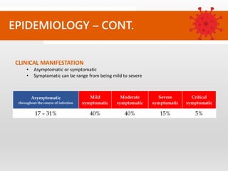
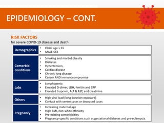
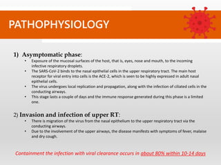
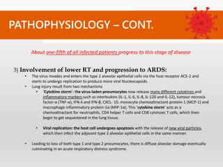
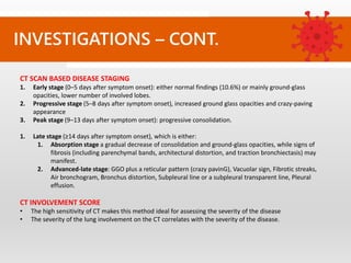
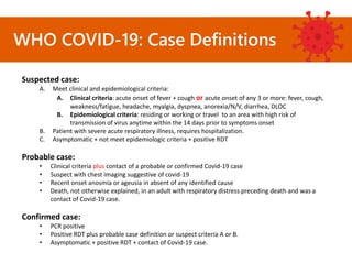
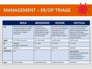
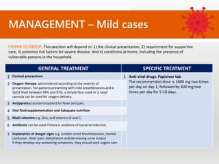
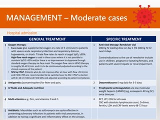
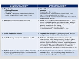
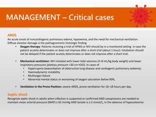
COVID – 19
CLINICAL DIAGNOSIS AND
MANAGEMENT
Dr. Mustafa Diaa Shayyal
MICROBIOLOGY
Coronavirus disease 2019 (COVID-19)
• An infectious disease caused by the severe acute respiratory syndrome
coronavirus 2 (SARSCoV-2)
• A newly emergent coronavirus, that was first recognized in Wuhan.
• SARS-CoV-2 is a positive-sense single-stranded RNA virus that is contagious
in humans.
EPIDEMIOLOGY
Transmission
• Respiratory droplets - which occurs when:
• A person is in close contact with someone who is actively coughing or sneezing
• Direct contact with infected persons
• Contact with contaminated objects and surfaces,
• Aerosols i.e. in enclosed spaces indoors, crowded and inadequately ventilated spaces.
• Fecal-oral route is possible
Incubation “presymptomatic” period
• Mean 5–7 days
• Range 2 – 14 days
Infectious period
• Presymptomatic period: 1–3 days before symptom onset
• Symptomatic period: infectious virus shedding up to 8 days from the onset of any
symptoms, and highest within the first 3 days from onset of symptoms.
EPIDEMIOLOGY – CONT.
CLINICAL MANIFESTATION
• Asymptomatic or symptomatic
• Symptomatic can be range from being mild to severe
Asymptomatic
throughout the course of infection
Mild
symptomatic
Moderate
symptomatic
Severe
symptomatic
Critical
symptomatic
17 – 31% 40% 40% 15% 5%
EPIDEMIOLOGY – CONT.
RISK FACTORS
for severe COVID-19 disease and death
Demographics
• Older age > 65
• MALE SEX
Comorbid
conditions
• Smoking and morbid obesity
• Diabetes
• Hypertension,
• Cardiac disease
• Chronic lung disease
• Cancer AND immunocompromise
Labs
• Lymphopenia
• Elevated D-dimer, LDH, ferritin and CRP
• Elevated troponin, ALT & AST, and creatinine
Others
• High viral load (long duration exposure)
• Contact with severe cases or deceased cases
Pregnancy
• Increasing maternal age
• High BMI, non-white ethnicity
• Pre-existing comorbidities
• Pregnancy-specific conditions such as gestational diabetes and pre-eclampsia.
PATHOPHYSIOLOGY
1) Asymptomatic phase:
• Exposure of the mucosal surfaces of the host, that is, eyes, nose and mouth, to the incoming
infective respiratory droplets.
• The SARS-CoV-2 binds to the nasal epithelial cells in the upper respiratory tract. The main host
receptor for viral entry into cells is the ACE-2, which is seen to be highly expressed in adult nasal
epithelial cells.
• The virus undergoes local replication and propagation, along with the infection of ciliated cells in the
conducting airways.
• This stage lasts a couple of days and the immune response generated during this phase is a limited
one.
2) Invasion and infection of upper RT:
• There is migration of the virus from the nasal epithelium to the upper respiratory tract via the
conducting airways.
• Due to the involvement of the upper airways, the disease manifests with symptoms of fever, malaise
and dry cough.
Containment the infection with viral clearance occurs in about 80% within 10-14 days
PATHOPHYSIOLOGY – CONT.
3) Involvement of lower RT and progression to ARDS:
• The virus invades and enters the type 2 alveolar epithelial cells via the host receptor ACE-2 and
starts to undergo replication to produce more viral Nucleocapsids.
• Lung injury result from two mechanisms
• ‘Cytokine storm’: the virus-laden pneumocytes now release many different cytokines and
inflammatory markers such as interleukins (IL-1, IL-6, IL-8, IL-120 and IL-12), tumour necrosis
factor-α (TNF-α), IFN-λ and IFN-β, CXCL- 10, monocyte chemoattractant protein-1 (MCP-1) and
macrophage inflammatory protein-1α (MIP-1α). This ‘cytokine storm’ acts as a
chemoattractant for neutrophils, CD4 helper T cells and CD8 cytotoxic T cells, which then
begin to get sequestered in the lung tissue.
• Viral replication: the host cell undergoes apoptosis with the release of new viral particles,
which then infect the adjacent type 2 alveolar epithelial cells in the same manner.
• Leading to loss of both type 1 and type 2 pneumocytes, there is diffuse alveolar damage eventually
culminating in an acute respiratory distress syndrome.
About one-fifth of all infected patients progress to this stage of disease
(1) ASYMPTOMATIC
PHASE
(2) UPPER
RESPIRATORY TRACT
INFECTION
(5) ARDS
(3) LOWER
RESPIRATORY TRACT
INFECTION
ASYMPTOMATIC
No symptoms.
Positive nasal swab test and Normal chest X-ray and CT scan
MILD
The most common:
• Fever (88%), Dry cough (59–82%), fatigue (44–70%).
The common:
• anorexia (38.8%), chest tightness (35.7%), shortness of breath (35%), and muscle soreness
(33%). Loss of smell (anosmia) (41.0%) and loss of taste (ageusia) (38.2%)
The less common:
• Headache (15.4%), pharyngalgia (13.1%), diarrhea (12.9%), shivering (10.9%), nausea and
vomiting (10.2%), and abdominal pain (4.4%)
MODERATE
Symptoms of pneumonia (persistent fever > 37.8 C and dry cough)
Without hypoxemia.
Crackles and wheezing in chest auscultation.
SEVERE
Pneumonia with dyspnea and hypoxemia (SpO2 < 92%)
Other symptoms diarrhea, vomiting and nausea.
CRITICAL
Severe difficulty of breathing and shortness of breath, chest pain
Plus symptoms of complications of acute respiratory distress syndrome, shock, coagulation
defects, encephalopathy, heart failure and acute kidney injury
CLINICAL PRESENTATION
CLINICAL PRESENTATION – CONT.
Body systems and organs affected
• Mental and Neurological manifestations:
• Anxiety, depression, delirium/encephalopathy, agitation
• Stroke, hypoxic ischaemic brain injury, seizures, coma, meningo-encephalitis
• Guillain-Barré syndrome.
• Cardiovascular manifestations:
• Acute myocardial injury (8%–12%),
• Acute pulmonary embolism
• Brady- or tachyarrhythmias (16.7%)
• Acute pericarditis, heart failure, cardiogenic shock, or myocarditis
• Digestive symptoms:
• Nausea and/or vomiting, diarrhea, anorexia, or loss of appetite.
• Rarer symptoms include abdominal pain, abdominal distension, tenesmus, dysgeusia,
gastrointestinal bleeding, or hematochezia.
• Skin manifestations:
• Maculopapular exanthem, papulovesicular rash,
• Urticaria, livedo reticularis lesions, or petechiae.
• Blood:
• Normal or decreased white blood cell counts,
• Lymphopenia, or thrombocytopenia.
Differential diagnosis
1. MERS, SARS: travel history, epidemiological data and PCR.
2. Community acquired pneumonia: blood or sputum culture.
3. Influenza infection: travel history, epidemiological data and PCR. More sore throat
than covid-19.
4. Common cold: more coryza and sore throat, and PCR
5. Pulmonary TB: History of symptoms is usually longer.. Presence of night sweats
and weight loss, Chest x-ray: fibronodular opacities in upper lobes with or without
cavitation; Sputum acid-fast bacillismear and sputum culture: positive.
LABORATORY TESTS
Confirmatory tests
1) RT-PCR:
• Highly specific (~95%) and low sensitivity (~71%).
• Negative test results do not preclude SARS-CoV-2 infection, and shall be interpreted in light of the clinical
picture and epidemiologic information.
• Accuracy of viral RNA swabs depending
• The site and quality of sampling. Best: broncho-alveolar lavage > sputum > nasal swabs > throat
swabs.
• The Stage of disease and degree of viral multiplication or clearance: Positivity usually after 2–8 days
of infection.
2) Serology testing for SARS-COV2
• Detect IgM and IgG antibodies against SARS-CoV-2 in serum, plasma and whole blood
• Assess prior exposure to virus and cannot be used in the diagnosis of current infection.
• Useful
• When the viral test is not available.
• Serology test + the clinical picture could guide in decision making.
• In some patients, virus shedding is reduced, making RT-qPCR results falsely negative.
3) Rapid antigen testing:
• Monoclonal antibodies are specifically directed against the SARS-CoV-2 nucleocapsid protein,
• Sensitivity of 84.1% and a specificity of 98.5%.
INVESTIGATIONS
INVESTIGATIONS – CONT.
LABORATORY TESTS
Severity predictor tests
Test Result Severe COVID-19 pneumonia
CBC N/decreased WBC counts,
lymphopenia or
Thrombocytopnia
Leukocytosis
Neutrophil count (>7 × 10⁹/L), Lymphocyte
count (<0.4 × 10⁹/L)
Inflammatory biomarkers
• LDH
• CRP and ESR
• D-dimer
• Serum ferritin
• Procalcitonin
• IL-6
CRP (>150 mg/L)
D-dimer levels (> 1112 ng/L)
Cytokine storm
• S. ferritin >300ug/L with doubling within 24
hours or > 600ug/L at presentation
• LDH > 250
• Serum IL-6 more than or equal to three
times upper normal limit
Serum troponin increased in myocardial injury or acute myocardial syndrome
Bleeding profile Increased PT/INR
High fibrin/fibrinogen degradation products
LFT, RFT and RBS increased in acute liver injury and acute renal injury
increased as result of stress, or treatment eg. Steroid
INVESTIGATIONS – CONT.
Images
1) CHEST X-RAY:
• Useless in the early stages of the disease.
• Useful in the follow-up of the disease e.g. ARDS
2) CHEST CT SCAN: CT has a higher sensitivity but lower specificity
and can play a role in the diagnosis and treatment of the
disease. The most commonly seen features are:
• Ground-Glass’ Opacity (GGO): Most common finding in
early stage. Characterized by Multifocal, bilateral and
peripheral, unifocal (in early phase), and most common in
inferior lobes.
• Crazy paving: thickened interlobular and intralobular lines
in combination with a ground glass pattern. Usually seen in
later stage.
• Traction bronchiectasis
• Vascular dilatation
CT SCAN BASED DISEASE STAGING
1. Early stage (0–5 days after symptom onset): either normal findings (10.6%) or mainly ground-glass
opacities, lower number of involved lobes.
2. Progressive stage (5–8 days after symptom onset), increased ground glass opacities and crazy-paving
appearance
3. Peak stage (9–13 days after symptom onset): progressive consolidation.
1. Late stage (≥14 days after symptom onset), which is either:
1. Absorption stage a gradual decrease of consolidation and ground-glass opacities, while signs of
fibrosis (including parenchymal bands, architectural distortion, and traction bronchiectasis) may
manifest.
2. Advanced-late stage: GGO plus a reticular pattern (crazy pavinG), Vacuolar sign, Fibrotic streaks,
Air bronchogram, Bronchus distortion, Subpleural line or a subpleural transparent line, Pleural
effusion.
CT INVOLVEMENT SCORE
• The high sensitivity of CT makes this method ideal for assessing the severity of the disease
• The severity of the lung involvement on the CT correlates with the severity of the disease.
INVESTIGATIONS – CONT.
WHO COVID-19: Case Definitions
Suspected case:
A. Meet clinical and epidemiological criteria:
A. Clinical criteria: acute onset of fever + cough or acute onset of any 3 or more: fever, cough,
weakness/fatigue, headache, myalgia, dyspnea, anorexia/N/V, diarrhea, DLOC
B. Epidemiological criteria: residing or working or travel to an area with high risk of
transmission of virus anytime within the 14 days prior to symptoms onset
B. Patient with severe acute respiratory illness, requires hospitalization.
C. Asymptomatic + not meet epidemiologic criteria + positive RDT
Probable case:
• Clinical criteria plus contact of a probable or confirmed Covid-19 case
• Suspect with chest imaging suggestive of covid-19
• Recent onset anosmia or ageusia in absent of any identified cause
• Death, not otherwise explained, in an adult with respiratory distress preceding death and was a
contact of Covid-19 case.
Confirmed case:
• PCR positive
• Positive RDT plus probable case definition or suspect criteria A or B.
• Asymptomatic + positive RDT + contact of Covid-19 case.
MANAGEMENT – ER/OP TRIAGE
MILD MODERATE SEVERE CRITICAL
C/F Symptomatic patients meeting
the case definition for COVID-
19 without evidence of viral
pneumonia or hypoxia.
Normal RR
Symptoms of
pneumonia (fever,
cough, dyspnoea, fast
breathing) but no signs
of severe pneumonia
RR > 24 /minute
Symptoms of pneumonia
plus one of the following:
respiratory rate > 30
breaths/min; severe
respiratory distress; or
SpO2 < 90% on room air.
Severe pneumonia plus one
or more of the following:
ARDS and Respiratory failure,
Sepsis and multi-organ
dysfunction, acute thrombosis
and Patients with cytokine
release syndrome.
SpO2 94 – 97% on room air 90 - 94 % on RA < 90 % on RA < 90 % on RA
CT NORMAL
No signs of pneumonia
CT > 25% CT > 50 % CT > 50 %
PCR POSITIVE POSITIVE POSITIVE POSITIVE
BIOMARKERS N/+ + ++ High elevated
Lab evidence of coagulopathy,
thrombocytopenia,
Acidosis, high lactate, or
Hyper-bilirubinemia
Care Home isolation Hospital admission Hospital admission ICU/ward ?
MANAGEMENT – Mild cases
GENERAL TREATMENT SPECIFIC TREATMENT
1 Contact precautions 1 Anti-viral drugs: Fapivivor tab
The recommended dose is 1600 mg two times
per day on day 1, followed by 600 mg two
times per day for 5-10 days.
2 Oxygen therapy: administered according to the severity of
presentation. For patients presenting with mild breathlessness and a
SpO2 level between 94% and 97%, a simple face mask or a nasal
cannula can be used for oxygen delivery.
3 Antipyretics (acetaminophen) for fever and pain,
4 Oral fluid supplementation and Adequate nutrition
5 Multi-vitamins e.g. Zinc, and vitamins D and C.
6 Antibiotic can be used if there is evidence of bacterial infection.
7 Explanation of danger signs e.g. sudden onset breathlessness, mental
confusion, chest pain, dehydration and decreasing urine output
If they develop any worsening symptoms, they should seek urgent care
Home isolation: This decision will depend on 1) the clinical presentation, 2) requirement for supportive
care, 3) potential risk factors for severe disease. And 4) conditions at home, including the presence of
vulnerable persons in the household.
MANAGEMENT – Moderate cases
Hospital admission
GENERAL TREATMENT SPECIFIC TREATMENT
1 Oxygen therapy:
• Face mask: give supplemental oxygen at a rate of 5 L/minute to patients
with severe acute respiratory infection and respiratory distress,
hypoxaemia, or shock. Titrate flow rates to reach a target SpO₂ ≥90%.
High-flow nasal oxygen is used in these cases where it is not possible to
maintain SpO2 >92% and/or there is no improvement in dyspnoea through
standard oxygen therapy via face mask. The oxygen flow rate in HFNO therapy
is roughly 30–40 L/min. and it is to be continuously adjusted according to the
clinical response of the patient.
• NIV by CPAP, Patients who do not improve after an hour with flow >50 L/min
and FiO2>70% are recommended to be switched over to NIV. CPAP is started
with 8–10 cm H2O and FiO2 60% and adjusted according to patient compliance.
1 Anti-viral therapy: Remdsiver vial
200mg IV loading dose on day 1 f/b 100mg IV for
next 4 days.
Contraindications to the use of remdesivir include
use in children, pregnant or lactating females, and
patients with severe hepatic or renal impairment.
2 Antipyretics (acetaminophen) for fever and pain, 2 Dexamethasone 6 mg daily for 3-5 days
3 IV fluids and Adequate nutrition 3 Prophylactic anticoagulation via low molecular
weight heparin (LMWH) (eg, enoxaparin 40 mg SC)
once time per.
4 Multi-vitamins e.g. Zinc, and vitamins D and C. 4 RFT, LFT, ECG for all cases
CBC with absolute lymphocyte count, D-dimer,
ferritin, LDH and CRP levels every 48-72 hour
5 Antibiotic: Macrolides such as azithromycin are quite effective in
preventing pulmonary infections in patients with viral pneumonias, in
addition to having a significant anti inflammatory effect on the airways.
MANAGEMENT – Severe cases
GENERAL TREATMENT SPECIFIC TREATMENT
1 Oxygen therapy:
• High-flow nasal oxygen
• NIV by CPAP
• Transfer to ICU as needed endotracheal intubation in
case of falling SpO2 levels despite oxygen therapy
1 Antiviral therapy
Remdesivir
Dosage (weight 40 kg or more)
Day 1 loading dose: 200 mg IV infused over 30-120 min, Then
Days 2-5 maintenance dose: 100 mg IV daily.
Vial dissolved by 20 ml sterile water then diluted by 80 or 230 ml NS
and given over 30 -120 min
2 Antipyretics (acetaminophen) for fever and pain, 2 Steroids can be used for a short period of time, that is, 3–5 days in
patients who show progressive deterioration of oxygen saturation,
increased activation of the pro inflammatory response and rapid
worsening of features on chest imaging.
Dexamethasone 6 mg per day up to 10 days,
Or methylprednisolone 1-2 mg /kg per day (3-5 days) .
3 IV fluids and Adequate nutrition 3 Prophylactic anticoagulation e.g. enoxaparin 40 mg SC two times
per day in view of the high risk of thromboembolism.
4 Multi-vitamins e.g. Zinc, selenium and vitamins D and C. 4 Tocilizumab (anti IL6) It may be considered in patients with
moderate disease having raised inflammatory markers (IL-6) with
progressively increasing oxygen demand and in mechanically
ventilated patients unresponsive to therapy. The dosage is 8 mg/kg
(maximum 800 mg at one time) given slowly in 100 mL NS over 1
hour, which can be repeated once after 12–24 hours if needed.
Active tuberculosis and neutropenia are contraindications to the use.
5 Antibiotic should be used as empirical and then deescalated
according to culture and sensitivity. if C/S not available initial
antibiotics should be reconsidered.
5 Convalescent plasma: may be considered in patients with severe
disease who do not show improvement (oxygen requirement is
progressively increasing) despite use of steroids.
MANAGEMENT – Critical cases
ARDS
An acute onset of noncardiogenic pulmonary edema, hypoxemia, and the need for mechanical ventilation.
Diffuse alveolar damage is the pathognomonic histologic finding.
• Oxygen therapy: Patients receiving a trial of HFNO or NIV should be in a monitored setting in case the
patient acutely deteriorates or does not improve after a short trial (about 1 hour). Intubation should
not be delayed if the patient acutely deteriorates or does not improve after a short trial.
• Mechanical ventilation: MV initiated with lower tidal volumes (4–8 mL/kg body weight) and lower
inspiratory pressures (plateau pressure <30 cm H2O). In cases of
• Hypercapnia (exacerbation of obstructive lung disease and cardiogenic pulmonary oedema),
• Haemodynamic instability
• Multiorgan failure
• Abnormal mental status or worsening of oxygen saturation below 90%,
• Ventilation in the Prone Position: severe ARDS, prone ventilation for 16–18 hours per day.
Septic shock
Recognize septic shock in adults when infection is suspected or confirmed AND vasopressors are needed to
maintain mean arterial pressure (MAP) ≥ 65 mmHg AND lactate is ≥ 2 mmol/L, in the absence of hypovolaemia
Monitoring and Investigations
1. Routine investigations: CBC, Renal FT, Liver FT, serum electrolyte…etc
2. Specific investigations: Serum ferritin, D-Dimer, procalcitonin, CPK, CRP, LDH, IL-6
when available.
3. Imaging: chest X-ray, CT, Echocardiography, US…etc.
4. Routine monitoring in critical care units including (ECG, SpO2, BP, HR, Temp.,…etc)
MANAGEMENT
Drug name MOA Cases Side effects
Azithromycin acts by binding to 50S ribosomal subunit of
susceptible microorganisms.
Moderate to
severe
QT prolongation, headache, dizziness,
cholestasis, hepatitis, Diarrhoea
Remdesivir Inhibits RdRp of RNA viruses. Moderate to
severe
Elevated liver enzymes, diarrhoea,
hypotension, acute kidney injury, atrial
fibrillation, deep venous thrombosis
In all patients, before initiating treatment as
clinically appropriate, perform renal and hepatic
laboratory testing and assess prothrombin time.
Favipiravir Inhibits RdRp of RNA viruses. It is an
inhibitor of viral RNAdependent RNA
polymerase, causing chain termination and
preventing RNA elongation.
Early to mild Nausea, vomiting, liver dysfunction
Tocilizumab Monoclonal antibody; blocks IL- 6 receptor
and inhibit IL-6 pathway.
Other IL-6 inhibitor e.g. siltuximab
Severe Nasopharyngitis, headache, hypertension,
elevated alanine aminotransferase, rash,
dizziness, leukopenia, liver injury
Precautions: Thrombocytopenia, neutropenia,
acute liver injury, renal failure
Convalescent
plasma
Convalescent plasma contains specific IgG
and IgM anti–SARSCoV-2 antibodies, which
can neutralize virus
Recent MA/SR show not significantly
associated with decrease
mortality/morbidity as compared with
placebo.
Severe and life-
threatening
Anaphylaxis
Drugs used in Covid-19
New anti-Covid-19 drugs
1. Anti-SARS CoV 2 Monoclonal antibodies e.g. Casirivimab/imdevimab and
Bamlanivimab/etesevimab: IV neutralizing human IG G-1 monoclonal antibodies.
Act by blocking virus entry into host cells. Emergency use authorization in US for
mild/moderate cases in children and adults.
2. Kinase inhibitors: Baricitinib an oral janus kinase inhibitor, it is though to prevent
the dysregulated production of proinflammatory cytokines in patients with
severe or critical disease. Emergency use authorization in US for mild/moderate
cases in children and adults.
3. Ivermectin: has been shown to inhibit the replication of SARS-CoV-2 in cell
cultures. However, pharmacokinetic and pharmacodynamic studies suggest that
achieving the plasma concentrations necessary for the antiviral efficacy detected
in vitro would require administration of doses up to 100-fold higher than those
approved for use in humans.
COMPLICATIONS
Short term complication
Frequency Complication
Common 1. ARDS and Acute respiratory failure.
2. Necrotizing pneumonia due to superinfection
3. Sepsis, septic shock and multiple organ failure
4. Coagulopathy: DIC, and venous thromboembolism.
5. Cardiovascular complications: acute pericarditis, left ventricular dysfunction, acute myocardial
injury, new or worsening arrhythmias and new or worsening heart failure.
6. Massive pulmonary embolism
7. Acute kidney injury
8. Bleeding
9. Hypoxic encephalopathy
Rare Rhabdomyolysis
Multisystem inflammatory syndrome
Aspergillosis
Pancreatitis
Autoimmune haemolytic anaemia
Neurological complications
COMPLICATIONS
Long term complication
• The understanding of the mid- and long-term sequelae of COVID-19 is emerging.
• Post-COVID syndrome or long COVID still lacks a consensus worldwide on terminology and
clinical definition.
PROGNOSIS
Predictors of severe disease may include:
• High virus load
• Elevated neutrophil-to-lymphocyte ratio
• Chest changes or changed extent of lesion on computed tomography
• Patient age and presence of comorbidities
The most common cause of death was
• Respiratory failure (46.91%),
• Septic shock (19.75%),
• Multiple organ failure (16.05%),
• Cardiac arrest (8.64%).
• Rarer death causes were acute coronary syndrome, malignant arrhythmia, or disseminated
intravascular coagulation (DIC).
Typical timing of admission
Fever
Cough
Dyspnea
Typical timing of recovery or death
among hospitalized patients
THANK YOU

Covid-19 Diagnosis and Mx

  • 1.
    COVID – 19 CLINICALDIAGNOSIS AND MANAGEMENT Dr. Mustafa Diaa Shayyal
  • 2.
    MICROBIOLOGY Coronavirus disease 2019(COVID-19) • An infectious disease caused by the severe acute respiratory syndrome coronavirus 2 (SARSCoV-2) • A newly emergent coronavirus, that was first recognized in Wuhan. • SARS-CoV-2 is a positive-sense single-stranded RNA virus that is contagious in humans.
  • 3.
    EPIDEMIOLOGY Transmission • Respiratory droplets- which occurs when: • A person is in close contact with someone who is actively coughing or sneezing • Direct contact with infected persons • Contact with contaminated objects and surfaces, • Aerosols i.e. in enclosed spaces indoors, crowded and inadequately ventilated spaces. • Fecal-oral route is possible Incubation “presymptomatic” period • Mean 5–7 days • Range 2 – 14 days Infectious period • Presymptomatic period: 1–3 days before symptom onset • Symptomatic period: infectious virus shedding up to 8 days from the onset of any symptoms, and highest within the first 3 days from onset of symptoms.
  • 4.
    EPIDEMIOLOGY – CONT. CLINICALMANIFESTATION • Asymptomatic or symptomatic • Symptomatic can be range from being mild to severe Asymptomatic throughout the course of infection Mild symptomatic Moderate symptomatic Severe symptomatic Critical symptomatic 17 – 31% 40% 40% 15% 5%
  • 5.
    EPIDEMIOLOGY – CONT. RISKFACTORS for severe COVID-19 disease and death Demographics • Older age > 65 • MALE SEX Comorbid conditions • Smoking and morbid obesity • Diabetes • Hypertension, • Cardiac disease • Chronic lung disease • Cancer AND immunocompromise Labs • Lymphopenia • Elevated D-dimer, LDH, ferritin and CRP • Elevated troponin, ALT & AST, and creatinine Others • High viral load (long duration exposure) • Contact with severe cases or deceased cases Pregnancy • Increasing maternal age • High BMI, non-white ethnicity • Pre-existing comorbidities • Pregnancy-specific conditions such as gestational diabetes and pre-eclampsia.
  • 6.
    PATHOPHYSIOLOGY 1) Asymptomatic phase: •Exposure of the mucosal surfaces of the host, that is, eyes, nose and mouth, to the incoming infective respiratory droplets. • The SARS-CoV-2 binds to the nasal epithelial cells in the upper respiratory tract. The main host receptor for viral entry into cells is the ACE-2, which is seen to be highly expressed in adult nasal epithelial cells. • The virus undergoes local replication and propagation, along with the infection of ciliated cells in the conducting airways. • This stage lasts a couple of days and the immune response generated during this phase is a limited one. 2) Invasion and infection of upper RT: • There is migration of the virus from the nasal epithelium to the upper respiratory tract via the conducting airways. • Due to the involvement of the upper airways, the disease manifests with symptoms of fever, malaise and dry cough. Containment the infection with viral clearance occurs in about 80% within 10-14 days
  • 7.
    PATHOPHYSIOLOGY – CONT. 3)Involvement of lower RT and progression to ARDS: • The virus invades and enters the type 2 alveolar epithelial cells via the host receptor ACE-2 and starts to undergo replication to produce more viral Nucleocapsids. • Lung injury result from two mechanisms • ‘Cytokine storm’: the virus-laden pneumocytes now release many different cytokines and inflammatory markers such as interleukins (IL-1, IL-6, IL-8, IL-120 and IL-12), tumour necrosis factor-α (TNF-α), IFN-λ and IFN-β, CXCL- 10, monocyte chemoattractant protein-1 (MCP-1) and macrophage inflammatory protein-1α (MIP-1α). This ‘cytokine storm’ acts as a chemoattractant for neutrophils, CD4 helper T cells and CD8 cytotoxic T cells, which then begin to get sequestered in the lung tissue. • Viral replication: the host cell undergoes apoptosis with the release of new viral particles, which then infect the adjacent type 2 alveolar epithelial cells in the same manner. • Leading to loss of both type 1 and type 2 pneumocytes, there is diffuse alveolar damage eventually culminating in an acute respiratory distress syndrome. About one-fifth of all infected patients progress to this stage of disease
  • 8.
    (1) ASYMPTOMATIC PHASE (2) UPPER RESPIRATORYTRACT INFECTION (5) ARDS (3) LOWER RESPIRATORY TRACT INFECTION
  • 9.
    ASYMPTOMATIC No symptoms. Positive nasalswab test and Normal chest X-ray and CT scan MILD The most common: • Fever (88%), Dry cough (59–82%), fatigue (44–70%). The common: • anorexia (38.8%), chest tightness (35.7%), shortness of breath (35%), and muscle soreness (33%). Loss of smell (anosmia) (41.0%) and loss of taste (ageusia) (38.2%) The less common: • Headache (15.4%), pharyngalgia (13.1%), diarrhea (12.9%), shivering (10.9%), nausea and vomiting (10.2%), and abdominal pain (4.4%) MODERATE Symptoms of pneumonia (persistent fever > 37.8 C and dry cough) Without hypoxemia. Crackles and wheezing in chest auscultation. SEVERE Pneumonia with dyspnea and hypoxemia (SpO2 < 92%) Other symptoms diarrhea, vomiting and nausea. CRITICAL Severe difficulty of breathing and shortness of breath, chest pain Plus symptoms of complications of acute respiratory distress syndrome, shock, coagulation defects, encephalopathy, heart failure and acute kidney injury CLINICAL PRESENTATION
  • 11.
    CLINICAL PRESENTATION –CONT. Body systems and organs affected • Mental and Neurological manifestations: • Anxiety, depression, delirium/encephalopathy, agitation • Stroke, hypoxic ischaemic brain injury, seizures, coma, meningo-encephalitis • Guillain-Barré syndrome. • Cardiovascular manifestations: • Acute myocardial injury (8%–12%), • Acute pulmonary embolism • Brady- or tachyarrhythmias (16.7%) • Acute pericarditis, heart failure, cardiogenic shock, or myocarditis • Digestive symptoms: • Nausea and/or vomiting, diarrhea, anorexia, or loss of appetite. • Rarer symptoms include abdominal pain, abdominal distension, tenesmus, dysgeusia, gastrointestinal bleeding, or hematochezia. • Skin manifestations: • Maculopapular exanthem, papulovesicular rash, • Urticaria, livedo reticularis lesions, or petechiae. • Blood: • Normal or decreased white blood cell counts, • Lymphopenia, or thrombocytopenia.
  • 12.
    Differential diagnosis 1. MERS,SARS: travel history, epidemiological data and PCR. 2. Community acquired pneumonia: blood or sputum culture. 3. Influenza infection: travel history, epidemiological data and PCR. More sore throat than covid-19. 4. Common cold: more coryza and sore throat, and PCR 5. Pulmonary TB: History of symptoms is usually longer.. Presence of night sweats and weight loss, Chest x-ray: fibronodular opacities in upper lobes with or without cavitation; Sputum acid-fast bacillismear and sputum culture: positive.
  • 13.
    LABORATORY TESTS Confirmatory tests 1)RT-PCR: • Highly specific (~95%) and low sensitivity (~71%). • Negative test results do not preclude SARS-CoV-2 infection, and shall be interpreted in light of the clinical picture and epidemiologic information. • Accuracy of viral RNA swabs depending • The site and quality of sampling. Best: broncho-alveolar lavage > sputum > nasal swabs > throat swabs. • The Stage of disease and degree of viral multiplication or clearance: Positivity usually after 2–8 days of infection. 2) Serology testing for SARS-COV2 • Detect IgM and IgG antibodies against SARS-CoV-2 in serum, plasma and whole blood • Assess prior exposure to virus and cannot be used in the diagnosis of current infection. • Useful • When the viral test is not available. • Serology test + the clinical picture could guide in decision making. • In some patients, virus shedding is reduced, making RT-qPCR results falsely negative. 3) Rapid antigen testing: • Monoclonal antibodies are specifically directed against the SARS-CoV-2 nucleocapsid protein, • Sensitivity of 84.1% and a specificity of 98.5%. INVESTIGATIONS
  • 14.
    INVESTIGATIONS – CONT. LABORATORYTESTS Severity predictor tests Test Result Severe COVID-19 pneumonia CBC N/decreased WBC counts, lymphopenia or Thrombocytopnia Leukocytosis Neutrophil count (>7 × 10⁹/L), Lymphocyte count (<0.4 × 10⁹/L) Inflammatory biomarkers • LDH • CRP and ESR • D-dimer • Serum ferritin • Procalcitonin • IL-6 CRP (>150 mg/L) D-dimer levels (> 1112 ng/L) Cytokine storm • S. ferritin >300ug/L with doubling within 24 hours or > 600ug/L at presentation • LDH > 250 • Serum IL-6 more than or equal to three times upper normal limit Serum troponin increased in myocardial injury or acute myocardial syndrome Bleeding profile Increased PT/INR High fibrin/fibrinogen degradation products LFT, RFT and RBS increased in acute liver injury and acute renal injury increased as result of stress, or treatment eg. Steroid
  • 15.
    INVESTIGATIONS – CONT. Images 1)CHEST X-RAY: • Useless in the early stages of the disease. • Useful in the follow-up of the disease e.g. ARDS 2) CHEST CT SCAN: CT has a higher sensitivity but lower specificity and can play a role in the diagnosis and treatment of the disease. The most commonly seen features are: • Ground-Glass’ Opacity (GGO): Most common finding in early stage. Characterized by Multifocal, bilateral and peripheral, unifocal (in early phase), and most common in inferior lobes. • Crazy paving: thickened interlobular and intralobular lines in combination with a ground glass pattern. Usually seen in later stage. • Traction bronchiectasis • Vascular dilatation
  • 16.
    CT SCAN BASEDDISEASE STAGING 1. Early stage (0–5 days after symptom onset): either normal findings (10.6%) or mainly ground-glass opacities, lower number of involved lobes. 2. Progressive stage (5–8 days after symptom onset), increased ground glass opacities and crazy-paving appearance 3. Peak stage (9–13 days after symptom onset): progressive consolidation. 1. Late stage (≥14 days after symptom onset), which is either: 1. Absorption stage a gradual decrease of consolidation and ground-glass opacities, while signs of fibrosis (including parenchymal bands, architectural distortion, and traction bronchiectasis) may manifest. 2. Advanced-late stage: GGO plus a reticular pattern (crazy pavinG), Vacuolar sign, Fibrotic streaks, Air bronchogram, Bronchus distortion, Subpleural line or a subpleural transparent line, Pleural effusion. CT INVOLVEMENT SCORE • The high sensitivity of CT makes this method ideal for assessing the severity of the disease • The severity of the lung involvement on the CT correlates with the severity of the disease. INVESTIGATIONS – CONT.
  • 17.
    WHO COVID-19: CaseDefinitions Suspected case: A. Meet clinical and epidemiological criteria: A. Clinical criteria: acute onset of fever + cough or acute onset of any 3 or more: fever, cough, weakness/fatigue, headache, myalgia, dyspnea, anorexia/N/V, diarrhea, DLOC B. Epidemiological criteria: residing or working or travel to an area with high risk of transmission of virus anytime within the 14 days prior to symptoms onset B. Patient with severe acute respiratory illness, requires hospitalization. C. Asymptomatic + not meet epidemiologic criteria + positive RDT Probable case: • Clinical criteria plus contact of a probable or confirmed Covid-19 case • Suspect with chest imaging suggestive of covid-19 • Recent onset anosmia or ageusia in absent of any identified cause • Death, not otherwise explained, in an adult with respiratory distress preceding death and was a contact of Covid-19 case. Confirmed case: • PCR positive • Positive RDT plus probable case definition or suspect criteria A or B. • Asymptomatic + positive RDT + contact of Covid-19 case.
  • 18.
    MANAGEMENT – ER/OPTRIAGE MILD MODERATE SEVERE CRITICAL C/F Symptomatic patients meeting the case definition for COVID- 19 without evidence of viral pneumonia or hypoxia. Normal RR Symptoms of pneumonia (fever, cough, dyspnoea, fast breathing) but no signs of severe pneumonia RR > 24 /minute Symptoms of pneumonia plus one of the following: respiratory rate > 30 breaths/min; severe respiratory distress; or SpO2 < 90% on room air. Severe pneumonia plus one or more of the following: ARDS and Respiratory failure, Sepsis and multi-organ dysfunction, acute thrombosis and Patients with cytokine release syndrome. SpO2 94 – 97% on room air 90 - 94 % on RA < 90 % on RA < 90 % on RA CT NORMAL No signs of pneumonia CT > 25% CT > 50 % CT > 50 % PCR POSITIVE POSITIVE POSITIVE POSITIVE BIOMARKERS N/+ + ++ High elevated Lab evidence of coagulopathy, thrombocytopenia, Acidosis, high lactate, or Hyper-bilirubinemia Care Home isolation Hospital admission Hospital admission ICU/ward ?
  • 19.
    MANAGEMENT – Mildcases GENERAL TREATMENT SPECIFIC TREATMENT 1 Contact precautions 1 Anti-viral drugs: Fapivivor tab The recommended dose is 1600 mg two times per day on day 1, followed by 600 mg two times per day for 5-10 days. 2 Oxygen therapy: administered according to the severity of presentation. For patients presenting with mild breathlessness and a SpO2 level between 94% and 97%, a simple face mask or a nasal cannula can be used for oxygen delivery. 3 Antipyretics (acetaminophen) for fever and pain, 4 Oral fluid supplementation and Adequate nutrition 5 Multi-vitamins e.g. Zinc, and vitamins D and C. 6 Antibiotic can be used if there is evidence of bacterial infection. 7 Explanation of danger signs e.g. sudden onset breathlessness, mental confusion, chest pain, dehydration and decreasing urine output If they develop any worsening symptoms, they should seek urgent care Home isolation: This decision will depend on 1) the clinical presentation, 2) requirement for supportive care, 3) potential risk factors for severe disease. And 4) conditions at home, including the presence of vulnerable persons in the household.
  • 20.
    MANAGEMENT – Moderatecases Hospital admission GENERAL TREATMENT SPECIFIC TREATMENT 1 Oxygen therapy: • Face mask: give supplemental oxygen at a rate of 5 L/minute to patients with severe acute respiratory infection and respiratory distress, hypoxaemia, or shock. Titrate flow rates to reach a target SpO₂ ≥90%. High-flow nasal oxygen is used in these cases where it is not possible to maintain SpO2 >92% and/or there is no improvement in dyspnoea through standard oxygen therapy via face mask. The oxygen flow rate in HFNO therapy is roughly 30–40 L/min. and it is to be continuously adjusted according to the clinical response of the patient. • NIV by CPAP, Patients who do not improve after an hour with flow >50 L/min and FiO2>70% are recommended to be switched over to NIV. CPAP is started with 8–10 cm H2O and FiO2 60% and adjusted according to patient compliance. 1 Anti-viral therapy: Remdsiver vial 200mg IV loading dose on day 1 f/b 100mg IV for next 4 days. Contraindications to the use of remdesivir include use in children, pregnant or lactating females, and patients with severe hepatic or renal impairment. 2 Antipyretics (acetaminophen) for fever and pain, 2 Dexamethasone 6 mg daily for 3-5 days 3 IV fluids and Adequate nutrition 3 Prophylactic anticoagulation via low molecular weight heparin (LMWH) (eg, enoxaparin 40 mg SC) once time per. 4 Multi-vitamins e.g. Zinc, and vitamins D and C. 4 RFT, LFT, ECG for all cases CBC with absolute lymphocyte count, D-dimer, ferritin, LDH and CRP levels every 48-72 hour 5 Antibiotic: Macrolides such as azithromycin are quite effective in preventing pulmonary infections in patients with viral pneumonias, in addition to having a significant anti inflammatory effect on the airways.
  • 21.
  • 22.
    GENERAL TREATMENT SPECIFICTREATMENT 1 Oxygen therapy: • High-flow nasal oxygen • NIV by CPAP • Transfer to ICU as needed endotracheal intubation in case of falling SpO2 levels despite oxygen therapy 1 Antiviral therapy Remdesivir Dosage (weight 40 kg or more) Day 1 loading dose: 200 mg IV infused over 30-120 min, Then Days 2-5 maintenance dose: 100 mg IV daily. Vial dissolved by 20 ml sterile water then diluted by 80 or 230 ml NS and given over 30 -120 min 2 Antipyretics (acetaminophen) for fever and pain, 2 Steroids can be used for a short period of time, that is, 3–5 days in patients who show progressive deterioration of oxygen saturation, increased activation of the pro inflammatory response and rapid worsening of features on chest imaging. Dexamethasone 6 mg per day up to 10 days, Or methylprednisolone 1-2 mg /kg per day (3-5 days) . 3 IV fluids and Adequate nutrition 3 Prophylactic anticoagulation e.g. enoxaparin 40 mg SC two times per day in view of the high risk of thromboembolism. 4 Multi-vitamins e.g. Zinc, selenium and vitamins D and C. 4 Tocilizumab (anti IL6) It may be considered in patients with moderate disease having raised inflammatory markers (IL-6) with progressively increasing oxygen demand and in mechanically ventilated patients unresponsive to therapy. The dosage is 8 mg/kg (maximum 800 mg at one time) given slowly in 100 mL NS over 1 hour, which can be repeated once after 12–24 hours if needed. Active tuberculosis and neutropenia are contraindications to the use. 5 Antibiotic should be used as empirical and then deescalated according to culture and sensitivity. if C/S not available initial antibiotics should be reconsidered. 5 Convalescent plasma: may be considered in patients with severe disease who do not show improvement (oxygen requirement is progressively increasing) despite use of steroids.
  • 23.
    MANAGEMENT – Criticalcases ARDS An acute onset of noncardiogenic pulmonary edema, hypoxemia, and the need for mechanical ventilation. Diffuse alveolar damage is the pathognomonic histologic finding. • Oxygen therapy: Patients receiving a trial of HFNO or NIV should be in a monitored setting in case the patient acutely deteriorates or does not improve after a short trial (about 1 hour). Intubation should not be delayed if the patient acutely deteriorates or does not improve after a short trial. • Mechanical ventilation: MV initiated with lower tidal volumes (4–8 mL/kg body weight) and lower inspiratory pressures (plateau pressure <30 cm H2O). In cases of • Hypercapnia (exacerbation of obstructive lung disease and cardiogenic pulmonary oedema), • Haemodynamic instability • Multiorgan failure • Abnormal mental status or worsening of oxygen saturation below 90%, • Ventilation in the Prone Position: severe ARDS, prone ventilation for 16–18 hours per day. Septic shock Recognize septic shock in adults when infection is suspected or confirmed AND vasopressors are needed to maintain mean arterial pressure (MAP) ≥ 65 mmHg AND lactate is ≥ 2 mmol/L, in the absence of hypovolaemia
  • 24.
    Monitoring and Investigations 1.Routine investigations: CBC, Renal FT, Liver FT, serum electrolyte…etc 2. Specific investigations: Serum ferritin, D-Dimer, procalcitonin, CPK, CRP, LDH, IL-6 when available. 3. Imaging: chest X-ray, CT, Echocardiography, US…etc. 4. Routine monitoring in critical care units including (ECG, SpO2, BP, HR, Temp.,…etc) MANAGEMENT
  • 25.
    Drug name MOACases Side effects Azithromycin acts by binding to 50S ribosomal subunit of susceptible microorganisms. Moderate to severe QT prolongation, headache, dizziness, cholestasis, hepatitis, Diarrhoea Remdesivir Inhibits RdRp of RNA viruses. Moderate to severe Elevated liver enzymes, diarrhoea, hypotension, acute kidney injury, atrial fibrillation, deep venous thrombosis In all patients, before initiating treatment as clinically appropriate, perform renal and hepatic laboratory testing and assess prothrombin time. Favipiravir Inhibits RdRp of RNA viruses. It is an inhibitor of viral RNAdependent RNA polymerase, causing chain termination and preventing RNA elongation. Early to mild Nausea, vomiting, liver dysfunction Tocilizumab Monoclonal antibody; blocks IL- 6 receptor and inhibit IL-6 pathway. Other IL-6 inhibitor e.g. siltuximab Severe Nasopharyngitis, headache, hypertension, elevated alanine aminotransferase, rash, dizziness, leukopenia, liver injury Precautions: Thrombocytopenia, neutropenia, acute liver injury, renal failure Convalescent plasma Convalescent plasma contains specific IgG and IgM anti–SARSCoV-2 antibodies, which can neutralize virus Recent MA/SR show not significantly associated with decrease mortality/morbidity as compared with placebo. Severe and life- threatening Anaphylaxis Drugs used in Covid-19
  • 26.
    New anti-Covid-19 drugs 1.Anti-SARS CoV 2 Monoclonal antibodies e.g. Casirivimab/imdevimab and Bamlanivimab/etesevimab: IV neutralizing human IG G-1 monoclonal antibodies. Act by blocking virus entry into host cells. Emergency use authorization in US for mild/moderate cases in children and adults. 2. Kinase inhibitors: Baricitinib an oral janus kinase inhibitor, it is though to prevent the dysregulated production of proinflammatory cytokines in patients with severe or critical disease. Emergency use authorization in US for mild/moderate cases in children and adults. 3. Ivermectin: has been shown to inhibit the replication of SARS-CoV-2 in cell cultures. However, pharmacokinetic and pharmacodynamic studies suggest that achieving the plasma concentrations necessary for the antiviral efficacy detected in vitro would require administration of doses up to 100-fold higher than those approved for use in humans.
  • 27.
    COMPLICATIONS Short term complication FrequencyComplication Common 1. ARDS and Acute respiratory failure. 2. Necrotizing pneumonia due to superinfection 3. Sepsis, septic shock and multiple organ failure 4. Coagulopathy: DIC, and venous thromboembolism. 5. Cardiovascular complications: acute pericarditis, left ventricular dysfunction, acute myocardial injury, new or worsening arrhythmias and new or worsening heart failure. 6. Massive pulmonary embolism 7. Acute kidney injury 8. Bleeding 9. Hypoxic encephalopathy Rare Rhabdomyolysis Multisystem inflammatory syndrome Aspergillosis Pancreatitis Autoimmune haemolytic anaemia Neurological complications
  • 28.
    COMPLICATIONS Long term complication •The understanding of the mid- and long-term sequelae of COVID-19 is emerging. • Post-COVID syndrome or long COVID still lacks a consensus worldwide on terminology and clinical definition.
  • 29.
    PROGNOSIS Predictors of severedisease may include: • High virus load • Elevated neutrophil-to-lymphocyte ratio • Chest changes or changed extent of lesion on computed tomography • Patient age and presence of comorbidities The most common cause of death was • Respiratory failure (46.91%), • Septic shock (19.75%), • Multiple organ failure (16.05%), • Cardiac arrest (8.64%). • Rarer death causes were acute coronary syndrome, malignant arrhythmia, or disseminated intravascular coagulation (DIC).
  • 30.
    Typical timing ofadmission Fever Cough Dyspnea Typical timing of recovery or death among hospitalized patients
  • 31.