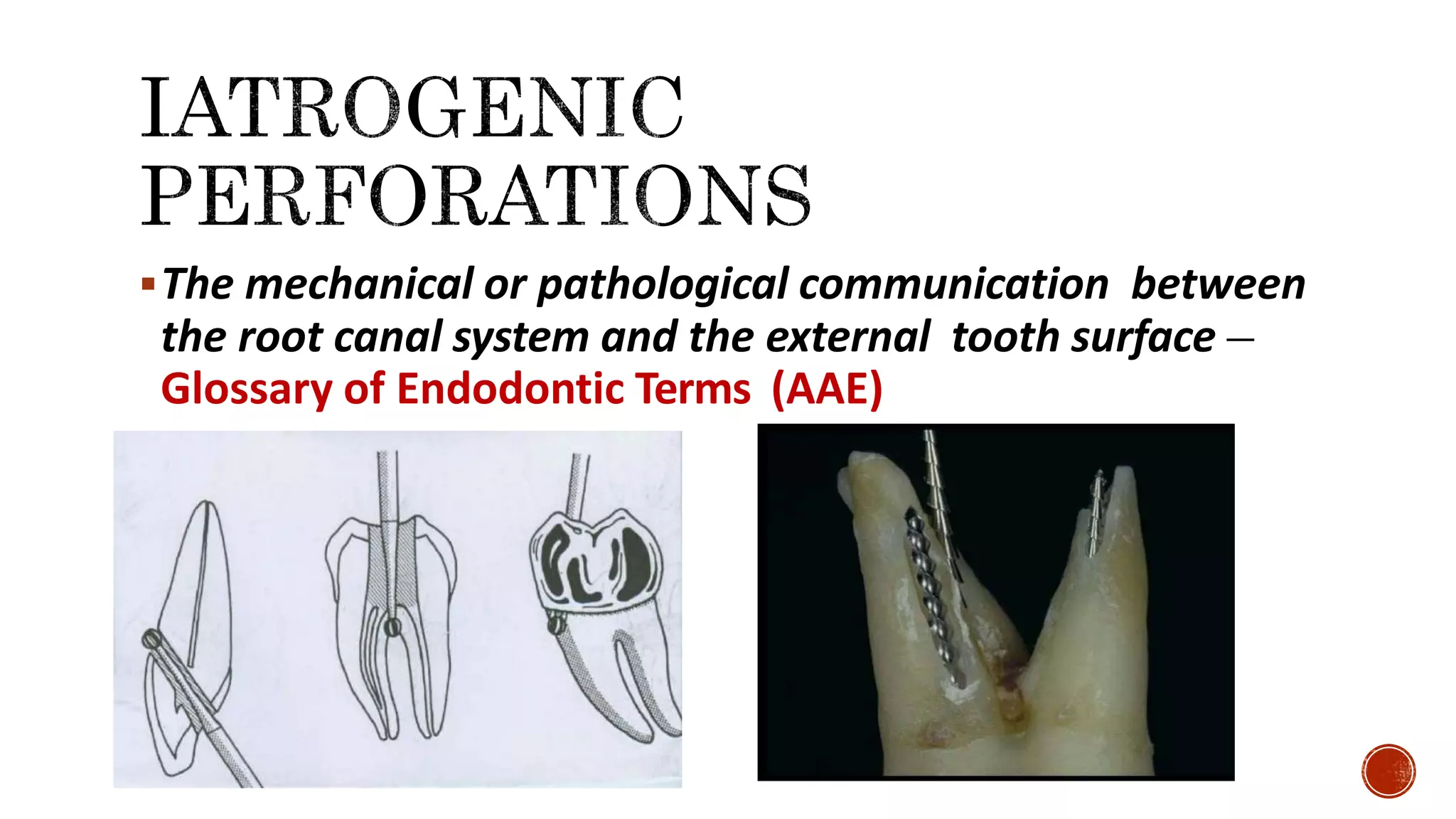
Dr Gurmeen Kaur
II MDS, Department of Conservative Dentistry and Endodontics,
A B Shetty Memorial Institute of Dental Sciences, Mangalore.
ļ‚§ Iatrogenic root perforations
ļ‚§ Predisposing Factors
ļ‚§ Classification of root perforations
ļ‚§ Considerations influencing perforation repair
ļ‚§ Location in relation to the gingival sulcus
ļ‚§ time that the perforation is open to contamination
ļ‚§ Extend & Size of Perforation
ļ‚§ Accessibility
ļ‚§ Esthetics
ļ‚§ Access Cavity Perforation
ļ‚§ In incisors
ļ‚§ Calcifies/narrow canals
ļ‚§ Correction
ļ‚§ Prognosis
ļ‚§ prevention
ļ‚§ Furcal perforations
ļ‚§ Avoided by
ļ‚§ Cervical Canal Perforation
ļ‚§ Cause
ļ‚§ Recognition
ļ‚§ Correction
ļ‚§ prevention
ļ‚§ Lateral wall Perforation
ļ‚§ Apical Perforation
ļ‚§ Curved canals-
ļ‚§ Over instrumentation
ļ‚§ Zipping
ļ‚§ Correction
ļ‚§ Straight canals
ļ‚§ Post perforation
ļ‚§ Management
ļ‚§ Prognosis
ļ‚§ Prevention
ļ‚§ Previously treated tooth with perforation
ļ‚§ Treatment by orthograde approach
ļ‚§ Sealing materials
ļ‚§ Internal matrix technique
ļ‚§ Treatment by surgical approach
ļ‚§ Parameters to be considered
ļ‚§ Guided tissue regeneration
ļ‚§ Intentional replantation
ļ‚§The mechanical or pathological communication between
the root canal system and the external tooth surface –
Glossary of Endodontic Terms (AAE)
PREDISPOSING FACTORS
• Inadequate knowledge of tooth anatomy
• Improper use of drilling instruments leading to gouging
• Forceful/unregulated canal instrumentation leading to ledge
formation, zipping with or without elbow formation and loss of
apical constriction
• Irregular tooth morphology ex inclined crown, curved root canals,
calcified pulp chamber.
Inclination of tooth Abnormal tooth morphology
Curved canals
Calcified pulp chamber
Gouging Ledge Zipping
ļ‚§ Fresh perforation – treated immediately or shortly after occurrence under aseptic
conditions, Good Prognosis.
ļ‚§ Old perforation – previously not treated with likely bacterial infection, Questionable
Prognosis.
ļ‚§ Small perforation (smaller than #20 endodontic instrument) – mechanical damage to
tissue is minimal with easy sealing opportunity, Good Prognosis.
ļ‚§ Large perforation – done during post preparation, with significant tissue damage and
obvious difficulty in providing an adequate seal, salivary contamination, or coronal
leakage along temporary restoration, Questionable Prognosis.
ļ‚§ Coronal perforation – coronal to the level of crestal bone and epithelial attachment with
minimal damage to the supporting tissues and easy access, Good Prognosis.
ļ‚§ Crestal perforation – at the level of the epithelial attachment into the crestal bone,
Questionable Prognosis.
ļ‚§ Apical perforation – apical to the crestal bone and the epithelial attachment, Good
Prognosis.
Fuss Z, Trope M. Root perforations: classification and treatment choices based on
prognostic factors. Endod Dent Traumatol 1996: 12: 255–264.
1. Coronal Perforation
2. Furcation Perforation
3. Lateral wall perforation (Strip Perforation)
4. Root canal perforation
a. Cervical Canal Perforation
b. Mid root Perforation
c. Apical Perforation
5. Post space perforation
• Location in relation to the gingival sulcus:
• Coronal / furcation perforation : threaten sulcular epithelium
• In general, more apical the perforation, more favourable the prognosis
• time that the perforation is open to contamination:
ļ‚§ factor in the degree of inflammatory change and breakdown in the periodontium.
ļ‚§ immediate closure carrying the best prognosis.
• Chronic perforations exhibiting a loss of sulcular attachment pose treatment challenges that potentially
escalate to surgical correction and effort directed toward guided tissue regeneration procedures.
• possibility of achieving a seal of the perforation:
• Extend & Size of Perforation:
• Size greatly affects the clinician’s ability to establish a hermetic seal.
• The area of a circular shaped perforation can be mathematically described as Ļ€ r2.
• Therefore doubling the perforation size with any bur or instrument increases the surface
area to seal four-fold.
• accessibility of the main canal:
• Esthetics:
• Perforations in the anterior region can definitely impact esthetics.
• Patients with high lip line - esthetically compromised by soft tissue defects such as cleft, ossious or
discrepancies in the incisogingival dimensions of a crown when compared with the adjacent teeth
• Happens during the search for canal orifices.
• Can occur either peripherally through the sides of the crown
• or through furcation.
ļ‚§ Above PDL attachment
• First indication: Presence of leakage into the access cavity
Into PDL
• Bleeding into the access cavity is often the first indication of an accidental
perforation.
• Cause: Failure to identify the angle of the crown to the root and the angle of the
tooth in the dental arch.
• Ex: Access through crowned teeth.
• Maxillary lateral incisors and mandibular first premolars.
• Using a surgical length bur
• Misidentification of canals
ļ‚§ Incisors Mandibular– weakened coronally by
excessive removal of tooth structure
ļ‚§ Mesiodistal width of pulp chamber< bur
ļ‚§ Lateral perforations– rare-- ease of visibility
& clear definition of external anatomy
ļ‚§ Labial perforations– common – especially
with calcifications
ļ‚§ Clinician must consider relationship between
incisal edge and location of pulp chamber
ļ‚§ Intact incisal edge– impossible to perforate
lingually
ļ‚§ Bur does not drop easily into chamber
ļ‚§ Change to smaller diameter burs
ļ‚§ Direct cutting action in apical- lingual version
ļ‚§ Canal orifice still does not materialize- remove bur and place it in access cavity–
radiograph
ļ‚§ Resultant film- reveal depth of cutting & angulation of cutting from mesial to
distal
• Coronal walls above the alveolar crest – can be repaired intracoronally
without surgical intervention.
• Perforations into periodontal ligament– should be done as early as possible
to minimize injury to the tooth’s supporting tissues.
• Materials used for these perforations
• - GIC, MTA, Super EBA, Tricalcium phosphate, Calcium
hydroxide paste, amalgam or haemostatic agents such as gel foam.
• Study by Alhadainy andAbdalla
• Calcium sulfate and hydroxyapatite, used as barriers, significantly
improved sealing ability of vitrebond and provide successful barriers
against its overextension.
Alhadainy, Hatem A. et al. Artificial floor technique used for the repair of furcation perforations: A microleakage
study. Journal of Endodontics, Volume 24, Issue 1, 33 - 35
• Mittal et al reported highest amount of leakage was associated with
amalgam followed glass-ionomer, composite, IRM and AH26.
• MTA showed better results, it can be placed in presence of blood since it
require moisture to cure.
ļ‚§Prognosis
• Depends on:
• Location
• Time
• Adequacy of seal
• Perforationsize
• Accessibility to main canals
Mittal M, Chandra S, Chandra S. An evaluation of plaster if Paris barriers used under various materials
to repair furcation perforations (in vitro study). J Endodon 1999;25:385–8
• Proper bur alignment with the long axis of the tooth
• Bur penetration for both depth and angulation can be confirmed
with radiographs
• Knowledge about the morphology
• Adequate access preparation
ļ‚§ Iatrogenic perforation of the furcation area in multi-rooted molars during
preparation of the access cavity can potentially lead to tooth extraction
ļ‚§ Worst possible outcome of any cleaning & shaping procedure
ļ‚§ Mid-curvature opening into PDL space
ļ‚§ Close to clinical crown- develop or continue microleakage from coronal restoration into the space
ļ‚§ Result from improper file manipulation or oversized radicular access preparation
ļ‚§Hallmark features:
1. Continuous dull localized pain on the tooth undergoing root
canal treatment
2. Localized inflammation on the buccal mucosa overlying the
furcal region
3. Tenderness on mastication
4. May or may not exhibit fluid discharge from the access cavity
• Non surgical-
– tooth extraction
– tooth preservation using calcium hydroxide, Cavit, amalgam,
glass ionomer, composite resin, mineral trioxide aggregate
(MTA) and calcium enriched mixture (CEM) cement
• Surgical – Endodontic surgery (example hemisection)
ļ‚§Steps of management
1. Evaluation of the extensiveness of the perforation. Very large openings
have very poor prognosis
2. Isolate and obtain adequate hemostasis
3. Flair the canal orifice and irrigate perforation site adequately
4. Place solid fillers (Gutta percha) into the canal to prevent blockage of
canals
5. Pack the repair material into the perforation and obtain proper marginal
adaptation
6. Provide adequate time for the material to set
7. Resume/ restart root canal preparation. Complete obturation as
necessary
(A)Preoperative radiograph showing the perforations present in mandibular right 1st and
2nd molars
(B)Clinical evaluation of pulpal floor perforation of 1st molar under endomicroscope.
Endodontic retreatment was carried out and the canals were dressed with calcium
hydroxide and obturated with gutta percha on the subsequent visit
(C)Blood sample was collected from the Cubital region of forearm and Platelet rich
fibrin (PRF) was separated from the blood. The PRF was collected in dappen dish
and mixed with hydroxyapatite graft material.
Rhythm Bains, Vivek K. Bains, Kapil Loomba, Kavita Verma, Afreena Nasir. Management of pulpal floor perforation and
grade II Furcation involvement using mineral trioxide aggregate and platelet rich fibrin: A clinical report; Contemp Clin
Dent. 2012 Sep; 3(Suppl 2): S223–S227. doi: 10.4103/0976-237X.101100 PMCID: PMC3514927
(D) Full thickness mucoperiosteal envelop flap was reflected from
the lingual aspect of tooth 45, 46 and 47. The area was debrided
and hydroxyapatite graft+ PRF was applied into the furcation.
The flap was readapted and stabilized.
(E)The perforation was repaired using MTA and then the tooth
was completely sealed using Type II glass ionomer cement
(F)Post operative radiograph after 4 months
ļ‚§ Incorporating anticurvature pressure when cutting instruments are pushed or
pulled in a curved canal system
ļ‚§ Anticurvature pressure- extremely effective when used with Gates Glidden drills
in early radicular access preparations
ļ‚§ Commonly advocated for conventional Hedstrom files for preparation of curved canals. H
files capable of creating midcanal perforation
ļ‚§ Little protection against perforation in apical regions of canal
ļ‚§ Never take large GG drill or Peezo drill deeply into root canal
ļ‚§ New drills of large size – 3 to 6- grab canal walls & pull themselves deeply into
canal before the clinician can stop handpiece
ļ‚§ Prevent by- run handpiece in reverse direction with new drills- drills tend to
back out of canal.
ļ‚§ More apical pressure- drill can be moved into canal & made to cut dentin
ļ‚§Locations
ļ‚§ Cervical
ļ‚§ Lateral
ļ‚§ Apical
ļ‚§Two errors of commission
ļ‚§ Starting a ledge & then drilling out through side of the root at a point of
canal obstruction or root curvature
ļ‚§ Using too large or long instrument & either perforating directly out
through the apical foramen or ā€œweaningā€ a hole in lateral surface of root
by over instrumentation
CERVICAL CANAL
PERFORATION
ļ‚§Cause
• Locating and widening the canal orifice.
• Inappropriate use of Gates-Glidden burs.
ļ‚§Recognition
• Sudden appearance of blood.
• Magnification with either loupes, an endoscope, or a microscope is very
useful.
• Confirmed : place a small file and take a radiograph of the tooth.
• Hemostatics to control bleeding.
• Small area : sealed from inside the tooth
• Large area : seal from inside, then surgical repair
• Materials used:
• Calcium Hydroxide, Collagen, Calcium Sulfate, Freeze-dried Bone,
MTA
ļ‚§ Where esthetics is a concern, a barrier along with composite restoration is
generally used.
• Super EBA have been used when esthetics not an issue.
• Presently MTAis rapidly becoming the barrier/ restorative of choice for
repairing non- esthetic coronal one-third defects because of its many
desirable attributes.
ļ‚§ Reviewing each tooth’s morphology prior to entering its pulp space.
• Thorough examination of pre-operative radiographs is the paramount step
to avoid this mishap.
• Checking the long axis of the tooth and aligning the long axis of the
access bur with the long axis of the tooth - tipped tooth.
• Following principles of access cavity preparation, adequate size and
location, both permitting direct access to the root canals.
• Commonly occurs in curved canals or in canals
which have ledge formed and further
instrumented
• Often accompanied by strip perforation as the
curved canal is straightened out
ļ‚§ Location learned by- placing paper point in canal until it is
removed with a bloody tip & measuring this distance on the
point
ļ‚§ Caused by over-instrumentation & stripping through a thin
wall
ļ‚§ Most likely to happen on ā€˜inside’ curve of canal severely curved canal
ļ‚§ Overzealous enlargement with rotary instruments- diameter
exceeds width of canal at its narrowest point perforation
ļ‚§ Most common in mesial roots of mandibular molars or area of mesial
concavity in maxillary first premolars
• By natureof occurrence, these defects are ovoid in shape and
typically represent relatively large surface area to seal.
• Accessto midrootperforation is most often difficult, and repair is
not predictable.
• Successful repair depends upon the adequacy of the seal established by the
repair material.
• The repair should be immediate, to protect the perforated site from saliva and
other contaminants.
• Barrier material of choice is MTA.
• Two-step method: canals obturated and then defect is repaired surgically
ļ‚§ Obturation- GP condensed with lateral pressure
ļ‚§ Prognosis improved if canal is immediately filled upon discovery
ļ‚§ Surgical correction- necessary in refractory cases
ļ‚§ Due to uncontrolled transportation & subsequent ledge
formation
ļ‚§ Attempt to reestablish canal length past ledge formation
file tip cutting straight through root structure & into PDL
space
ļ‚§ Failure to follow apical curvature of canals- perforations of
maxillary lateral incisors or palatal roots of maxillary
molars
ļ‚§ Important to use curved & proper sized instruments in
curved canals
ļ‚§ Important to return to natural canal to complete
debridement & step back preparation
ļ‚§ Done by bypassing the perforation with severely curved
instruments
• Hall mark clinical feature: loss of previously tactile resistance
at apical stop accompanied by patient complaining of pain.
• Bleeding at tip of paper point can be a telltale sign of apical
perforation
• Clinical Detection- In all cases patient may complain of sudden pain in a
previously painless appointment session with taste of irrigating solution. The
clinician may also detect bleeding into the canal and chamber.
• Obtain Proper Isolation
• Immediate Hemostasis- using any biocompatible material that will not obstruct
final obturation of canal (example. Calcium Hydroxide, Calcium Sulphate etc.)
• Radiographic Detection- by Intraoral Periapical Radiograph or Cone Beam
Commuted Tomography (CBCT)
• Overinstumentation:
• Re-establish the WL and enlarge with larger instrument.
• Apical barrier: Ca(OH)2, MTA, Dentin Chips, Hydroxyapatite
• Apical Perforation :
• Negotiate
• Perforation site as the new apical opening and obturation is done to seal of the foramen.
• Surgery is necessary, if a lesion present apically.
• SurgicalApproach:
• A combined intracoronal and surgical approach involves repairing the defect intracoronally, then
reflecting a surgical flap to remove the inevitable overextension of the repair material from the
periodontal space.
• In case of failing furcation repairs,
• Bicuspidation
• Hemi-Section
• Intentional Replantation
ļ‚§ Curve in instrument
ļ‚§ Proper placement in canal
ļ‚§ Two canals- one natural– other iatrogenic
ļ‚§ Obturation of both & main body of canal– vertical compressive techniques
with softened gutta percha or chlorapercha
Must coincide with canal
curvature
(confirmation with radiograph)
ļ‚§ Owing to incorrect tooth length
ļ‚§ Destroys resistance form cavity at CDJ
ļ‚§ Corrected by reestablishing tooth length short of original length &
then enlarging canal with larger instruments to this point
ļ‚§ Initial gutta percha placed in retention form cavity will not be forced
out the apex
ļ‚§ Compromised solution> preferable to surgical correction
ļ‚§Iatrogenic perforation during the
preparation and flaring of obturated canals
with the intention of placing prosthesis
(dowels) of post endodontic restoration
ļ‚§ Kvinnsland et al found 47% of perforations occurred during
endodontic treatment and 53% were due to
restorative/prosthodontic treatment
ļ‚§ (Kvinnsland I, Oswald RJ, Halse A, GrĆønningsaeter AG. A clinical and roentgenological study of
55 cases of root perforation. Endod J. 1989;22 (2):75-84)
• Excessively large posts increased risk of perforation &
tooth fracture
• Detection:
• Sudden presence of blood in the canal
• Radiographic evidence
• Presence of sinus tract stroma eluding to the base of a
post.
ļ‚§Management
• Sealing of the perforation if possible
• Least effect if perforation is within bone
• If in gingival sulcus, then periodontal breakdown occurs
• There is a 15% failure rate in areas other than furcation. (Rud J et
al)
PREVENTION
• Good knowledge of root canal anatomy
• Planning the post space preparation based on radiographic
information
• Preparing the space at the time the root canal is obturated.
• GG drills / Peeso reamers can be used
• Post should not exceed 1/3rd of mesial-distal width of tooth & should
(A)A radiograph taken in 1999 showed periapical and lateral radiolucencies. The
original treatment had occurred in 1984, and the treating dentist had filled
the perforation with gutta-percha.
(B)A radiograph of tooth #7 in 2003 before endodontic surgery. The lateral and
apical radiolucencies did not appear to have reduced in size.
(C)Surgical access was made with a full-thickness flap, the area was
debrided, and the perforation site was visualized.
• Marga Ree, DDS, MSc, Richard Schwartz, DDS. Management of Perforations: Four Cases from Two Private Practices
with Medium- to Long-term Recalls (2012); Journal of endodontics, ISSN: 1878-3554, Vol: 38, Issue: 10, Page: 1422-7
(D)The perforation and root-end preparation were filled with MTA.
(E)At the 1-year recall, there was a reduction of both
radiolucencies.
(F)At the 8-year recall, periapical radiographs showed no evidence of
endodontic disease
ļ‚§ Improperly sealed- very poor prognosis
ļ‚§ Apical 2/3rd of root- surgically treatable
ļ‚§ Furcation- possible to pack a matrix of hydroxyapatite & seal perforation
with GIC
ļ‚§ Bone loss has already occurred-
ļ‚§ Hemisection
ļ‚§ Root amputation
ļ‚§ Extraction
ļ‚§Rationale: Same as that of conservative endodontic therapy,
i.e. prevention and treatment of periradicular inflammation.
ļ‚§Measures aimed
ļ‚§ control infection of the perforation site,
ļ‚§ if already infected, disinfect the site
ļ‚§ best possible seal against penetration of bacterial elements.
ļ‚§Fresh perforations  hemorrhage.
ļ‚§First step : control hemorrhage by pressure or irrigation;
ļ‚§Subsequently, perforation should be adequately sealed
ļ‚§ For large perforations
• Used to prevent overfilling of repair material into the periodontium
• A material used as matrix should be biocompatible, easy to manipulate and
stimulate osteogenesis.
ļ‚§ The defect should then be directly accessible and visualized
ļ‚§ internal matrix must be sterile, possible to manipulate, and should not produce
inflammation
ļ‚§ hydroxylapatite, decalcified freeze-dried bone, resorbable collagen with MTA
ļ‚§ high surface pH of MTA supports repair and hard tissue formation in a similar
fashion as calcium hydroxide.
ļ‚§ Calcium oxide in MTA reacts with tissue fluids to form calcium hydroxide, which
in turn may encourage hard tissue deposition.
ļ‚§ large perforations, perforations as a result of resorption, failure of healing after
non-surgical repair, non-surgically inaccessible perforations, extensive coronal
restorations, when concomitant management of the periodontium is indicated,
and large overfilling of the defect
ļ‚§ Apical perforation: Resection of the apical root to sound root structure with an
adequate filling is recommended
ļ‚§ Crestal perforations: loss of the epithelial attachment and pocket formation
ļ‚§ Rud et al: after sealing root perforation elsewhere with dentin-bonded resin-
composite (Retroplast), bone regenerated and a periodontal ligament space was
partly formed with a lamina dura against the material.
ļ‚§ amount of remaining bone,
ļ‚§ Accessibility to the perforation
ļ‚§ extent of osseous destruction,
ļ‚§ duration of the defect,
ļ‚§ periodontal disease status,
ļ‚§ soft tissue attachment level,
ļ‚§ patient’s oral hygiene, and
ļ‚§ surgeon’s expertise in tissue management
ļ‚§ Hemostasis:
ļ‚§ profound anesthesia with a vasoconstriction agent (infiltration of 2% Lidocaine with 1 : 50 000
epinephrine),
ļ‚§ cotton pellets soaked in epinephrine, Gelfoam
ļ‚§ calcium sulfate and CollaCote collagen sponges saturated with 2.25% racemic epinephrine.
ļ‚§ Class I cavity is prepared and the preferred filling material is placed
ļ‚§ Acts by serving as a barrier for apical migration of epithelium.
ļ‚§ both costly and technically demanding
Intentional replantation
• defect is too large for repair and when the perforation is inaccessible without
excessive bone removal
• The success rate reported in clinical follow-ups ranges from 80% to 90% for
carefully performed procedures with proper case selection
• Inflammatory root resorption and ankylosis
• The overall success rate was 72.5% after non- surgical repair of perforations.
• The use of MTA increased the success rate to 80.9%.
• The presence of preexisting radiolucencies adjacent to the perforation sites was
associated with decreased success rates.
• The favorable success rate associated with the non-surgical repair of
perforations >80% when using bioactive materials
Siew K, Lee AH and Cheung GS. Treatment outcome of repaired root perforation: a systematic review and meta-
analysis ( J Endod. 2015;41(11):1795-1804)
Efficacy of sealing material
ļ‚§ Sealability
ļ‚§ Biocompatibility - ability to support osteogenesis and cementogenesis.
ļ‚§ Relatively inexpensive, radiopaque, and bacteriostatic
Materials used over the years-
ļ‚§ amalgam
ļ‚§ phosphate cement
ļ‚§ gutta-percha
ļ‚§ zinc oxide eugenol
ļ‚§ SuperEBA
ļ‚§ dentin chips
ļ‚§ calcium hydroxide
ļ‚§ Cavit
ļ‚§ Tricalcium phosphate
ļ‚§ Hydroxylapatite
ļ‚§ glass ionomer cement
ļ‚§ resin-ionomer
ļ‚§ mineral trioxide aggregate (MTA)
ļ‚§ tin foil, indium foil
ļ‚§ Biodentin
Zinc oxide Eugenol Super Ethoxy benzoic Acid
Cavit Bisfil-2BIntermediate Restorative
Material
Tricalcium Phosphate Hydroxyapatite Powder
Portland Cement
Mineral Trioxide Aggregate
Biodentine Endosequence
Bioaggregate Calcium Enriched Mixture
ļ‚§ Fuss Z, Trope M. Root perforations: classification and treatment choices based on prognostic factors.
Endod Dent Traumatol 1996: 12: 255–264
ļ‚§ Irving H. Sinai. Endodontic perforations: their prognosis and treatment. JADA, Vol. 95, July 1977
ļ‚§ IGOR TSESIS & ZVI FUSS. Diagnosis and treatment of accidental root perforations. Endodontic Topics
2006, 13, 95–107
ļ‚§ Alhadainy, Hatem A. et al. Artificial floor technique used for the repair of furcation perforations: A
microleakage study. Journal of Endodontics, Volume 24, Issue 1, 33 – 35
ļ‚§ Mittal M, Chandra S, Chandra S. An evaluation of plaster if Paris barriers used under various materials
to repair furcation perforations (in vitro study). J Endodon 1999;25:385–8
ļ‚§ Rhythm Bains, Vivek K. Bains, Kapil Loomba, Kavita Verma, Afreena Nasir. Management of pulpal floor perforation and
grade II Furcation involvement using mineral trioxide aggregate and platelet rich fibrin: A clinical report; Contemp Clin
Dent. 2012 Sep; 3(Suppl 2): S223–S227. doi: 10.4103/0976-237X.101100 PMCID: PMC3514927
ļ‚§ (Kvinnsland I, Oswald RJ, Halse A, GrĆønningsaeter AG. A clinical and roentgenological study of 55 cases of
root perforation. Endod J. 1989;22 (2):75-84)
ļ‚§ Endodontic Mishaps in detail. Iraqi Dental Academy. iraqidental.wordpress.com/2016/09/12/endodontic-
mishaps-in-detail/
ļ‚§ Gunjan Gautam. "Management of Iatrogenic Lateral Root Perforation At The Alveolar Crestal Level Using
Biodentine: A Case Report With 2 Year Follow Up." IOSR Journal of Dental and Medical Sciences (IOSR-
JDMS) 16.12 (2017): 67-69
ļ‚§ Marga Ree, DDS, MSc, Richard Schwartz, DDS. Management of Perforations: Four Cases from Two Private
Practices with Medium- to Long-term Recalls (2012); Journal of endodontics, ISSN: 1878-3554, Vol: 38,
Issue: 10, Page: 1422-7
ļ‚§ Siew K, Lee AH and Cheung GS. Treatment outcome of repaired root perforation: a systematic review and
meta-analysis ( J Endod. 2015;41(11):1795-1804)
ļ‚§ John I. Ingles, Leif K. Bakland. Ingle’s Endodontics, 6th edition. BC DeckerInc
ļ‚§ Cohen’s Pathways of Pulp, 11th edition

Perforation in Endodontics

  • 1.
    Dr Gurmeen Kaur IIMDS, Department of Conservative Dentistry and Endodontics, A B Shetty Memorial Institute of Dental Sciences, Mangalore.
  • 2.
    ļ‚§ Iatrogenic rootperforations ļ‚§ Predisposing Factors ļ‚§ Classification of root perforations ļ‚§ Considerations influencing perforation repair ļ‚§ Location in relation to the gingival sulcus ļ‚§ time that the perforation is open to contamination ļ‚§ Extend & Size of Perforation ļ‚§ Accessibility ļ‚§ Esthetics ļ‚§ Access Cavity Perforation ļ‚§ In incisors ļ‚§ Calcifies/narrow canals ļ‚§ Correction ļ‚§ Prognosis ļ‚§ prevention ļ‚§ Furcal perforations ļ‚§ Avoided by
  • 3.
    ļ‚§ Cervical CanalPerforation ļ‚§ Cause ļ‚§ Recognition ļ‚§ Correction ļ‚§ prevention ļ‚§ Lateral wall Perforation ļ‚§ Apical Perforation ļ‚§ Curved canals- ļ‚§ Over instrumentation ļ‚§ Zipping ļ‚§ Correction ļ‚§ Straight canals
  • 4.
    ļ‚§ Post perforation ļ‚§Management ļ‚§ Prognosis ļ‚§ Prevention ļ‚§ Previously treated tooth with perforation ļ‚§ Treatment by orthograde approach ļ‚§ Sealing materials ļ‚§ Internal matrix technique ļ‚§ Treatment by surgical approach ļ‚§ Parameters to be considered ļ‚§ Guided tissue regeneration ļ‚§ Intentional replantation
  • 5.
    ļ‚§The mechanical orpathological communication between the root canal system and the external tooth surface – Glossary of Endodontic Terms (AAE)
  • 6.
    PREDISPOSING FACTORS • Inadequateknowledge of tooth anatomy • Improper use of drilling instruments leading to gouging • Forceful/unregulated canal instrumentation leading to ledge formation, zipping with or without elbow formation and loss of apical constriction • Irregular tooth morphology ex inclined crown, curved root canals, calcified pulp chamber.
  • 7.
    Inclination of toothAbnormal tooth morphology Curved canals Calcified pulp chamber
  • 8.
  • 10.
    ļ‚§ Fresh perforation– treated immediately or shortly after occurrence under aseptic conditions, Good Prognosis. ļ‚§ Old perforation – previously not treated with likely bacterial infection, Questionable Prognosis. ļ‚§ Small perforation (smaller than #20 endodontic instrument) – mechanical damage to tissue is minimal with easy sealing opportunity, Good Prognosis. ļ‚§ Large perforation – done during post preparation, with significant tissue damage and obvious difficulty in providing an adequate seal, salivary contamination, or coronal leakage along temporary restoration, Questionable Prognosis. ļ‚§ Coronal perforation – coronal to the level of crestal bone and epithelial attachment with minimal damage to the supporting tissues and easy access, Good Prognosis. ļ‚§ Crestal perforation – at the level of the epithelial attachment into the crestal bone, Questionable Prognosis. ļ‚§ Apical perforation – apical to the crestal bone and the epithelial attachment, Good Prognosis. Fuss Z, Trope M. Root perforations: classification and treatment choices based on prognostic factors. Endod Dent Traumatol 1996: 12: 255–264.
  • 11.
    1. Coronal Perforation 2.Furcation Perforation 3. Lateral wall perforation (Strip Perforation) 4. Root canal perforation a. Cervical Canal Perforation b. Mid root Perforation c. Apical Perforation 5. Post space perforation
  • 12.
    • Location inrelation to the gingival sulcus: • Coronal / furcation perforation : threaten sulcular epithelium • In general, more apical the perforation, more favourable the prognosis • time that the perforation is open to contamination: ļ‚§ factor in the degree of inflammatory change and breakdown in the periodontium. ļ‚§ immediate closure carrying the best prognosis. • Chronic perforations exhibiting a loss of sulcular attachment pose treatment challenges that potentially escalate to surgical correction and effort directed toward guided tissue regeneration procedures.
  • 13.
    • possibility ofachieving a seal of the perforation: • Extend & Size of Perforation: • Size greatly affects the clinician’s ability to establish a hermetic seal. • The area of a circular shaped perforation can be mathematically described as Ļ€ r2. • Therefore doubling the perforation size with any bur or instrument increases the surface area to seal four-fold. • accessibility of the main canal: • Esthetics: • Perforations in the anterior region can definitely impact esthetics. • Patients with high lip line - esthetically compromised by soft tissue defects such as cleft, ossious or discrepancies in the incisogingival dimensions of a crown when compared with the adjacent teeth
  • 14.
    • Happens duringthe search for canal orifices. • Can occur either peripherally through the sides of the crown • or through furcation. ļ‚§ Above PDL attachment • First indication: Presence of leakage into the access cavity Into PDL • Bleeding into the access cavity is often the first indication of an accidental perforation. • Cause: Failure to identify the angle of the crown to the root and the angle of the tooth in the dental arch. • Ex: Access through crowned teeth. • Maxillary lateral incisors and mandibular first premolars. • Using a surgical length bur • Misidentification of canals
  • 15.
    ļ‚§ Incisors Mandibular–weakened coronally by excessive removal of tooth structure ļ‚§ Mesiodistal width of pulp chamber< bur ļ‚§ Lateral perforations– rare-- ease of visibility & clear definition of external anatomy ļ‚§ Labial perforations– common – especially with calcifications ļ‚§ Clinician must consider relationship between incisal edge and location of pulp chamber ļ‚§ Intact incisal edge– impossible to perforate lingually
  • 16.
    ļ‚§ Bur doesnot drop easily into chamber ļ‚§ Change to smaller diameter burs ļ‚§ Direct cutting action in apical- lingual version ļ‚§ Canal orifice still does not materialize- remove bur and place it in access cavity– radiograph ļ‚§ Resultant film- reveal depth of cutting & angulation of cutting from mesial to distal
  • 17.
    • Coronal wallsabove the alveolar crest – can be repaired intracoronally without surgical intervention. • Perforations into periodontal ligament– should be done as early as possible to minimize injury to the tooth’s supporting tissues. • Materials used for these perforations • - GIC, MTA, Super EBA, Tricalcium phosphate, Calcium hydroxide paste, amalgam or haemostatic agents such as gel foam. • Study by Alhadainy andAbdalla • Calcium sulfate and hydroxyapatite, used as barriers, significantly improved sealing ability of vitrebond and provide successful barriers against its overextension. Alhadainy, Hatem A. et al. Artificial floor technique used for the repair of furcation perforations: A microleakage study. Journal of Endodontics, Volume 24, Issue 1, 33 - 35
  • 18.
    • Mittal etal reported highest amount of leakage was associated with amalgam followed glass-ionomer, composite, IRM and AH26. • MTA showed better results, it can be placed in presence of blood since it require moisture to cure. ļ‚§Prognosis • Depends on: • Location • Time • Adequacy of seal • Perforationsize • Accessibility to main canals Mittal M, Chandra S, Chandra S. An evaluation of plaster if Paris barriers used under various materials to repair furcation perforations (in vitro study). J Endodon 1999;25:385–8
  • 19.
    • Proper buralignment with the long axis of the tooth • Bur penetration for both depth and angulation can be confirmed with radiographs • Knowledge about the morphology • Adequate access preparation
  • 20.
    ļ‚§ Iatrogenic perforationof the furcation area in multi-rooted molars during preparation of the access cavity can potentially lead to tooth extraction ļ‚§ Worst possible outcome of any cleaning & shaping procedure ļ‚§ Mid-curvature opening into PDL space ļ‚§ Close to clinical crown- develop or continue microleakage from coronal restoration into the space ļ‚§ Result from improper file manipulation or oversized radicular access preparation
  • 21.
    ļ‚§Hallmark features: 1. Continuousdull localized pain on the tooth undergoing root canal treatment 2. Localized inflammation on the buccal mucosa overlying the furcal region 3. Tenderness on mastication 4. May or may not exhibit fluid discharge from the access cavity
  • 22.
    • Non surgical- –tooth extraction – tooth preservation using calcium hydroxide, Cavit, amalgam, glass ionomer, composite resin, mineral trioxide aggregate (MTA) and calcium enriched mixture (CEM) cement • Surgical – Endodontic surgery (example hemisection)
  • 23.
    ļ‚§Steps of management 1.Evaluation of the extensiveness of the perforation. Very large openings have very poor prognosis 2. Isolate and obtain adequate hemostasis 3. Flair the canal orifice and irrigate perforation site adequately 4. Place solid fillers (Gutta percha) into the canal to prevent blockage of canals 5. Pack the repair material into the perforation and obtain proper marginal adaptation 6. Provide adequate time for the material to set 7. Resume/ restart root canal preparation. Complete obturation as necessary
  • 24.
    (A)Preoperative radiograph showingthe perforations present in mandibular right 1st and 2nd molars (B)Clinical evaluation of pulpal floor perforation of 1st molar under endomicroscope. Endodontic retreatment was carried out and the canals were dressed with calcium hydroxide and obturated with gutta percha on the subsequent visit (C)Blood sample was collected from the Cubital region of forearm and Platelet rich fibrin (PRF) was separated from the blood. The PRF was collected in dappen dish and mixed with hydroxyapatite graft material. Rhythm Bains, Vivek K. Bains, Kapil Loomba, Kavita Verma, Afreena Nasir. Management of pulpal floor perforation and grade II Furcation involvement using mineral trioxide aggregate and platelet rich fibrin: A clinical report; Contemp Clin Dent. 2012 Sep; 3(Suppl 2): S223–S227. doi: 10.4103/0976-237X.101100 PMCID: PMC3514927
  • 25.
    (D) Full thicknessmucoperiosteal envelop flap was reflected from the lingual aspect of tooth 45, 46 and 47. The area was debrided and hydroxyapatite graft+ PRF was applied into the furcation. The flap was readapted and stabilized. (E)The perforation was repaired using MTA and then the tooth was completely sealed using Type II glass ionomer cement (F)Post operative radiograph after 4 months
  • 26.
    ļ‚§ Incorporating anticurvaturepressure when cutting instruments are pushed or pulled in a curved canal system ļ‚§ Anticurvature pressure- extremely effective when used with Gates Glidden drills in early radicular access preparations ļ‚§ Commonly advocated for conventional Hedstrom files for preparation of curved canals. H files capable of creating midcanal perforation ļ‚§ Little protection against perforation in apical regions of canal ļ‚§ Never take large GG drill or Peezo drill deeply into root canal ļ‚§ New drills of large size – 3 to 6- grab canal walls & pull themselves deeply into canal before the clinician can stop handpiece ļ‚§ Prevent by- run handpiece in reverse direction with new drills- drills tend to back out of canal. ļ‚§ More apical pressure- drill can be moved into canal & made to cut dentin
  • 27.
    ļ‚§Locations ļ‚§ Cervical ļ‚§ Lateral ļ‚§Apical ļ‚§Two errors of commission ļ‚§ Starting a ledge & then drilling out through side of the root at a point of canal obstruction or root curvature ļ‚§ Using too large or long instrument & either perforating directly out through the apical foramen or ā€œweaningā€ a hole in lateral surface of root by over instrumentation
  • 28.
    CERVICAL CANAL PERFORATION ļ‚§Cause • Locatingand widening the canal orifice. • Inappropriate use of Gates-Glidden burs. ļ‚§Recognition • Sudden appearance of blood. • Magnification with either loupes, an endoscope, or a microscope is very useful. • Confirmed : place a small file and take a radiograph of the tooth.
  • 29.
    • Hemostatics tocontrol bleeding. • Small area : sealed from inside the tooth • Large area : seal from inside, then surgical repair • Materials used: • Calcium Hydroxide, Collagen, Calcium Sulfate, Freeze-dried Bone, MTA ļ‚§ Where esthetics is a concern, a barrier along with composite restoration is generally used. • Super EBA have been used when esthetics not an issue. • Presently MTAis rapidly becoming the barrier/ restorative of choice for repairing non- esthetic coronal one-third defects because of its many desirable attributes.
  • 30.
    ļ‚§ Reviewing eachtooth’s morphology prior to entering its pulp space. • Thorough examination of pre-operative radiographs is the paramount step to avoid this mishap. • Checking the long axis of the tooth and aligning the long axis of the access bur with the long axis of the tooth - tipped tooth. • Following principles of access cavity preparation, adequate size and location, both permitting direct access to the root canals.
  • 31.
    • Commonly occursin curved canals or in canals which have ledge formed and further instrumented • Often accompanied by strip perforation as the curved canal is straightened out
  • 32.
    ļ‚§ Location learnedby- placing paper point in canal until it is removed with a bloody tip & measuring this distance on the point ļ‚§ Caused by over-instrumentation & stripping through a thin wall ļ‚§ Most likely to happen on ā€˜inside’ curve of canal severely curved canal ļ‚§ Overzealous enlargement with rotary instruments- diameter exceeds width of canal at its narrowest point perforation ļ‚§ Most common in mesial roots of mandibular molars or area of mesial concavity in maxillary first premolars
  • 34.
    • By natureofoccurrence, these defects are ovoid in shape and typically represent relatively large surface area to seal. • Accessto midrootperforation is most often difficult, and repair is not predictable. • Successful repair depends upon the adequacy of the seal established by the repair material. • The repair should be immediate, to protect the perforated site from saliva and other contaminants. • Barrier material of choice is MTA. • Two-step method: canals obturated and then defect is repaired surgically ļ‚§ Obturation- GP condensed with lateral pressure ļ‚§ Prognosis improved if canal is immediately filled upon discovery ļ‚§ Surgical correction- necessary in refractory cases
  • 35.
    ļ‚§ Due touncontrolled transportation & subsequent ledge formation ļ‚§ Attempt to reestablish canal length past ledge formation file tip cutting straight through root structure & into PDL space ļ‚§ Failure to follow apical curvature of canals- perforations of maxillary lateral incisors or palatal roots of maxillary molars ļ‚§ Important to use curved & proper sized instruments in curved canals ļ‚§ Important to return to natural canal to complete debridement & step back preparation ļ‚§ Done by bypassing the perforation with severely curved instruments
  • 36.
    • Hall markclinical feature: loss of previously tactile resistance at apical stop accompanied by patient complaining of pain. • Bleeding at tip of paper point can be a telltale sign of apical perforation • Clinical Detection- In all cases patient may complain of sudden pain in a previously painless appointment session with taste of irrigating solution. The clinician may also detect bleeding into the canal and chamber. • Obtain Proper Isolation • Immediate Hemostasis- using any biocompatible material that will not obstruct final obturation of canal (example. Calcium Hydroxide, Calcium Sulphate etc.) • Radiographic Detection- by Intraoral Periapical Radiograph or Cone Beam Commuted Tomography (CBCT)
  • 37.
    • Overinstumentation: • Re-establishthe WL and enlarge with larger instrument. • Apical barrier: Ca(OH)2, MTA, Dentin Chips, Hydroxyapatite • Apical Perforation : • Negotiate • Perforation site as the new apical opening and obturation is done to seal of the foramen. • Surgery is necessary, if a lesion present apically. • SurgicalApproach: • A combined intracoronal and surgical approach involves repairing the defect intracoronally, then reflecting a surgical flap to remove the inevitable overextension of the repair material from the periodontal space. • In case of failing furcation repairs, • Bicuspidation • Hemi-Section • Intentional Replantation
  • 38.
    ļ‚§ Curve ininstrument ļ‚§ Proper placement in canal ļ‚§ Two canals- one natural– other iatrogenic ļ‚§ Obturation of both & main body of canal– vertical compressive techniques with softened gutta percha or chlorapercha Must coincide with canal curvature (confirmation with radiograph)
  • 39.
    ļ‚§ Owing toincorrect tooth length ļ‚§ Destroys resistance form cavity at CDJ ļ‚§ Corrected by reestablishing tooth length short of original length & then enlarging canal with larger instruments to this point ļ‚§ Initial gutta percha placed in retention form cavity will not be forced out the apex ļ‚§ Compromised solution> preferable to surgical correction
  • 40.
    ļ‚§Iatrogenic perforation duringthe preparation and flaring of obturated canals with the intention of placing prosthesis (dowels) of post endodontic restoration ļ‚§ Kvinnsland et al found 47% of perforations occurred during endodontic treatment and 53% were due to restorative/prosthodontic treatment ļ‚§ (Kvinnsland I, Oswald RJ, Halse A, GrĆønningsaeter AG. A clinical and roentgenological study of 55 cases of root perforation. Endod J. 1989;22 (2):75-84)
  • 41.
    • Excessively largeposts increased risk of perforation & tooth fracture • Detection: • Sudden presence of blood in the canal • Radiographic evidence • Presence of sinus tract stroma eluding to the base of a post. ļ‚§Management • Sealing of the perforation if possible
  • 42.
    • Least effectif perforation is within bone • If in gingival sulcus, then periodontal breakdown occurs • There is a 15% failure rate in areas other than furcation. (Rud J et al) PREVENTION • Good knowledge of root canal anatomy • Planning the post space preparation based on radiographic information • Preparing the space at the time the root canal is obturated. • GG drills / Peeso reamers can be used • Post should not exceed 1/3rd of mesial-distal width of tooth & should
  • 43.
    (A)A radiograph takenin 1999 showed periapical and lateral radiolucencies. The original treatment had occurred in 1984, and the treating dentist had filled the perforation with gutta-percha. (B)A radiograph of tooth #7 in 2003 before endodontic surgery. The lateral and apical radiolucencies did not appear to have reduced in size. (C)Surgical access was made with a full-thickness flap, the area was debrided, and the perforation site was visualized. • Marga Ree, DDS, MSc, Richard Schwartz, DDS. Management of Perforations: Four Cases from Two Private Practices with Medium- to Long-term Recalls (2012); Journal of endodontics, ISSN: 1878-3554, Vol: 38, Issue: 10, Page: 1422-7
  • 44.
    (D)The perforation androot-end preparation were filled with MTA. (E)At the 1-year recall, there was a reduction of both radiolucencies. (F)At the 8-year recall, periapical radiographs showed no evidence of endodontic disease
  • 45.
    ļ‚§ Improperly sealed-very poor prognosis ļ‚§ Apical 2/3rd of root- surgically treatable ļ‚§ Furcation- possible to pack a matrix of hydroxyapatite & seal perforation with GIC ļ‚§ Bone loss has already occurred- ļ‚§ Hemisection ļ‚§ Root amputation ļ‚§ Extraction
  • 46.
    ļ‚§Rationale: Same asthat of conservative endodontic therapy, i.e. prevention and treatment of periradicular inflammation. ļ‚§Measures aimed ļ‚§ control infection of the perforation site, ļ‚§ if already infected, disinfect the site ļ‚§ best possible seal against penetration of bacterial elements. ļ‚§Fresh perforations  hemorrhage. ļ‚§First step : control hemorrhage by pressure or irrigation; ļ‚§Subsequently, perforation should be adequately sealed
  • 47.
    ļ‚§ For largeperforations • Used to prevent overfilling of repair material into the periodontium • A material used as matrix should be biocompatible, easy to manipulate and stimulate osteogenesis. ļ‚§ The defect should then be directly accessible and visualized ļ‚§ internal matrix must be sterile, possible to manipulate, and should not produce inflammation ļ‚§ hydroxylapatite, decalcified freeze-dried bone, resorbable collagen with MTA ļ‚§ high surface pH of MTA supports repair and hard tissue formation in a similar fashion as calcium hydroxide. ļ‚§ Calcium oxide in MTA reacts with tissue fluids to form calcium hydroxide, which in turn may encourage hard tissue deposition.
  • 48.
    ļ‚§ large perforations,perforations as a result of resorption, failure of healing after non-surgical repair, non-surgically inaccessible perforations, extensive coronal restorations, when concomitant management of the periodontium is indicated, and large overfilling of the defect ļ‚§ Apical perforation: Resection of the apical root to sound root structure with an adequate filling is recommended ļ‚§ Crestal perforations: loss of the epithelial attachment and pocket formation ļ‚§ Rud et al: after sealing root perforation elsewhere with dentin-bonded resin- composite (Retroplast), bone regenerated and a periodontal ligament space was partly formed with a lamina dura against the material.
  • 49.
    ļ‚§ amount ofremaining bone, ļ‚§ Accessibility to the perforation ļ‚§ extent of osseous destruction, ļ‚§ duration of the defect, ļ‚§ periodontal disease status, ļ‚§ soft tissue attachment level, ļ‚§ patient’s oral hygiene, and ļ‚§ surgeon’s expertise in tissue management ļ‚§ Hemostasis: ļ‚§ profound anesthesia with a vasoconstriction agent (infiltration of 2% Lidocaine with 1 : 50 000 epinephrine), ļ‚§ cotton pellets soaked in epinephrine, Gelfoam ļ‚§ calcium sulfate and CollaCote collagen sponges saturated with 2.25% racemic epinephrine. ļ‚§ Class I cavity is prepared and the preferred filling material is placed
  • 50.
    ļ‚§ Acts byserving as a barrier for apical migration of epithelium. ļ‚§ both costly and technically demanding Intentional replantation • defect is too large for repair and when the perforation is inaccessible without excessive bone removal • The success rate reported in clinical follow-ups ranges from 80% to 90% for carefully performed procedures with proper case selection • Inflammatory root resorption and ankylosis
  • 51.
    • The overallsuccess rate was 72.5% after non- surgical repair of perforations. • The use of MTA increased the success rate to 80.9%. • The presence of preexisting radiolucencies adjacent to the perforation sites was associated with decreased success rates. • The favorable success rate associated with the non-surgical repair of perforations >80% when using bioactive materials Siew K, Lee AH and Cheung GS. Treatment outcome of repaired root perforation: a systematic review and meta- analysis ( J Endod. 2015;41(11):1795-1804)
  • 53.
    Efficacy of sealingmaterial ļ‚§ Sealability ļ‚§ Biocompatibility - ability to support osteogenesis and cementogenesis. ļ‚§ Relatively inexpensive, radiopaque, and bacteriostatic Materials used over the years- ļ‚§ amalgam ļ‚§ phosphate cement ļ‚§ gutta-percha ļ‚§ zinc oxide eugenol ļ‚§ SuperEBA ļ‚§ dentin chips ļ‚§ calcium hydroxide ļ‚§ Cavit ļ‚§ Tricalcium phosphate ļ‚§ Hydroxylapatite ļ‚§ glass ionomer cement ļ‚§ resin-ionomer ļ‚§ mineral trioxide aggregate (MTA) ļ‚§ tin foil, indium foil ļ‚§ Biodentin
  • 54.
    Zinc oxide EugenolSuper Ethoxy benzoic Acid Cavit Bisfil-2BIntermediate Restorative Material
  • 55.
    Tricalcium Phosphate HydroxyapatitePowder Portland Cement Mineral Trioxide Aggregate
  • 56.
  • 57.
    ļ‚§ Fuss Z,Trope M. Root perforations: classification and treatment choices based on prognostic factors. Endod Dent Traumatol 1996: 12: 255–264 ļ‚§ Irving H. Sinai. Endodontic perforations: their prognosis and treatment. JADA, Vol. 95, July 1977 ļ‚§ IGOR TSESIS & ZVI FUSS. Diagnosis and treatment of accidental root perforations. Endodontic Topics 2006, 13, 95–107 ļ‚§ Alhadainy, Hatem A. et al. Artificial floor technique used for the repair of furcation perforations: A microleakage study. Journal of Endodontics, Volume 24, Issue 1, 33 – 35 ļ‚§ Mittal M, Chandra S, Chandra S. An evaluation of plaster if Paris barriers used under various materials to repair furcation perforations (in vitro study). J Endodon 1999;25:385–8 ļ‚§ Rhythm Bains, Vivek K. Bains, Kapil Loomba, Kavita Verma, Afreena Nasir. Management of pulpal floor perforation and grade II Furcation involvement using mineral trioxide aggregate and platelet rich fibrin: A clinical report; Contemp Clin Dent. 2012 Sep; 3(Suppl 2): S223–S227. doi: 10.4103/0976-237X.101100 PMCID: PMC3514927 ļ‚§ (Kvinnsland I, Oswald RJ, Halse A, GrĆønningsaeter AG. A clinical and roentgenological study of 55 cases of root perforation. Endod J. 1989;22 (2):75-84) ļ‚§ Endodontic Mishaps in detail. Iraqi Dental Academy. iraqidental.wordpress.com/2016/09/12/endodontic- mishaps-in-detail/
  • 58.
    ļ‚§ Gunjan Gautam."Management of Iatrogenic Lateral Root Perforation At The Alveolar Crestal Level Using Biodentine: A Case Report With 2 Year Follow Up." IOSR Journal of Dental and Medical Sciences (IOSR- JDMS) 16.12 (2017): 67-69 ļ‚§ Marga Ree, DDS, MSc, Richard Schwartz, DDS. Management of Perforations: Four Cases from Two Private Practices with Medium- to Long-term Recalls (2012); Journal of endodontics, ISSN: 1878-3554, Vol: 38, Issue: 10, Page: 1422-7 ļ‚§ Siew K, Lee AH and Cheung GS. Treatment outcome of repaired root perforation: a systematic review and meta-analysis ( J Endod. 2015;41(11):1795-1804) ļ‚§ John I. Ingles, Leif K. Bakland. Ingle’s Endodontics, 6th edition. BC DeckerInc ļ‚§ Cohen’s Pathways of Pulp, 11th edition