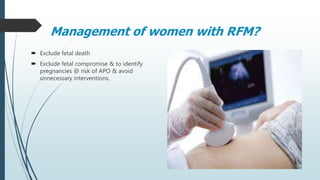
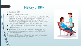
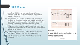
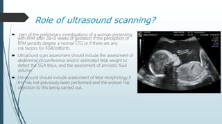
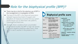
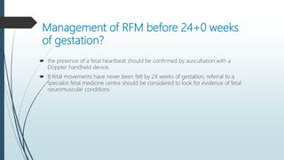
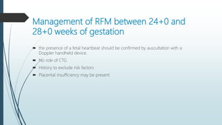
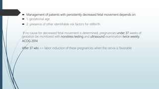
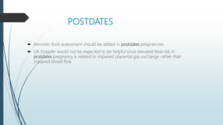
Reduced Fetal
MovementsRCOG GTG # 57
By. Dr Sana Lodhi
Introduction
 Fetal movements have been defined as any discrete kick,flutter, swish or roll.
 Decreased fetal movements affect 5–15% of pregnancies.
 • Time of FM perception:
Mean: 18-20wks
PG: 20-22wks
MP: 16-20wks
 By term, avg no of FM/hr is 31 (16-45), with the longest period between
movements ranging from 50 to 75 minutes.
 Fetal movements tend to plateau at 32 weeks of gestation, there is no reduction in
the frequency of fetal movements in the late third trimester.
Physiology
 FM follow a circadian pattern & are an expression of fetal
wellbeing. It has been suggested that a gradual decline
during the third trimester is due to improved fetal
coordination and reduced amniotic fluid volume, coupled
with increased fetal size.
 Decreased fetal movements are regarded as a marker for
suboptimal intrauterine conditions.
 Conserve energy & reduce O2 consumption.
 Diurnal variation: The afternoon and evening periods are
periods of peak activity.
Factors which influence a woman’s
perception of this activity
 Busy mother
 Lying>sitting>standing
 Anterior placenta (<28wks gestation)
 Drugs e.g. Alcohol, Sedatives, Benzodiazepines, methadones,
other opioids
 Corticosteroids > decreased FM & FHR variability (detected
by CTG over 2 days)
 CO2 & Cigerrete smoking
 Malformations
 Anencephaly ( normal or ed )
 Fetal presentation (no effect)
 Fetal position
How can fetal movements be assessed?
 Maternal perception ( Subjective)
 Doppler or real-time ultrasound (Objective):
Duration of recording is restricted to 20–30 minutes with the mother in a semi-
recumbent position but results not correlate strongly to perinatal outcome
Should fetal movements be counted routinely
in a formal manner
 There is insufficient evidence to recommend formal fetal movement
counting using specified alarm limits.
 Women should be advised to be aware of their baby’s individual
pattern of movements. If they are concerned about a reduction in or
cessation of fetal movements after 28+0weeks of gestation, they
should contact their maternity unit.
 Women who are concerned about RFM should not wait until the
next day for assessment of fetal wellbeing.
 If women are unsure whether movements are reduced after 28+0
weeks of gestation, they should be advised to lie on their left side
and focus on fetal movements for 2 hours. If they do not feel 10 or
more discrete movements in 2 hours, they should contact their
midwife or maternity unit immediately.
 Clinicians should be aware that instructing women to monitor fetal
movements is potentially associated with increased maternal anxiety.
Management of women with RFM?
 Exclude fetal death
 Exclude fetal compromise & to identify
pregnancies @ risk of APO & avoid
unnecessary interventions.
History of RFM
 Duration of RFM
 Previous H/O RFM or 1st time
 Stillbirth risk evaluation such as multiple consultation for RFM,
knowns FGR, HTN,diabetes, extremes of maternal age,
primiparity,smoking,placental insufficiency, congenital
malformation,obesity, racial/ethnic factors,poor past obstetric
history, genetic factors & issues with access to care
 CLINICAL EXAMINATION: auscultation of FH using Doppler
handheld device
 Assessment of fetal size with the aim of detecting SGA
fetuses(abdominal palpation, measurement of SFH and
ultrasound biometry.
 Measure BP & test urine for proteinuria as pre-eclampsia is
associated with placental dysfunction
Role of CTG
 After fetal viability has been confirmed & history
confirms RFM , do CTG to exclude fetal compromise
(>28wks gestation)
 The presence of a normal fetal heart rate pattern (i.e.
showing accelerations of fetal heart rate coinciding with
fetal movements) is indicative of a healthy fetus with a
properly functioning autonomic nervous system
 if the term fetus does not experience a fetal heart rate
acceleration for more than 80 minutes, fetal compromise
is likely to be present.
 Cardiotocography is useful in the detection of acute
hypoxia but is a poor test for chronic hypoxia.
 CTG does not reduce rates of stillbirth or perinatal
morbidity
Role of ultrasound scanning?
 part of the preliminary investigations of a woman presenting
with RFM after 28+0 weeks of gestation if the perception of
RFM persists despite a normal CTG or if there are any
risk factors for FGR/stillbirth.
 Ultrasound scan assessment should include the assessment of
abdominal circumference and/or estimated fetal weight to
detect the SGA fetus, and the assessment of amniotic fluid
volume.
 Ultrasound should include assessment of fetal morphology if
this has not previously been performed and the woman has
objection to this being carried out.
Role for the biophysical profile (BPP)?
 There may be a role for the selective use of BPP in
the management or investigation of RFM.
 The basis of the BPP is the observed association
between hypoxia (low levels of oxygen) and
alterations of measures of central nervous system
performance such as fetal heart rate patterns,
movement and fetal tone.
 however, there is evidence from uncontrolled
observational studies that BPP in high-risk women
has good negative predictive value; that is, fetal
death is rare in women in the presence of a
BPP
Optimal surveillance method for women who have
presented with RFM in
whom investigations are normal?
 Women should be reassured that 70% of pregnancies with a single episode of
RFM are uncomplicated
 no role of formal fetal movement counting (kick charts) in those with normal
investigation
 Advise women to contact in case of another episode of RFM
Optimal management of the woman who
presents recurrently with reduced
RFM?
 her case should be reviewed to exclude predisposing causes
 ultrasound scan assessment should be undertaken as part of the investigations.
 increased risk of poor perinatal outcome in women presenting with recurrent
RFM.
Management of RFM before 24+0 weeks
of gestation?
 the presence of a fetal heartbeat should be confirmed by auscultation with a
Doppler handheld device.
 If fetal movements have never been felt by 24 weeks of gestation, referral to a
specialist fetal medicine centre should be considered to look for evidence of fetal
neuromuscular conditions
Management of RFM between 24+0 and
28+0 weeks of gestation
 the presence of a fetal heartbeat should be confirmed by auscultation with a
Doppler handheld device.
 No role of CTG
 History to exclude risk factors
 Placental insufficiency may be present
 Management of patients with persistently decreased fetal movement depends on:
 1. gestational age
 2. presence of other identifiable risk factors for stillbirth.
If no cause for decreased fetal movement is determined, pregnancies under 37 weeks of
gestation be monitored with nonstress testing and ultrasound examination twice weekly.
ACOG 2014
After 37 wks >> labor induction of these pregnancies when the cervix is favorable
POSTDATES
 Amniotic fluid assessment should be added in postdates pregnancies.
 UA Doppler would not be expected to be helpful since elevated fetal risk in
postdates pregnancy is related to impaired placental gas exchange rather than
impaired blood flow
Reduced fetal movements

Reduced fetal movements

  • 1.
    Reduced Fetal MovementsRCOG GTG# 57 By. Dr Sana Lodhi
  • 2.
    Introduction  Fetal movementshave been defined as any discrete kick,flutter, swish or roll.  Decreased fetal movements affect 5–15% of pregnancies.  • Time of FM perception: Mean: 18-20wks PG: 20-22wks MP: 16-20wks  By term, avg no of FM/hr is 31 (16-45), with the longest period between movements ranging from 50 to 75 minutes.  Fetal movements tend to plateau at 32 weeks of gestation, there is no reduction in the frequency of fetal movements in the late third trimester.
  • 3.
    Physiology  FM followa circadian pattern & are an expression of fetal wellbeing. It has been suggested that a gradual decline during the third trimester is due to improved fetal coordination and reduced amniotic fluid volume, coupled with increased fetal size.  Decreased fetal movements are regarded as a marker for suboptimal intrauterine conditions.  Conserve energy & reduce O2 consumption.  Diurnal variation: The afternoon and evening periods are periods of peak activity.
  • 4.
    Factors which influencea woman’s perception of this activity  Busy mother  Lying>sitting>standing  Anterior placenta (<28wks gestation)  Drugs e.g. Alcohol, Sedatives, Benzodiazepines, methadones, other opioids  Corticosteroids > decreased FM & FHR variability (detected by CTG over 2 days)  CO2 & Cigerrete smoking  Malformations  Anencephaly ( normal or ed )  Fetal presentation (no effect)  Fetal position
  • 5.
    How can fetalmovements be assessed?  Maternal perception ( Subjective)  Doppler or real-time ultrasound (Objective): Duration of recording is restricted to 20–30 minutes with the mother in a semi- recumbent position but results not correlate strongly to perinatal outcome
  • 6.
    Should fetal movementsbe counted routinely in a formal manner  There is insufficient evidence to recommend formal fetal movement counting using specified alarm limits.  Women should be advised to be aware of their baby’s individual pattern of movements. If they are concerned about a reduction in or cessation of fetal movements after 28+0weeks of gestation, they should contact their maternity unit.  Women who are concerned about RFM should not wait until the next day for assessment of fetal wellbeing.  If women are unsure whether movements are reduced after 28+0 weeks of gestation, they should be advised to lie on their left side and focus on fetal movements for 2 hours. If they do not feel 10 or more discrete movements in 2 hours, they should contact their midwife or maternity unit immediately.  Clinicians should be aware that instructing women to monitor fetal movements is potentially associated with increased maternal anxiety.
  • 7.
    Management of womenwith RFM?  Exclude fetal death  Exclude fetal compromise & to identify pregnancies @ risk of APO & avoid unnecessary interventions.
  • 8.
    History of RFM Duration of RFM  Previous H/O RFM or 1st time  Stillbirth risk evaluation such as multiple consultation for RFM, knowns FGR, HTN,diabetes, extremes of maternal age, primiparity,smoking,placental insufficiency, congenital malformation,obesity, racial/ethnic factors,poor past obstetric history, genetic factors & issues with access to care  CLINICAL EXAMINATION: auscultation of FH using Doppler handheld device  Assessment of fetal size with the aim of detecting SGA fetuses(abdominal palpation, measurement of SFH and ultrasound biometry.  Measure BP & test urine for proteinuria as pre-eclampsia is associated with placental dysfunction
  • 9.
    Role of CTG After fetal viability has been confirmed & history confirms RFM , do CTG to exclude fetal compromise (>28wks gestation)  The presence of a normal fetal heart rate pattern (i.e. showing accelerations of fetal heart rate coinciding with fetal movements) is indicative of a healthy fetus with a properly functioning autonomic nervous system  if the term fetus does not experience a fetal heart rate acceleration for more than 80 minutes, fetal compromise is likely to be present.  Cardiotocography is useful in the detection of acute hypoxia but is a poor test for chronic hypoxia.  CTG does not reduce rates of stillbirth or perinatal morbidity
  • 10.
    Role of ultrasoundscanning?  part of the preliminary investigations of a woman presenting with RFM after 28+0 weeks of gestation if the perception of RFM persists despite a normal CTG or if there are any risk factors for FGR/stillbirth.  Ultrasound scan assessment should include the assessment of abdominal circumference and/or estimated fetal weight to detect the SGA fetus, and the assessment of amniotic fluid volume.  Ultrasound should include assessment of fetal morphology if this has not previously been performed and the woman has objection to this being carried out.
  • 11.
    Role for thebiophysical profile (BPP)?  There may be a role for the selective use of BPP in the management or investigation of RFM.  The basis of the BPP is the observed association between hypoxia (low levels of oxygen) and alterations of measures of central nervous system performance such as fetal heart rate patterns, movement and fetal tone.  however, there is evidence from uncontrolled observational studies that BPP in high-risk women has good negative predictive value; that is, fetal death is rare in women in the presence of a BPP
  • 12.
    Optimal surveillance methodfor women who have presented with RFM in whom investigations are normal?  Women should be reassured that 70% of pregnancies with a single episode of RFM are uncomplicated  no role of formal fetal movement counting (kick charts) in those with normal investigation  Advise women to contact in case of another episode of RFM
  • 13.
    Optimal management ofthe woman who presents recurrently with reduced RFM?  her case should be reviewed to exclude predisposing causes  ultrasound scan assessment should be undertaken as part of the investigations.  increased risk of poor perinatal outcome in women presenting with recurrent RFM.
  • 14.
    Management of RFMbefore 24+0 weeks of gestation?  the presence of a fetal heartbeat should be confirmed by auscultation with a Doppler handheld device.  If fetal movements have never been felt by 24 weeks of gestation, referral to a specialist fetal medicine centre should be considered to look for evidence of fetal neuromuscular conditions
  • 15.
    Management of RFMbetween 24+0 and 28+0 weeks of gestation  the presence of a fetal heartbeat should be confirmed by auscultation with a Doppler handheld device.  No role of CTG  History to exclude risk factors  Placental insufficiency may be present
  • 16.
     Management ofpatients with persistently decreased fetal movement depends on:  1. gestational age  2. presence of other identifiable risk factors for stillbirth. If no cause for decreased fetal movement is determined, pregnancies under 37 weeks of gestation be monitored with nonstress testing and ultrasound examination twice weekly. ACOG 2014 After 37 wks >> labor induction of these pregnancies when the cervix is favorable
  • 17.
    POSTDATES  Amniotic fluidassessment should be added in postdates pregnancies.  UA Doppler would not be expected to be helpful since elevated fetal risk in postdates pregnancy is related to impaired placental gas exchange rather than impaired blood flow

Editor's Notes

  • #4 conditions.The fetus responds to chronic hypoxia by conserving energy and the subsequent reduction of fetal movements is an adaptive mechanism to reduce oxygen consumption. It is recognised that intrauterine death is preceded by cessation of fetal movements for 24 hours. From as early as 20 weeks of gestation, fetal movements show diurnal changes. The afternoon and evening periods are periods of peak activity.14,15 Fetal movements are usually absent during fetal ‘sleep’cycles,which occur regularly throughout the day and night and usually last for 20–40 minutes.5,16 These sleep cycles rarely exceed 90 minutes in the normal, healthy fetus.