This document provides guidelines for the optimal management of women who experience reduced fetal movements (RFM). It discusses:
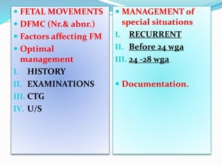
1) Performing a history and examination to check for fetal viability, assess risk factors, and exclude complications like preeclampsia.
2) Using cardiotocography and ultrasound scanning when needed to check for fetal wellbeing and growth issues.
3) Providing additional surveillance and testing for women with recurrent RFM or RFM before 28 weeks, while reassuring low-risk women.
4) Documenting all assessments, advice, and plans for follow-up in the patient's records. The goal is to evaluate the fetus, identify at-risk pregnancies, and reassure women when the