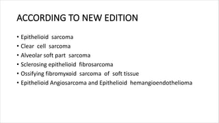
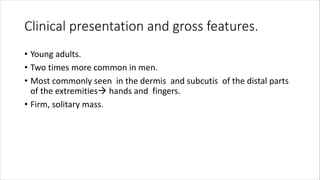
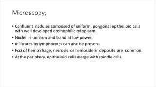
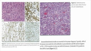
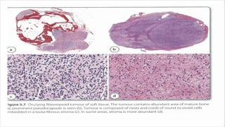
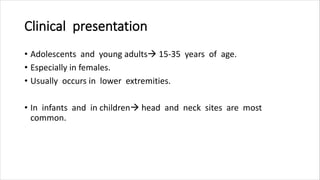
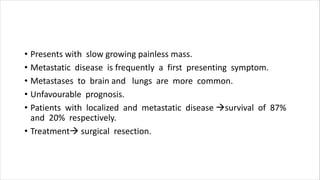
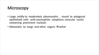
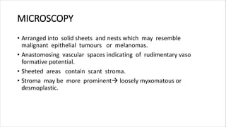
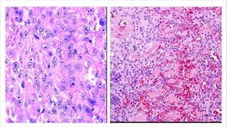
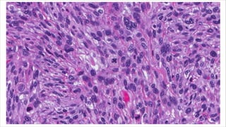
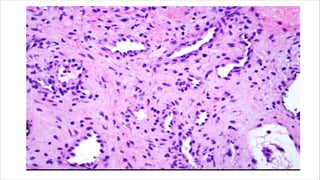
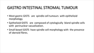
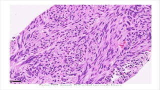
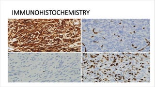
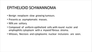
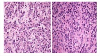
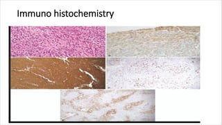
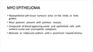
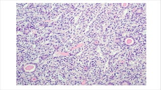
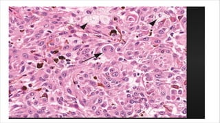
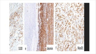
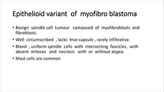
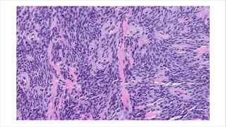
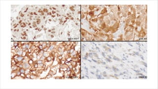
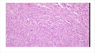
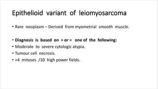
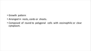
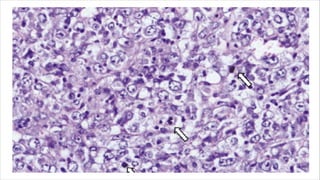
SOFT TISSUE TUMOURS WITH
EPITHELIOID MORPHOLOGY
RECENT ADVANCES-Vol :26- Chapter 5
DR.D.MANJU
PATHOLOGY POSTGRADUATE
INTRODUCTION
The purpose is to highlight some epithelioid mesenchymal tumours
which can be encountered in practice. As numerous soft tissue
neoplasms can exhibit an epithelioid variant, this discussion is by no
means exhaustive.
ACCORDING TO NEW EDITION
• Epithelioid sarcoma
• Clear cell sarcoma
• Alveolar soft part sarcoma
• Sclerosing epithelioid fibrosarcoma
• Ossifying fibromyxoid sarcoma of soft tissue
• Epithelioid Angiosarcoma and Epithelioid hemangioendothelioma
Epithelioid sarcoma
• It is a rare malignant soft tissue tumour composed of multinodular
aggregates of epithelioid cell surrounded by stromal mesenchymal
stem cells.
• The usual type.
• The proximal type.
Clinical presentation and gross features.
• Young adults.
• Two times more common in men.
• Most commonly seen in the dermis and subcutis of the distal parts
of the extremities hands and fingers.
• Firm, solitary mass.
Microscopy;
• Confluent nodules composed of uniform, polygonal epithelioid cells
with well developed eosinophilic cytoplasm.
• Nuclei is uniform and bland at low power.
• Infiltrates by lymphocytes can also be present.
• Foci of hemorrhage, necrosis or hemosiderin deposits are common.
• At the periphery, epithelioid cells merge with spindle cells.
Differential diagnosis:-
• Non Neoplastic granulomatous diseases.
• Non keratinizing squamous cell carcinoma and vascular tumours.
• Granulomatous inflammationpositive for CD163 and negative for
AE1/AE3.
Clear cell sarcoma of soft tissue
• Malignant tumour composed of glycogen rich clear cells.
• Also termed as malignant melanoma of soft tissue parts.
• Share some features of clear cell sarcoma of GIT.
Clinical presentation.
• 20-40 years of age (Median age is 39 years)
• Arises from tendon sheaths and aponeurosis.
• Occurs in lower limbs, especially ankles.
Microscopy
• Epithelioid to spindle cell morphology vesicular nuclei ,
prominent nucleoli & abundant clear cytoplasm.
• Multinucleated tumour giant cells and melanin pigment may also be
seen.
Differential diagnosis
• Melanoma and other soft tissue sarcomas of epithelioid or spindle
cell morphology.
• Synovial sarcoma and malignant peripheral nerve sheath tumour.
Ossifying fibromyxoid tumour of soft tissue
• Rare neoplasm.
• Occur in young adults.
• Commonly seen in males.
• Arises in the extremities.
• Occurs in head and neck ,trunk , mediastinum.
Microscopy
• Surrounded by pseudo capsule.
• Composed of round to ovoid and spindled cells with myxoid stroma
or fibrous stroma with vesicular nuclei, small nucleoli and small
eosinophilic cytoplasm.
• Sometimes have partial amounts of bone.
• Low cellularity, low nuclear grade and mitotic activity.
• Tumour cells express S100.
• Positive for cytokeratin, desmin ,CD56,CD57, neuron specific enolase
GFAP and SMA.
ALVEOLAR SOFT PART SARCOMA
• Alveolar soft part sarcoma
• Uncommon neoplasm <1%
Clinical presentation
• Adolescents and young adults 15-35 years of age.
• Especially in females.
• Usually occurs in lower extremities.
• In infants and in children head and neck sites are most
common.
• Presents with slow growing painless mass.
• Metastatic disease is frequently a first presenting symptom.
• Metastases to brain and lungs are more common.
• Unfavourable prognosis.
• Patients with localized and metastatic disease survival of 87%
and 20% respectively.
• Treatment surgical resection.
MICROSCOPY
• Composed of nests or aggregates of cells separated by
fibrovascular septa
• Cells show degeneration and loss of cell cohesiveness
pseudoalveolar pattern
• Some cases especially in infants and children show more solid
sheets of malignant cells with loss of nesting , lack of pseudo
alveolar pattern
DIFFERENTIAL DIAGNOSIS;
• Extraskeletal myxoid chondrosarcoma.
• Proximal variant of ES.
• Metastatic renal cell carcinoma
• Granular cell tumour.
Sclerosing epithelioid fibrosarcoma
• Diagnostically problematic.
• Most commonly presents as a mass in deep soft tissue in lower
extremities.
• Other sites:- upper extremities, trunk, head & neck regions.
• Age at diagnosis45 years of age.
• Characterised by local recurrences and distant metastases.
• Well circumscribed and lobulated neoplasm with myxoid changes
or calcification.
• Infiltrative in nature.
• The cells have round to ovoid nuclei, vesicular nuclei
inconspicious nucleoli and scant or pale cytoplasm.
MICROSCOPY
• Large or polygonal with eosinophilic granular cytoplasm
• Often with prominent nucleoli
• Nuclear pleomorphism including multinucleation and with rare
mitoses.
• Immunochemistry:- Positive for TFE3, cathepsin K, and desmin.
• Arranged into nests or cords and embedded into hyalinized
stroma.
• Spindled cells with varying amounts of hyalinized and myxoid
stroma.
• Arcuate vessels and hyalinized collagen rosettes seen.
Immunochemistry:-
• Positive for low grade fibromyxoid stroma.
• EMA positive, cytokeratin, S100, SMA, desmin and CD34 stains are
negative.
Epithelioid angiosarcoma
• Rare variant of angiosarcoma
• Composed of predominantly endothelial cells that have
epithelioid features.
• Occurs frequently in older adults
• Peak incidence in seventh decade of life.
• Seen in deep soft tissue of extremities and are commonly
intramuscular.
Microscopy
• Large, mildly to moderately pleomorphic , round to polygonal
epithelioid cells with eosinophilic cytoplasm, vesicular nuclei
containing prominent nucleoli.
• Metastatic to lungs and other organs Lethal
MICROSCOPY
• Arranged into solid sheets and nests which may resemble
malignant epithelial tumours or melanomas.
• Anastomosing vascular spaces indicating of rudimentary vaso
formative potential.
• Sheeted areas contain scant stroma.
• Stroma may be more prominent loosely myxomatous or
desmoplastic.
Immunochemistry
• Hybrid nature of tumour cells which express epithelial or
mesenchymal cells markers.
• Mitoses and areas of necrosis are seen.
Differential diagnosis
• Includes carcinomas and vascular tumours.
• Epithelioid tumours have lower mitotic activity and areas of
hemorrhage and necrosis are not prominent.
• Nuclei are less hyperchromatic and without prominent nucleoli.
• IHC : Positive for CD31,CD34 and ERG.
Epithelioid hemangioendothelioma
• Another vascular tumour with epithelioid morphology.
• Often occurs in young adults
• Found around a vessel in the superficial soft tissue
• Other sites: pleura, lungs,bone and liver.
• Present as a single mass or have a multicentric presentation.
Microscopy
• Less vasoformative structure.
• Composed of individual cords or small nests of epithelioid cells in
myxo hyalinized stroma.
• Painful mass
• Centered in a vascular space that extends through the vessel into
the adjacent soft tissue.
• The cells have eosinophilic cytoplasm with vesicular nuclei.
• High risk tumours  > or = 3 mitoses /50 HPF or >=3cm in the
greatest dimension.
• <3cm in greatest dimension & <3mitoses/HPF and the low risk
group have no tumour specific mortality.
Immunohistochemistry
• Positive : CD31 and ERG.
• CD34 is considered to be less sensitive.
• WWTR1 –CAM TA1 fusion transcripts have been identified in
identifying the accuracy and consistency of diagnosis.
Epithelioid variant of other soft tissue
tumours
• Leiomyosarcoma
• Pleomorphic liposarcoma
• Myofibroblastoma
• Myxofibrosarcoma
• Schwannoma
• Malignant peripheral nerve sheath tumour
• GIST
• Melanoma
OTHER TUMOURS WITH EPITHELIOID
MORPHOLOGY
• Epithelioid subtype of myxofibrosarcoma.
• Predominantly of atypical epithelioid tumour cells
• Abundant eosinophilic cytoplasm and round vesicular nuclei
arranged in small cohesive clusters in the myxoid areas forming
sheets in the hypercellular areas mimicking metastatic carcinoma or
melanoma.
MYO PERICYTOMA-INTRA VASCULAR
• Distinctive perivascular myoid neoplasm.
• Arises in the dermis and subcutis and it rarely involves in deep soft
tissue.
• The distal extremities are affected.
• Encapsulated and well circumscribed nodular lesions
• Composed of bland oval to spindle shaped cells myoid tumour cells
with characteristic multilayered concentric growth around tumour
cells.
GASTRO INTESTINAL STROMAL TUMOUR
• Most gastric GISTS are spindle cell tumours with epithelioid
morphology.
• Epithelioid GISTS are composed of cytologically bland spindle cells
with perinuclear vacuolization.
• Small bowel GISTS have spindle cell morphology with the presence
of skenoid fibres.
IMMUNOHISTOCHEMISTRY
EPITHELIOID SCHWANNOMA
• Benign neoplasm- slow growing tumours.
• Presents as asymptomatic masses.
• 90% are solitary.
• Composed of uniform epithelioid cells with round nuclei and
amphophilic cytoplasm with a myxoid fibrous stroma.
• Mitoses, Necrosis and cytoplasmic nuclear inclusions are seen.
Immuno histochemistry
MYO EPITHELIOMA
• Myoepithelial soft tissue tumours arise on the limbs or limb
girdles.
• Most patients present with painless masses.
• Composed of bland appearing ovoid and epithelioid cells with
uniform nuclei and eosinophilic cytoplasm
• Reticular or trabecular pattern with a prominent myxoid stroma.
Epithelioid variant of Melanoma
• Melanocytic tumour affects infants and children.
• Extremities are affected commonly .
• Head and neck and trunk are also get affected.
• Composed of small to large epithelioid cells with vesicular nuclei
with central nucleoli.
• Rare or absent mitoses and necrosis.
Epithelioid variant of myofibro blastoma
• Benign spindle cell tumour composed of myofibroblasts and
fibroblasts.
• Well circumscribed , lacks true capsule , rarely infiltrative.
• Bland , uniform spindle cells with intersecting fascicles, with
absent mitoses and necrosis with or without atypia.
• Mast cells are common
Epithelioid variant of pleomorphic
liposarcoma
• Rare variant of liposarcomas.
• Peak incidence in seventh decade of life.
• Occurs in extremities.
• Aggressive often exhibiting local recurrence and metastases.
Epithelioid variant of Malignant peripheral
nerve sheath tumour
• Rare subtype not associated with NF1
• Typically have strong expression of S100 and SOX10.
• Aggressive behaviour with frequent metastases and local
recurrence.
• Composed of uniform spindle cells with hyperchromatic, thin, wavy
or focally buckled nuclei.
• Composed of pleomorphic lipoblasts, subtle to prominent
cytoplasmic vacuolization.
• Multinucleated giant cells with signet ring lipoblasts .
• Tumour necrosis , with mitotic figures /10 high power fields.
• Neutrophils within giant cells are present.
Epithelioid variant of leiomyosarcoma
• Rare neoplasm – Derived from myometrial smooth muscle.
• Diagnosis is based on > or = one of the following:
• Moderate to severe cytologic atypia.
• Tumour cell necrosis.
• >4 mitoses /10 high power fields.
• Growth pattern
• Arranged in nests, cords or sheets.
• Composed of round to polygonal cells with eosinophilic or clear
cytoplasm.
RECENT ADVANCES- SOFT TISSUE TUMOURS.pdf

RECENT ADVANCES- SOFT TISSUE TUMOURS.pdf

  • 1.
    SOFT TISSUE TUMOURSWITH EPITHELIOID MORPHOLOGY RECENT ADVANCES-Vol :26- Chapter 5 DR.D.MANJU PATHOLOGY POSTGRADUATE
  • 2.
    INTRODUCTION The purpose isto highlight some epithelioid mesenchymal tumours which can be encountered in practice. As numerous soft tissue neoplasms can exhibit an epithelioid variant, this discussion is by no means exhaustive.
  • 3.
    ACCORDING TO NEWEDITION • Epithelioid sarcoma • Clear cell sarcoma • Alveolar soft part sarcoma • Sclerosing epithelioid fibrosarcoma • Ossifying fibromyxoid sarcoma of soft tissue • Epithelioid Angiosarcoma and Epithelioid hemangioendothelioma
  • 4.
    Epithelioid sarcoma • Itis a rare malignant soft tissue tumour composed of multinodular aggregates of epithelioid cell surrounded by stromal mesenchymal stem cells. • The usual type. • The proximal type.
  • 5.
    Clinical presentation andgross features. • Young adults. • Two times more common in men. • Most commonly seen in the dermis and subcutis of the distal parts of the extremities hands and fingers. • Firm, solitary mass.
  • 6.
    Microscopy; • Confluent nodulescomposed of uniform, polygonal epithelioid cells with well developed eosinophilic cytoplasm. • Nuclei is uniform and bland at low power. • Infiltrates by lymphocytes can also be present. • Foci of hemorrhage, necrosis or hemosiderin deposits are common. • At the periphery, epithelioid cells merge with spindle cells.
  • 8.
    Differential diagnosis:- • NonNeoplastic granulomatous diseases. • Non keratinizing squamous cell carcinoma and vascular tumours. • Granulomatous inflammationpositive for CD163 and negative for AE1/AE3.
  • 9.
    Clear cell sarcomaof soft tissue • Malignant tumour composed of glycogen rich clear cells. • Also termed as malignant melanoma of soft tissue parts. • Share some features of clear cell sarcoma of GIT.
  • 10.
    Clinical presentation. • 20-40years of age (Median age is 39 years) • Arises from tendon sheaths and aponeurosis. • Occurs in lower limbs, especially ankles.
  • 11.
    Microscopy • Epithelioid tospindle cell morphology vesicular nuclei , prominent nucleoli & abundant clear cytoplasm. • Multinucleated tumour giant cells and melanin pigment may also be seen.
  • 14.
    Differential diagnosis • Melanomaand other soft tissue sarcomas of epithelioid or spindle cell morphology. • Synovial sarcoma and malignant peripheral nerve sheath tumour.
  • 15.
    Ossifying fibromyxoid tumourof soft tissue • Rare neoplasm. • Occur in young adults. • Commonly seen in males. • Arises in the extremities. • Occurs in head and neck ,trunk , mediastinum.
  • 16.
    Microscopy • Surrounded bypseudo capsule. • Composed of round to ovoid and spindled cells with myxoid stroma or fibrous stroma with vesicular nuclei, small nucleoli and small eosinophilic cytoplasm. • Sometimes have partial amounts of bone.
  • 17.
    • Low cellularity,low nuclear grade and mitotic activity. • Tumour cells express S100. • Positive for cytokeratin, desmin ,CD56,CD57, neuron specific enolase GFAP and SMA.
  • 20.
    ALVEOLAR SOFT PARTSARCOMA • Alveolar soft part sarcoma • Uncommon neoplasm <1%
  • 21.
    Clinical presentation • Adolescentsand young adults 15-35 years of age. • Especially in females. • Usually occurs in lower extremities. • In infants and in children head and neck sites are most common.
  • 22.
    • Presents withslow growing painless mass. • Metastatic disease is frequently a first presenting symptom. • Metastases to brain and lungs are more common. • Unfavourable prognosis. • Patients with localized and metastatic disease survival of 87% and 20% respectively. • Treatment surgical resection.
  • 23.
    MICROSCOPY • Composed ofnests or aggregates of cells separated by fibrovascular septa • Cells show degeneration and loss of cell cohesiveness pseudoalveolar pattern • Some cases especially in infants and children show more solid sheets of malignant cells with loss of nesting , lack of pseudo alveolar pattern
  • 26.
    DIFFERENTIAL DIAGNOSIS; • Extraskeletalmyxoid chondrosarcoma. • Proximal variant of ES. • Metastatic renal cell carcinoma • Granular cell tumour.
  • 27.
    Sclerosing epithelioid fibrosarcoma •Diagnostically problematic. • Most commonly presents as a mass in deep soft tissue in lower extremities. • Other sites:- upper extremities, trunk, head & neck regions. • Age at diagnosis45 years of age.
  • 28.
    • Characterised bylocal recurrences and distant metastases. • Well circumscribed and lobulated neoplasm with myxoid changes or calcification. • Infiltrative in nature. • The cells have round to ovoid nuclei, vesicular nuclei inconspicious nucleoli and scant or pale cytoplasm.
  • 29.
    MICROSCOPY • Large orpolygonal with eosinophilic granular cytoplasm • Often with prominent nucleoli • Nuclear pleomorphism including multinucleation and with rare mitoses. • Immunochemistry:- Positive for TFE3, cathepsin K, and desmin.
  • 30.
    • Arranged intonests or cords and embedded into hyalinized stroma. • Spindled cells with varying amounts of hyalinized and myxoid stroma. • Arcuate vessels and hyalinized collagen rosettes seen.
  • 31.
    Immunochemistry:- • Positive forlow grade fibromyxoid stroma. • EMA positive, cytokeratin, S100, SMA, desmin and CD34 stains are negative.
  • 34.
    Epithelioid angiosarcoma • Rarevariant of angiosarcoma • Composed of predominantly endothelial cells that have epithelioid features. • Occurs frequently in older adults • Peak incidence in seventh decade of life. • Seen in deep soft tissue of extremities and are commonly intramuscular.
  • 35.
    Microscopy • Large, mildlyto moderately pleomorphic , round to polygonal epithelioid cells with eosinophilic cytoplasm, vesicular nuclei containing prominent nucleoli. • Metastatic to lungs and other organs Lethal
  • 36.
    MICROSCOPY • Arranged intosolid sheets and nests which may resemble malignant epithelial tumours or melanomas. • Anastomosing vascular spaces indicating of rudimentary vaso formative potential. • Sheeted areas contain scant stroma. • Stroma may be more prominent loosely myxomatous or desmoplastic.
  • 38.
    Immunochemistry • Hybrid natureof tumour cells which express epithelial or mesenchymal cells markers. • Mitoses and areas of necrosis are seen.
  • 39.
    Differential diagnosis • Includescarcinomas and vascular tumours. • Epithelioid tumours have lower mitotic activity and areas of hemorrhage and necrosis are not prominent. • Nuclei are less hyperchromatic and without prominent nucleoli. • IHC : Positive for CD31,CD34 and ERG.
  • 40.
    Epithelioid hemangioendothelioma • Anothervascular tumour with epithelioid morphology. • Often occurs in young adults • Found around a vessel in the superficial soft tissue • Other sites: pleura, lungs,bone and liver. • Present as a single mass or have a multicentric presentation.
  • 41.
    Microscopy • Less vasoformativestructure. • Composed of individual cords or small nests of epithelioid cells in myxo hyalinized stroma. • Painful mass • Centered in a vascular space that extends through the vessel into the adjacent soft tissue.
  • 43.
    • The cellshave eosinophilic cytoplasm with vesicular nuclei. • High risk tumours  > or = 3 mitoses /50 HPF or >=3cm in the greatest dimension. • <3cm in greatest dimension & <3mitoses/HPF and the low risk group have no tumour specific mortality.
  • 44.
    Immunohistochemistry • Positive :CD31 and ERG. • CD34 is considered to be less sensitive. • WWTR1 –CAM TA1 fusion transcripts have been identified in identifying the accuracy and consistency of diagnosis.
  • 45.
    Epithelioid variant ofother soft tissue tumours • Leiomyosarcoma • Pleomorphic liposarcoma • Myofibroblastoma • Myxofibrosarcoma • Schwannoma • Malignant peripheral nerve sheath tumour • GIST • Melanoma
  • 46.
    OTHER TUMOURS WITHEPITHELIOID MORPHOLOGY • Epithelioid subtype of myxofibrosarcoma. • Predominantly of atypical epithelioid tumour cells • Abundant eosinophilic cytoplasm and round vesicular nuclei arranged in small cohesive clusters in the myxoid areas forming sheets in the hypercellular areas mimicking metastatic carcinoma or melanoma.
  • 48.
    MYO PERICYTOMA-INTRA VASCULAR •Distinctive perivascular myoid neoplasm. • Arises in the dermis and subcutis and it rarely involves in deep soft tissue. • The distal extremities are affected. • Encapsulated and well circumscribed nodular lesions • Composed of bland oval to spindle shaped cells myoid tumour cells with characteristic multilayered concentric growth around tumour cells.
  • 50.
    GASTRO INTESTINAL STROMALTUMOUR • Most gastric GISTS are spindle cell tumours with epithelioid morphology. • Epithelioid GISTS are composed of cytologically bland spindle cells with perinuclear vacuolization. • Small bowel GISTS have spindle cell morphology with the presence of skenoid fibres.
  • 52.
  • 53.
    EPITHELIOID SCHWANNOMA • Benignneoplasm- slow growing tumours. • Presents as asymptomatic masses. • 90% are solitary. • Composed of uniform epithelioid cells with round nuclei and amphophilic cytoplasm with a myxoid fibrous stroma. • Mitoses, Necrosis and cytoplasmic nuclear inclusions are seen.
  • 55.
  • 56.
    MYO EPITHELIOMA • Myoepithelialsoft tissue tumours arise on the limbs or limb girdles. • Most patients present with painless masses. • Composed of bland appearing ovoid and epithelioid cells with uniform nuclei and eosinophilic cytoplasm • Reticular or trabecular pattern with a prominent myxoid stroma.
  • 59.
    Epithelioid variant ofMelanoma • Melanocytic tumour affects infants and children. • Extremities are affected commonly . • Head and neck and trunk are also get affected. • Composed of small to large epithelioid cells with vesicular nuclei with central nucleoli. • Rare or absent mitoses and necrosis.
  • 62.
    Epithelioid variant ofmyofibro blastoma • Benign spindle cell tumour composed of myofibroblasts and fibroblasts. • Well circumscribed , lacks true capsule , rarely infiltrative. • Bland , uniform spindle cells with intersecting fascicles, with absent mitoses and necrosis with or without atypia. • Mast cells are common
  • 64.
    Epithelioid variant ofpleomorphic liposarcoma • Rare variant of liposarcomas. • Peak incidence in seventh decade of life. • Occurs in extremities. • Aggressive often exhibiting local recurrence and metastases.
  • 67.
    Epithelioid variant ofMalignant peripheral nerve sheath tumour • Rare subtype not associated with NF1 • Typically have strong expression of S100 and SOX10. • Aggressive behaviour with frequent metastases and local recurrence. • Composed of uniform spindle cells with hyperchromatic, thin, wavy or focally buckled nuclei.
  • 68.
    • Composed ofpleomorphic lipoblasts, subtle to prominent cytoplasmic vacuolization. • Multinucleated giant cells with signet ring lipoblasts . • Tumour necrosis , with mitotic figures /10 high power fields. • Neutrophils within giant cells are present.
  • 70.
    Epithelioid variant ofleiomyosarcoma • Rare neoplasm – Derived from myometrial smooth muscle. • Diagnosis is based on > or = one of the following: • Moderate to severe cytologic atypia. • Tumour cell necrosis. • >4 mitoses /10 high power fields.
  • 71.
    • Growth pattern •Arranged in nests, cords or sheets. • Composed of round to polygonal cells with eosinophilic or clear cytoplasm.