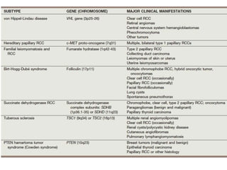
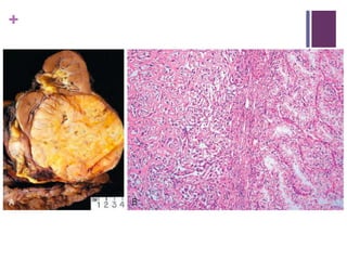
This document summarizes renal cell carcinoma (RCC), including its incidence, risk factors, pathology, subtypes, clinical presentation, and prognosis. RCC accounts for 2-3% of adult cancers and is the most lethal urologic cancer. Clear cell RCC is the most common subtype, accounting for 70-80% of cases. Other subtypes include papillary RCC, chromophobe RCC, collecting duct carcinoma, and unclassified RCC. Tumor size, grade, stage, and histologic subtype are important prognostic factors. Over 60% of RCCs are now detected incidentally with improved imaging techniques.
This is a presentation on the topic of cytology of the breast, prepared by Dr Ashish Jawarkar, he is MD in pathology and a teacher at Parul institute of Medical sciences and research Vadodara.
this PPT is all about case base approach to kidney tumors. clinical approach and their radiological findings. indication and contra-indications of Kidney FNAC of Kidney lesions.
Basavarajeeyam is a Sreshta Sangraha grantha (Compiled book ), written by Neelkanta kotturu Basavaraja Virachita. It contains 25 Prakaranas, First 24 Chapters related to Rogas& 25th to Rasadravyas.
New Drug Discovery and Development .....NEHA GUPTA
The "New Drug Discovery and Development" process involves the identification, design, testing, and manufacturing of novel pharmaceutical compounds with the aim of introducing new and improved treatments for various medical conditions. This comprehensive endeavor encompasses various stages, including target identification, preclinical studies, clinical trials, regulatory approval, and post-market surveillance. It involves multidisciplinary collaboration among scientists, researchers, clinicians, regulatory experts, and pharmaceutical companies to bring innovative therapies to market and address unmet medical needs.
These simplified slides by Dr. Sidra Arshad present an overview of the non-respiratory functions of the respiratory tract.
Learning objectives:
1. Enlist the non-respiratory functions of the respiratory tract
2. Briefly explain how these functions are carried out
3. Discuss the significance of dead space
4. Differentiate between minute ventilation and alveolar ventilation
5. Describe the cough and sneeze reflexes
Study Resources:
1. Chapter 39, Guyton and Hall Textbook of Medical Physiology, 14th edition
2. Chapter 34, Ganong’s Review of Medical Physiology, 26th edition
3. Chapter 17, Human Physiology by Lauralee Sherwood, 9th edition
4. Non-respiratory functions of the lungs https://academic.oup.com/bjaed/article/13/3/98/278874
CDSCO and Phamacovigilance {Regulatory body in India}NEHA GUPTA
The Central Drugs Standard Control Organization (CDSCO) is India's national regulatory body for pharmaceuticals and medical devices. Operating under the Directorate General of Health Services, Ministry of Health & Family Welfare, Government of India, the CDSCO is responsible for approving new drugs, conducting clinical trials, setting standards for drugs, controlling the quality of imported drugs, and coordinating the activities of State Drug Control Organizations by providing expert advice.
Pharmacovigilance, on the other hand, is the science and activities related to the detection, assessment, understanding, and prevention of adverse effects or any other drug-related problems. The primary aim of pharmacovigilance is to ensure the safety and efficacy of medicines, thereby protecting public health.
In India, pharmacovigilance activities are monitored by the Pharmacovigilance Programme of India (PvPI), which works closely with CDSCO to collect, analyze, and act upon data regarding adverse drug reactions (ADRs). Together, they play a critical role in ensuring that the benefits of drugs outweigh their risks, maintaining high standards of patient safety, and promoting the rational use of medicines.
These lecture slides, by Dr Sidra Arshad, offer a quick overview of the physiological basis of a normal electrocardiogram.
Learning objectives:
1. Define an electrocardiogram (ECG) and electrocardiography
2. Describe how dipoles generated by the heart produce the waveforms of the ECG
3. Describe the components of a normal electrocardiogram of a typical bipolar lead (limb II)
4. Differentiate between intervals and segments
5. Enlist some common indications for obtaining an ECG
6. Describe the flow of current around the heart during the cardiac cycle
7. Discuss the placement and polarity of the leads of electrocardiograph
8. Describe the normal electrocardiograms recorded from the limb leads and explain the physiological basis of the different records that are obtained
9. Define mean electrical vector (axis) of the heart and give the normal range
10. Define the mean QRS vector
11. Describe the axes of leads (hexagonal reference system)
12. Comprehend the vectorial analysis of the normal ECG
13. Determine the mean electrical axis of the ventricular QRS and appreciate the mean axis deviation
14. Explain the concepts of current of injury, J point, and their significance
Study Resources:
1. Chapter 11, Guyton and Hall Textbook of Medical Physiology, 14th edition
2. Chapter 9, Human Physiology - From Cells to Systems, Lauralee Sherwood, 9th edition
3. Chapter 29, Ganong’s Review of Medical Physiology, 26th edition
4. Electrocardiogram, StatPearls - https://www.ncbi.nlm.nih.gov/books/NBK549803/
5. ECG in Medical Practice by ABM Abdullah, 4th edition
6. Chapter 3, Cardiology Explained, https://www.ncbi.nlm.nih.gov/books/NBK2214/
7. ECG Basics, http://www.nataliescasebook.com/tag/e-c-g-basics
micro teaching on communication m.sc nursing.pdfAnurag Sharma
Microteaching is a unique model of practice teaching. It is a viable instrument for the. desired change in the teaching behavior or the behavior potential which, in specified types of real. classroom situations, tends to facilitate the achievement of specified types of objectives.
Ozempic: Preoperative Management of Patients on GLP-1 Receptor Agonists Saeid Safari
Preoperative Management of Patients on GLP-1 Receptor Agonists like Ozempic and Semiglutide
ASA GUIDELINE
NYSORA Guideline
2 Case Reports of Gastric Ultrasound
Title: Sense of Taste
Presenter: Dr. Faiza, Assistant Professor of Physiology
Qualifications:
MBBS (Best Graduate, AIMC Lahore)
FCPS Physiology
ICMT, CHPE, DHPE (STMU)
MPH (GC University, Faisalabad)
MBA (Virtual University of Pakistan)
Learning Objectives:
Describe the structure and function of taste buds.
Describe the relationship between the taste threshold and taste index of common substances.
Explain the chemical basis and signal transduction of taste perception for each type of primary taste sensation.
Recognize different abnormalities of taste perception and their causes.
Key Topics:
Significance of Taste Sensation:
Differentiation between pleasant and harmful food
Influence on behavior
Selection of food based on metabolic needs
Receptors of Taste:
Taste buds on the tongue
Influence of sense of smell, texture of food, and pain stimulation (e.g., by pepper)
Primary and Secondary Taste Sensations:
Primary taste sensations: Sweet, Sour, Salty, Bitter, Umami
Chemical basis and signal transduction mechanisms for each taste
Taste Threshold and Index:
Taste threshold values for Sweet (sucrose), Salty (NaCl), Sour (HCl), and Bitter (Quinine)
Taste index relationship: Inversely proportional to taste threshold
Taste Blindness:
Inability to taste certain substances, particularly thiourea compounds
Example: Phenylthiocarbamide
Structure and Function of Taste Buds:
Composition: Epithelial cells, Sustentacular/Supporting cells, Taste cells, Basal cells
Features: Taste pores, Taste hairs/microvilli, and Taste nerve fibers
Location of Taste Buds:
Found in papillae of the tongue (Fungiform, Circumvallate, Foliate)
Also present on the palate, tonsillar pillars, epiglottis, and proximal esophagus
Mechanism of Taste Stimulation:
Interaction of taste substances with receptors on microvilli
Signal transduction pathways for Umami, Sweet, Bitter, Sour, and Salty tastes
Taste Sensitivity and Adaptation:
Decrease in sensitivity with age
Rapid adaptation of taste sensation
Role of Saliva in Taste:
Dissolution of tastants to reach receptors
Washing away the stimulus
Taste Preferences and Aversions:
Mechanisms behind taste preference and aversion
Influence of receptors and neural pathways
Impact of Sensory Nerve Damage:
Degeneration of taste buds if the sensory nerve fiber is cut
Abnormalities of Taste Detection:
Conditions: Ageusia, Hypogeusia, Dysgeusia (parageusia)
Causes: Nerve damage, neurological disorders, infections, poor oral hygiene, adverse drug effects, deficiencies, aging, tobacco use, altered neurotransmitter levels
Neurotransmitters and Taste Threshold:
Effects of serotonin (5-HT) and norepinephrine (NE) on taste sensitivity
Supertasters:
25% of the population with heightened sensitivity to taste, especially bitterness
Increased number of fungiform papillae
Muktapishti is a traditional Ayurvedic preparation made from Shoditha Mukta (Purified Pearl), is believed to help regulate thyroid function and reduce symptoms of hyperthyroidism due to its cooling and balancing properties. Clinical evidence on its efficacy remains limited, necessitating further research to validate its therapeutic benefits.
2. +
RCC in numbers
Accounts for 2% to 3% of all adult malignant neoplasms
The most lethal of the common urologic cancers.
Five-year relative survival rates for patients diagnosed in 2002
to 2008 were 71% for kidney cancer, 78% for bladder cancer
(excluding carcinoma in situ), and 99% for prostate cancer
(Siegel et al, 2013).
3. +
Approximately 65,000 new diagnoses of RCC are made each
year in the United States, and 13,000 patients die of disease
(Siegel et al,2013).
Overall, approximately 12 new cases are diagnosed per
100,000 population per year, with a male-to-female
predominance of 3:2 (Siegel et al, 2013).
This is primarily a disease of older adults, with typical
presentation between 50 and 70 years of age.
4. +
However, diagnosis of renal cancer has increased more rapidly
in those less than 40 years of age than any other age group
(Nepple et al, 2012).
Incidence rates are 10% to 20% higher and 5-year survival
rates 5% lower in African-Americans for unknown reasons
(Lipworth et al, 2006; Stafford et al, 2008; Chow et al,2013;
Siegel et al, 2013).
5. +
The majority of cases of RCC are believed to be sporadic; only
2% to 3% are proven to be familial.
6. +
The incidence of RCC has increased since the 1970s by an
average of 3% to 4% per year, largely related to the more
prevalent use of ultrasonography of abdominal complaints
7. +
Although Wilms tumor is much more common in younger
children, RCC is as common as Wilms tumor during the second
decade of life.
9. +
RCCs were traditionally thought to arise primarily from the
proximal convoluted tubules, and this is probably true for the
clear cell and papillary variants.
However, other histologic subtypes of RCC, such as
chromophobe RCC and collecting duct carcinoma, are derived
from the more distal components of the nephron
10. +
The most general accepted environmental risk factor for RCC
is tobacco exposure ; Relative risk---1.4 to 2.5.
Obesity is now accepted as another major risk factor for RCC,
with an increased relative risk of 1.07 for each additional unit of
body mass index.
11. +
Hypertension appears to be the 3rd major etiologic
factor for RCC.
The proposed mechanisms are hypertension induced renal
injury and inflammation or metabolic or functional changes in
the renal tubules that may increase susceptibility to
carcinogens
12. +
A family history of RCC may also be a factor;
One study showed a relative risk of 2.9 for individuals with a
first- or second-degree relative with RCC.
13. +
Other potential iatrogenic causes include regular usage of
nonsteroidal anti-inflammatory drugs, which is associated with
a relative risk of 1.51,
while aspirin and acetaminophen were not associated with any
increased risk (Cho et al, 2011).
14. +
Retroperitoneal radiation therapy, typically administered for
Wilms tumor or testicular cancer, appears to be a risk factor for
RCC, although the relative risks are low (Romanenko et al,
2000).
15. +
An increased incidence of RCC is also observed in patients
with end-stage renal disease and certain familial
syndromes.
21. +
Most RCCs are round to ovoid and circumscribed by a
pseudocapsule of compressed parenchyma and fibrous tissue
rather than a true histologic capsule.
Unlike upper tract urothelial carcinomas, most RCCs are not
grossly infiltrative, with the notable exception of collecting duct
carcinoma and sarcomatoid variants.
22. +
Size Matters…
Tumor size has averaged between 4 and 8 cm in most series
but can vary from a few millimeters to large enough to fill the
entire abdomen.
Tumors smaller than 3 cm were previously classified a benign
adenomas, but some small tumors have been associated with
metastases.
With the exception of oncocytomas and some small (<5-mm)
low-grade papillary adenomas, there are no reliable histologic
or ultrastructural criteria to differentiate benign from malignant
renal epithelial tumors.
23. +
When they are bivalved, RCCs consist of yellow, tan, or brown
tumor interspersed with fibrotic, necrotic, or hemorrhagic areas;
few are uniform in gross appearance.
Cystic degeneration is found in 10% to 25% of RCCs and
appears to be associated with a better prognosis compared
with purely solid RCC.
Calcification can be stippled or plaque like and is found in 10%
to 20% of RCCs.
24. +
Nuclear features can be highly variable.
Grading has been based primarily on nuclear size and shape
and the presence or absence of prominent nucleoli.
25. +
Fuhrman’s system is an independent prognostic factor for RCC
generally and for clear cell RCC in particular.
Recent evidence suggests that Fuhrman grade is also a
significant predictor of outcome for papillary RCC, but features
other than nuclear characteristics may form the basis of a
preferred scheme for chromophobe RCC
27. +
Frank invasion and perforation of the renal capsule, renal
sinus, or collecting system are found in approximately 20% of
cases, although displacement of these structures is a more
common finding.
28. +
One unique feature of RCC is its predilection for involvement of
the venous system, which is found in 10% of RCCs, more often
than in any other tumor type.
This is most commonly manifested in the form of a contiguous
tumor thrombus that can extend into the inferior vena cava
(IVC) as high as the right atrium.
Many such tumor thrombi are highly vascularized by arterial
blood flow, and some directly invade the wall of the renal vein
or vena cava, which correlates with compromised prognosis
29. +
Most sporadic RCCs are unilateral and unifocal.
Bilateral involvement can be synchronous or asynchronous and
is found in 2% to 4% of sporadic RCCs, although it is
considerably more common in patients with familial forms of
RCC, such as von Hippel-Lindau disease.
Multicentricity, which is found in 10% to 20% of cases, is more
common in association with papillary histology and familial
RCC.
30. +
Microsatellite analysis suggests a clonal origin for most
multifocal RCC within the same kidney, but tumor in the
contralateral kidney is likely to be an independent growth if it is
synchronous or a metastasis if it is asynchronous.
31. +
Clear Cell RCC
Clear cell RCC accounts for 70% to 80% of all RCCs, formerly
known as “conventional” RCC.
These tumors are typically yellow when they are bivalved and
are highly vascular, containing a network of delicate vascular
sinusoids interspersed between sheets or acini of tumor cells.
On microscopic examination, clear cell RCC can include clear
cell, granular cell, or mixed types.
33. +
In general, patients with clear cell RCC have a worse
prognosis compared with papillary or chromophobe RCC,
even after stratification for stage and grade.
34. +
Papillary Renal Cell Carcinoma
Also designated chromophilic RCC in previous classification
schemes
The 2nd most common histologic subtype .
It represents 10% to 15%of all RCCs.
36. +
Chromophobe
Renal Cell Carcinoma
First described in 1985
A distinctive histologic subtype of RCC that represents 5% of all
RCCs.
Appears to be derived from the cortical portion of the collecting
duct
37. +
A perinuclear clearing or “halo” is typically found
Electron microscopic findings consist of numerous 150- to 300-
nm microvesicles, which are the single most distinctive and
defining feature of chromophobe cell carcinoma.
These microvesicles characteristically stain positive for Hale
colloidal iron, indicating the presence of a mucopolysaccharide
unique to chromophobe RCC.
39. +
Most studies of the clinical behavior of chromophobe RCC
suggest a better prognosis for localized chromophobe RCC
than for clear cell RCC
but a poor outcome in the subset of patients with sarcomatoid
features or metastatic disease.
40. +
Collecting Duct Carcinoma
Carcinoma of the collecting ducts of Bellini is a relatively rare
subtype of RCC, accounting for less than 1% of all RCCs
Most reported cases of collecting duct carcinoma have been
high grade, advanced stage, and unresponsive to
conventional therapies
41. +
Renal Medullary Carcinoma
Renal medullary carcinoma is a subtype of RCC that occurs
almost exclusively in patients with the sickle cell trait.
It is typically diagnosed in young African-Americans, often in
the third decade of life, and many cases are both locally
advanced and metastatic at the time of diagnosis.
This tumor shares many histologic features with collecting duct
carcinoma, and some consider it a subtype of collecting duct
carcinoma or at least a closely related tumor.
42. +
Sarcomatoid Differentiation
Sarcomatoid differentiation is found in 1%-5% of RCCs
Most commonly in association with clear cell RCC or
chromophobe RCC.
Sarcomatoid differentiation is characterized by
spindle cell histology,
positive staining for vimentin,
infiltrative growth pattern,
aggressive local and metastatic behaviour &
poor prognosis.
44. +
Unclassified
Renal Cell Carcinoma
Unclassified RCC represents a small minority of cases (1% to
5%) of presumed RCC
Features remain indeterminate even after careful analysis.
Poorly differentiated
Associated with a highly aggressive biologic behaviour &
A particularly poor prognosis.
45. +
Clinical Presentation
Because of the sequestered location of the kidney within the
retroperitoneum, many renal masses remain asymptomatic and
nonpalpable until they are locally advanced.
With the more pervasive use of noninvasive imaging for the
evaluation of a variety of nonspecific symptom complexes,
more than 60% of RCCs are now detected incidentally
47. +
A less common but important presentation of RCC is that of
spontaneous perirenal hemorrhage, in which the underlying
mass may be obscured.
More than 50% of patients with perirenal hematoma of unclear
etiology have an occult renal tumor, most often AML or RCC.
48. +
Paraneoplastic syndromes are found in 10% to 20% of patients
with RCC, and few tumors are associated with the diversity of
such syndromes.
In fact, RCC was previously referred to as the internist’s
tumor because of the predominance of systemic rather than
local manifestations.
Now, a more appropriate name would be the radiologist’s
tumor, given the frequency of incidental detection
50. +
Hypercalcemia has been reported in up to 13% of patients with
RCC and can be due to either paraneoplastic phenomena or
osteolytic metastatic involvement of the bone.
51. +
The signs and symptoms of hypercalcemia are often
nonspecific and include nausea, anorexia fatigue, and
decreased deep tendon reflexes.
Medical management predominates and includes vigorous
hydration followed by diuresis with furosemide and the
selective use of bisphosphonates, corticosteroids, or calcitonin.
52. +
Bisphosphonate therapy is now established as a standard of
care for patients with hypercalcemia of malignancy, as long as
renal function is adequate.
Zoledronic acid, 4 mg intravenously every 4 weeks, appears to
be particularly effective in patients with RCC but must be
withheld in the presence of renal insufficiency
53. +
Hypertension and polycythemia are other important
paraneoplastic syndromes commonly found in patients with
RCC
54. +
One of the more fascinating paraneoplastic syndromes
associated with RCC is nonmetastatic hepatic dysfunction, or
Stauffer syndrome, which has been reported in 3% to
20% of cases.
Almost all patients with Stauffer syndrome have an elevated serum
alkaline phosphatase level,
67% have elevated prothrombin time or hypoalbuminemia &
20% to 30% have elevated serum bilirubin or transaminase
55. +
In general, treatment of paraneoplastic syndromes associated
with RCC has required surgical excision or systemic therapy
and, except for hypercalcemia, medical therapies have not
proved helpful.
56. +
SCREENING
A number of factors make screening for RCC appealing.
Most important, RCC remains primarily a surgical disease
requiring early diagnosis to optimize the opportunity for cure.
57. +
The primary factor that limits the widespread implementation of
screening for RCC is the relatively low incidence of RCC in the
general population (approximately 12 cases per 100,000
population per year)
58. +
For now, however, the focus of screening for RCC must be on
well-defined target populations, such as patients with end-stage
renal disease and acquired renal cystic disease, tuberous
sclerosis, and familial RCC.