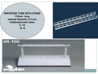
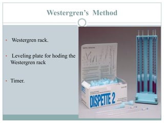
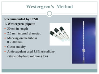
Rashida Ansari
Heamatopathology Lab
 Erythrocyte sedimentation rate (ESR) is a non-
specific test for inflammation.
 It is easy to perform, widely available and
inexpensive making it a widely used screening test
 It is also used a monitoring tool for response to
treatment in conditions in which it is raised
(Tuberculosis, Autoimmune diseases etc (
INTRODUCTION
ESR
 Consist of allowing a specific amount of blood
to sit in a vertical position for a period of time
(usually one hour).
 The distance, in millimeters, that the red cells
fall during this time period is the erythrocyte
sedimentation rate and is reported in mm at
one hour.
Stages of ESR
Three stages:
 Rouleaux formation
(10 minutes)
Red cells stack together
like pair of coins
 Sinking of rouleaux
(40 minutes)
 Cell packing :
(10 minutes)
The ESR is affected by three factors:
1. Erythrocytes
2. Plasma Composition
3. Mechanical/Technical Factors
Erythrocytes
 Macrocytes tend to sediment rapidly than
microcytes
 Alterations of ratio of red cells to plasma
 Decreased red cell mass in anaemia increases
ESR
 Increased red cell mass in polycythemia
decreases ESR
 Sickle cells and spherocytes are unable to form
rouleaux and therefore ESR is low.
Plasma Composition
 In normal blood, the RBCs remain more or less separated.
They are negatively charged and, therefore repel each
other. ( Zeta Potential)
 Increased plasma protein concentration, most notable
fibrinogen, immunoglobulins and acute phase proteins ( C-
reactive protein, ceruloplasmin, -1 globulin) , cause a
reduction in the negative charge of the RBCs and facilitates
formation of rouleaux.
 Removal of fibrinogen by defibrination and increase in the
albumin retard ESR
Mechanical/Technical factors
 It is important that the ESR tube be exactly perpendicular. A
tilt of 30⁰ can cause errors up to 30%.
 The rack, holding the tubes should not be subject to any
movement or vibration.
 The sedimentation rate increases as the temperature
increases.
 ESR tubes with a narrower than standard bore will generally
yield lower sedimentation rates.
Significance of the ESR
• The ESR represents a nonspecific response to tissue
damage and inflammation.
• It primarily reflects changes in the plasma proteins that
accompany most the acute and chronic infections, tumors,
and degenerative diseases.
• It may be used to follow the progress of certain diseases
such as tuberculosis and rheumatoid arthritis.
• Important criterion in establishing the diagnosis of
temporal arteritis and polymyalgia rheumatica.
 An elevated ESR may be found in
1. Pregnancy (after the third month).
2. Acute and chronic infections.
3. Rheumatic fever.
4. Rheumatoid arthritis.
5. Myocardial infection.
6. Nephrosis.
7. Acute hepatitis.
8. Menstruation.
9. Tuberculosis.
10. Hypothyroidism.
11. Hyperthyroidism.
 Adults over 60 years of age frequently have a slightly
higher ESR value due primarily to decreased
concentrations of plasma albumin.
 A decreased ESR will be present in:
1. Polycythemia.
2. Congestive heart failure.
3. Hypofibrinogenemia.
4. The presence of red blood cell abnormalities
(poikilocytosis, spherocytes, and sickle cells).
Methods for estimation of ESR
 MANUAL - Wintrobe’s Method
Westergen’s Method
Zeta Sedimentation ratio
Micro ESR Method
 AUTOMATED – Monitor 100
Ves-matic 60
Sedimatic
Sediscan
WINTROBE METHOD
WINTROBE TUBE WITH STAND
110mm long
Internal diameter 2.5 mm
Calibrated both sides
0- 10
10 -0
Westergren’s Method
• Westergren rack.
• Leveling plate for hoding the
Westergren rack
• Timer.
Westergren’s Method
Recommended by ICSH
1. Westergren pipette
 30 cm in length
 2.5 mm internal diameter,
 Marking on the tube is from 0 to 0 –
0 - 200 mm.
 Clean and dry
 Anticoagulant used 3.8% trisodium-
citrate dihydrate solution (1:4)
Specimen
 Venous blood collected in trisodium citrate solution in 4:1
proportion
 Test should be carried out within four hours of blood collection
if specimen is kept at room temperature
 Mix the blood and anticoagulant thouroughly
 There should be no clots and air bubbles in the blood.
Procedure
 Fill the Westergren pipette by inserting in a vacutainer tube
containing 1.6 ml blood and 0.4ml anticoagulant
 Keep the pipette upright in the ESR stand lying on the leveled
surface
 Read the upper level of RBC column exactly after one hour
 N.R. Adult Males : 0 to 10 mm in 1 hour &
Adult Females : 0 to 20 mm in 1 hour
Automated ESR method
Automated ESR analyzers
Vasmatic 20
WORKING PRINCIPLE
Blood is collected in a special cuvette
Sample is mixed by instrument
Sample is allowed to stand at 18° slant
from vertical
Opto-electrical sensor measure ESR
Data are elaborated and printed
ESR STAT
PLUS
centrifugation
based method
Provides result
in 5 minutes
WORKING PRINCIPLE
Sample is placed in centrifuge
Infrared laser tracks the erythrocytes plasma interface & takes multiple measurements
Linear portion of sedimentation is identified
Software algorithm to determine ESR result
Sedimate 15
Advantages of automated method
 Automated ESR analyzer is designed to accurately and
precisely measure the sedimentation rate of erythrocytes in
ESR tubes
 Less sample volume.
 Barcode Scanner
 Results in 20-30 minutes
 Built-in Printer
 No external influence of temperature, tilting of tubes.
 Higher number of samples can be processed simultaneously
Automated ESR method
 On comparing manual and automated methods for measuring
ESR, discrepancy was noted for HIGH ESR values.
 This was however, not evident for normal ESR values.
 Hence it is recommended that a correction factor be applied while
using automated equipment.
THANK YOU

Rashida Ansari ESR.pdf

  • 1.
  • 2.
     Erythrocyte sedimentationrate (ESR) is a non- specific test for inflammation.  It is easy to perform, widely available and inexpensive making it a widely used screening test  It is also used a monitoring tool for response to treatment in conditions in which it is raised (Tuberculosis, Autoimmune diseases etc ( INTRODUCTION
  • 3.
    ESR  Consist ofallowing a specific amount of blood to sit in a vertical position for a period of time (usually one hour).  The distance, in millimeters, that the red cells fall during this time period is the erythrocyte sedimentation rate and is reported in mm at one hour.
  • 4.
    Stages of ESR Threestages:  Rouleaux formation (10 minutes) Red cells stack together like pair of coins  Sinking of rouleaux (40 minutes)  Cell packing : (10 minutes)
  • 5.
    The ESR isaffected by three factors: 1. Erythrocytes 2. Plasma Composition 3. Mechanical/Technical Factors
  • 6.
    Erythrocytes  Macrocytes tendto sediment rapidly than microcytes  Alterations of ratio of red cells to plasma  Decreased red cell mass in anaemia increases ESR  Increased red cell mass in polycythemia decreases ESR  Sickle cells and spherocytes are unable to form rouleaux and therefore ESR is low.
  • 7.
    Plasma Composition  Innormal blood, the RBCs remain more or less separated. They are negatively charged and, therefore repel each other. ( Zeta Potential)  Increased plasma protein concentration, most notable fibrinogen, immunoglobulins and acute phase proteins ( C- reactive protein, ceruloplasmin, -1 globulin) , cause a reduction in the negative charge of the RBCs and facilitates formation of rouleaux.  Removal of fibrinogen by defibrination and increase in the albumin retard ESR
  • 8.
    Mechanical/Technical factors  Itis important that the ESR tube be exactly perpendicular. A tilt of 30⁰ can cause errors up to 30%.  The rack, holding the tubes should not be subject to any movement or vibration.  The sedimentation rate increases as the temperature increases.  ESR tubes with a narrower than standard bore will generally yield lower sedimentation rates.
  • 9.
    Significance of theESR • The ESR represents a nonspecific response to tissue damage and inflammation. • It primarily reflects changes in the plasma proteins that accompany most the acute and chronic infections, tumors, and degenerative diseases. • It may be used to follow the progress of certain diseases such as tuberculosis and rheumatoid arthritis. • Important criterion in establishing the diagnosis of temporal arteritis and polymyalgia rheumatica.
  • 10.
     An elevatedESR may be found in 1. Pregnancy (after the third month). 2. Acute and chronic infections. 3. Rheumatic fever. 4. Rheumatoid arthritis. 5. Myocardial infection. 6. Nephrosis. 7. Acute hepatitis. 8. Menstruation. 9. Tuberculosis. 10. Hypothyroidism. 11. Hyperthyroidism.
  • 11.
     Adults over60 years of age frequently have a slightly higher ESR value due primarily to decreased concentrations of plasma albumin.  A decreased ESR will be present in: 1. Polycythemia. 2. Congestive heart failure. 3. Hypofibrinogenemia. 4. The presence of red blood cell abnormalities (poikilocytosis, spherocytes, and sickle cells).
  • 12.
    Methods for estimationof ESR  MANUAL - Wintrobe’s Method Westergen’s Method Zeta Sedimentation ratio Micro ESR Method  AUTOMATED – Monitor 100 Ves-matic 60 Sedimatic Sediscan
  • 13.
  • 14.
    WINTROBE TUBE WITHSTAND 110mm long Internal diameter 2.5 mm Calibrated both sides 0- 10 10 -0
  • 16.
    Westergren’s Method • Westergrenrack. • Leveling plate for hoding the Westergren rack • Timer.
  • 17.
    Westergren’s Method Recommended byICSH 1. Westergren pipette  30 cm in length  2.5 mm internal diameter,  Marking on the tube is from 0 to 0 – 0 - 200 mm.  Clean and dry  Anticoagulant used 3.8% trisodium- citrate dihydrate solution (1:4)
  • 18.
    Specimen  Venous bloodcollected in trisodium citrate solution in 4:1 proportion  Test should be carried out within four hours of blood collection if specimen is kept at room temperature  Mix the blood and anticoagulant thouroughly  There should be no clots and air bubbles in the blood.
  • 19.
    Procedure  Fill theWestergren pipette by inserting in a vacutainer tube containing 1.6 ml blood and 0.4ml anticoagulant  Keep the pipette upright in the ESR stand lying on the leveled surface  Read the upper level of RBC column exactly after one hour  N.R. Adult Males : 0 to 10 mm in 1 hour & Adult Females : 0 to 20 mm in 1 hour
  • 22.
  • 24.
  • 25.
    WORKING PRINCIPLE Blood iscollected in a special cuvette Sample is mixed by instrument Sample is allowed to stand at 18° slant from vertical Opto-electrical sensor measure ESR Data are elaborated and printed
  • 27.
  • 28.
    WORKING PRINCIPLE Sample isplaced in centrifuge Infrared laser tracks the erythrocytes plasma interface & takes multiple measurements Linear portion of sedimentation is identified Software algorithm to determine ESR result
  • 30.
  • 33.
    Advantages of automatedmethod  Automated ESR analyzer is designed to accurately and precisely measure the sedimentation rate of erythrocytes in ESR tubes  Less sample volume.  Barcode Scanner  Results in 20-30 minutes  Built-in Printer  No external influence of temperature, tilting of tubes.  Higher number of samples can be processed simultaneously
  • 34.
    Automated ESR method On comparing manual and automated methods for measuring ESR, discrepancy was noted for HIGH ESR values.  This was however, not evident for normal ESR values.  Hence it is recommended that a correction factor be applied while using automated equipment.
  • 35.