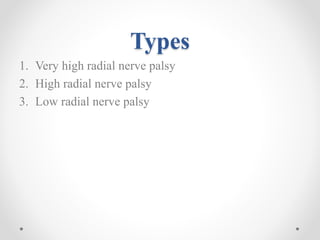
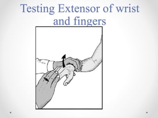
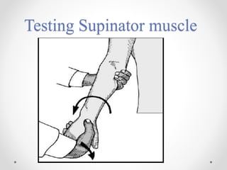
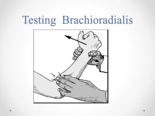
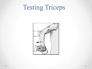
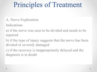
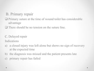
Radial nerve
Palsy
Anatomy of radial nerve
 Continuation of posterior cord of brachial plexus(C5-T1)
 In the axilla, gives off a branch to the long head of triceps and
enters the arm.
Course of radial nerve
IN THE AXILLA
 Radial nerve lies anterior to subscapularis , teres major ,
lattismus dorsi.
 Sensory supply : posterior cutaneous nerve of arm
 Radial nerve leaves axilla via triangular space
 Motor supply : long head of triceps
 Radial nerve comes to lie along spiral groove on posterior aspect
of humeral shaft along with profunda brachii artery
 Motor supply : medial and lateral head of triceps , anconeus
 Sensory supply : posterior cutaneous nerve of forearm, lower
cutaneous nerve of the arm
• Radial nerve then leaves the spiral groove by piercing the
lateral intermuscular septum to enter anterior compartment of
arm, 10-12 cm above the lateral epicondyle (lies b/w brachialis
and brachioradialis)
• Motor supply : brachialis (lateral part), BR, ECRL
• Anterior to lateral epicondyle: Radial nerve divides into 2
branches:
1. Posterior Interosseous nerve (PIN)
2. Superficial radial sensory nerve
Posterior interosseus nerve
• Gain access to posterior forearm by dividing posterior and
laterally through ligament of frohse and the interval b/w the
two heads of supinator.
• Arcade of frohse is a fibrous arch originating lateral
epicondyle superior part of superficial layer of supinator.
• Supply to all extensor compartment muscle.
• Superficial radial nerve courses through the forearm
immediately deep to the BR
• It emerges b/w tendon of BR and ECRL proximal to radial
styloid
• Passes over tendons of snuffbox
• Terminates as cutaneous branches to dorsum of hand and
lateral 3.5 digits short of nailbeds.
Types
1. Very high radial nerve palsy
2. High radial nerve palsy
3. Low radial nerve palsy
Very high radial nerve palsy
 Level of injury : at the axilla
 Causes : crutch palsy (chronic compression in the axilla)
Trauma or operation around the shoulder
Aneurysms of axillary vessels
 Clinical features
Motor deficit : loss of extension of forearm
weakness of supination
Loss of extension of hand and finger
Sensory deficit: lateral arm
Posterior forearm
Radial half of dorsum of hand
Dorsal aspect of radial three and half digits (excluding
their nail beds)
High radial nerve palsy
 Level of injury: in the radial groove
 Causes : Fracture shaft of humerus
Prolonged application of tourniquet
 Clinical features
Motor deficit : Weakness of supination
Loss of extension of hand and fingers
Sensory deficit : Posterior forearm
Radial half of dorsum of hand
Dorsal aspect of radial three and half digits
(excluding their nail bed )
Low radial nerve palsy
 Level of injury : below the elbow
 Causes : dislocation of elbow
Fracture neck of radius
 Clinical features
Motor deficit : Extension of hand
Extension of finger
Sensory deficit : None
Examinations
(a) Is there any an obvious wrist drop
(b) Is there wasting of the forearm muscles
(c) Is there wasting of the triceps
Testing Extensor of wrist
and fingers
Testing Supinator muscle
Testing Brachioradialis
Testing Triceps
Testing for sensory loss
Other tests
1. Nerve conduction study
2. Electromyography
3. Tinel sign
4. Sweat test
5. Skin resistance test
Principles of Treatment
A. Nerve Exploration
Indications
a) if the nerve was seen to be divided and needs to be
repaired
b) if the type of injury suggests that the nerve has been
divided or severely damaged
c) if the recovery is inappropriately delayed and the
diagnosis is in doubt
B. Primary repair
 Primary suture at the time of wound toilet has considerable
advantage
 There should be no tension on the suture line.
C. Delayed repair
Indications
a) a closed injury was left alone but shows no sign of recovery
at the expected time
b) the diagnosis was missed and the patient presents late
c) primary repair has failed
D. Nerve grafting
 Sural nerve is the most commonly used
 Up to 40cm can be obtained from each leg
 The graft should be long enough to lie without any tension and
it should be routed through a well vascularized bed
 The graft is attached at each end either by fine sutures or with
fibrin glue
E. Nerve transfer
The principle of nerve transfer is that a less essential function is
sacrificed to reinstate a more vital one
Indication :
a) very proximal nerve injury such as root avulsion
b) the distance to the target organ or length of graft required precludes
any chance of recovery
F. Tendon transfer
Is consider if axon regeneration at about 1mm per day do not reaches
the muscle within 18-24 months of injury
Prognosis
1. Delay to repair : best outcomes with immediate repair
2.Age of patent: children do better than adults
3.Nature of nerve injury :clean cut > crush > traction
4.Length of injured segment :graft of 10cm is unlikely to work
5.Distal > proximal level of lesion : higher the lesion the worst
the prognosis
6. Associated vascular injury : both nerve and its target organ
requires adequate blood supply
7. Associated direct muscle damage :prevent good muscle re-
innervation and in some circumstances (eg hamstring injury in
sciatic nerve lesion) make nerve repair technically difficult
8. Type of nerve : pure motor or pure sensory recover better than
mixed because there is less likelihood of axonal confusion
9.Surgical techniques
Radial nerve palsy, orthopaedics

Radial nerve palsy, orthopaedics

  • 1.
  • 2.
    Anatomy of radialnerve  Continuation of posterior cord of brachial plexus(C5-T1)  In the axilla, gives off a branch to the long head of triceps and enters the arm.
  • 5.
    Course of radialnerve IN THE AXILLA  Radial nerve lies anterior to subscapularis , teres major , lattismus dorsi.  Sensory supply : posterior cutaneous nerve of arm  Radial nerve leaves axilla via triangular space  Motor supply : long head of triceps  Radial nerve comes to lie along spiral groove on posterior aspect of humeral shaft along with profunda brachii artery  Motor supply : medial and lateral head of triceps , anconeus  Sensory supply : posterior cutaneous nerve of forearm, lower cutaneous nerve of the arm
  • 6.
    • Radial nervethen leaves the spiral groove by piercing the lateral intermuscular septum to enter anterior compartment of arm, 10-12 cm above the lateral epicondyle (lies b/w brachialis and brachioradialis) • Motor supply : brachialis (lateral part), BR, ECRL • Anterior to lateral epicondyle: Radial nerve divides into 2 branches: 1. Posterior Interosseous nerve (PIN) 2. Superficial radial sensory nerve
  • 7.
    Posterior interosseus nerve •Gain access to posterior forearm by dividing posterior and laterally through ligament of frohse and the interval b/w the two heads of supinator. • Arcade of frohse is a fibrous arch originating lateral epicondyle superior part of superficial layer of supinator. • Supply to all extensor compartment muscle.
  • 8.
    • Superficial radialnerve courses through the forearm immediately deep to the BR • It emerges b/w tendon of BR and ECRL proximal to radial styloid • Passes over tendons of snuffbox • Terminates as cutaneous branches to dorsum of hand and lateral 3.5 digits short of nailbeds.
  • 11.
    Types 1. Very highradial nerve palsy 2. High radial nerve palsy 3. Low radial nerve palsy
  • 12.
    Very high radialnerve palsy  Level of injury : at the axilla  Causes : crutch palsy (chronic compression in the axilla) Trauma or operation around the shoulder Aneurysms of axillary vessels  Clinical features Motor deficit : loss of extension of forearm weakness of supination Loss of extension of hand and finger Sensory deficit: lateral arm Posterior forearm Radial half of dorsum of hand Dorsal aspect of radial three and half digits (excluding their nail beds)
  • 13.
    High radial nervepalsy  Level of injury: in the radial groove  Causes : Fracture shaft of humerus Prolonged application of tourniquet  Clinical features Motor deficit : Weakness of supination Loss of extension of hand and fingers Sensory deficit : Posterior forearm Radial half of dorsum of hand Dorsal aspect of radial three and half digits (excluding their nail bed )
  • 14.
    Low radial nervepalsy  Level of injury : below the elbow  Causes : dislocation of elbow Fracture neck of radius  Clinical features Motor deficit : Extension of hand Extension of finger Sensory deficit : None
  • 15.
    Examinations (a) Is thereany an obvious wrist drop (b) Is there wasting of the forearm muscles (c) Is there wasting of the triceps
  • 16.
    Testing Extensor ofwrist and fingers
  • 17.
  • 18.
  • 19.
  • 20.
  • 21.
    Other tests 1. Nerveconduction study 2. Electromyography 3. Tinel sign 4. Sweat test 5. Skin resistance test
  • 22.
    Principles of Treatment A.Nerve Exploration Indications a) if the nerve was seen to be divided and needs to be repaired b) if the type of injury suggests that the nerve has been divided or severely damaged c) if the recovery is inappropriately delayed and the diagnosis is in doubt
  • 23.
    B. Primary repair Primary suture at the time of wound toilet has considerable advantage  There should be no tension on the suture line. C. Delayed repair Indications a) a closed injury was left alone but shows no sign of recovery at the expected time b) the diagnosis was missed and the patient presents late c) primary repair has failed
  • 24.
    D. Nerve grafting Sural nerve is the most commonly used  Up to 40cm can be obtained from each leg  The graft should be long enough to lie without any tension and it should be routed through a well vascularized bed  The graft is attached at each end either by fine sutures or with fibrin glue
  • 25.
    E. Nerve transfer Theprinciple of nerve transfer is that a less essential function is sacrificed to reinstate a more vital one Indication : a) very proximal nerve injury such as root avulsion b) the distance to the target organ or length of graft required precludes any chance of recovery F. Tendon transfer Is consider if axon regeneration at about 1mm per day do not reaches the muscle within 18-24 months of injury
  • 26.
    Prognosis 1. Delay torepair : best outcomes with immediate repair 2.Age of patent: children do better than adults 3.Nature of nerve injury :clean cut > crush > traction 4.Length of injured segment :graft of 10cm is unlikely to work 5.Distal > proximal level of lesion : higher the lesion the worst the prognosis 6. Associated vascular injury : both nerve and its target organ requires adequate blood supply
  • 27.
    7. Associated directmuscle damage :prevent good muscle re- innervation and in some circumstances (eg hamstring injury in sciatic nerve lesion) make nerve repair technically difficult 8. Type of nerve : pure motor or pure sensory recover better than mixed because there is less likelihood of axonal confusion 9.Surgical techniques