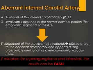
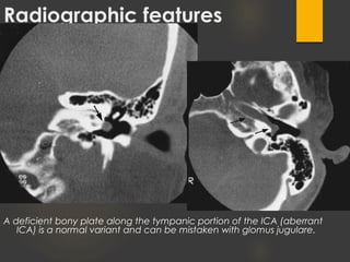
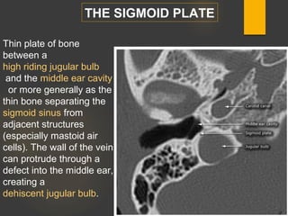
Tinnitus is the perception of sound when no external sound is present. It can be subjective, heard only by the patient, or objective, able to be heard by an examiner. Pulsatile tinnitus is a rhythmic type of tinnitus that is synchronous with the heartbeat. Imaging findings associated with pulsatile tinnitus include aberrant internal carotid arteries, persistent stapedial arteries, high or dehiscent jugular bulbs, sigmoid sinus wall anomalies, and vascular tumors or malformations. A detailed history, physical exam, audiologic evaluation, and imaging are needed to evaluate tinnitus and identify potential underlying causes.
Ct anatomy of paranasal sinuses( PNS) pk.pdf pptDr pradeep Kumar
This presentation includes cross sectional anatomy like axial,saggital and coronal images of paranasal sinuses and most important variation of paranasal sinus.This help alot. Must read topic for radiology resident. Thanks
Ct anatomy of paranasal sinuses( PNS) pk.pdf pptDr pradeep Kumar
This presentation includes cross sectional anatomy like axial,saggital and coronal images of paranasal sinuses and most important variation of paranasal sinus.This help alot. Must read topic for radiology resident. Thanks
USMLE CVS 006 007 Development of the heart anatomy .pdfAHMED ASHOUR
The development of the heart is a complex and highly regulated process that begins early in embryonic life.
Understanding the stages of heart development is crucial for recognizing
and addressing congenital heart defects that may occur during this intricate process.
Stapedial artery persistence is an uncommon issue to encounter in ordinary ear, nose, and throat practice.
If they are, they typically show up as a pulsatile mass or as a sneaky discovery. Due to the condition’s rarity, most
individuals are asymptomatic, but occasionally it can induce vertigo, pulsatile tinnitus, or conductive hearing loss
in certain people. We hereby present a case of incidental discovery of a persistent stapedial artery in a patient with
complaints of nasal obstruction.
Stroke as the first manifestation of Takayasu's arteritisApollo Hospitals
Takayasu's arteritis is an idiopathic inflammatory disease of the large elastic arteries occurring in the young resulting in occlusive or ectatic changes mainly in the aorta and its immediate branches as well as the pulmonary artery and its branches. The disease is common in women in the second and third decades of life. Stroke as the first manifestation of Takayasu's disease is relatively rare. However 10–20% patients with Takayasu's arteritis can have a primary cerebrovascular presentation with headaches, seizures, transient ischemic attacks, strokes or intra-cerebral hemorrhage. We report a case of a 39-year-old lady who developed a stroke and was diagnosed as Takayasu's arteritis. This patient had fulfilled three of six criteria for Takayasu's arteritis based on The American College of Rheumatology. She responded to steroids and immune suppressive therapy.
Stroke as the first presentation of Takayasu's arteritis is relatively rare and only a few instances have been reported in the literature. Our patient had bilateral carotid occlusion and collaterals between the vertebral artery and external carotid arteries. She presented with right hemiplegia and after diagnosis she was promptly treated with prednisolone, methotrexate and other supportive measure. The patient had good clinical recovery.
(May 29th, 2024) Advancements in Intravital Microscopy- Insights for Preclini...Scintica Instrumentation
Intravital microscopy (IVM) is a powerful tool utilized to study cellular behavior over time and space in vivo. Much of our understanding of cell biology has been accomplished using various in vitro and ex vivo methods; however, these studies do not necessarily reflect the natural dynamics of biological processes. Unlike traditional cell culture or fixed tissue imaging, IVM allows for the ultra-fast high-resolution imaging of cellular processes over time and space and were studied in its natural environment. Real-time visualization of biological processes in the context of an intact organism helps maintain physiological relevance and provide insights into the progression of disease, response to treatments or developmental processes.
In this webinar we give an overview of advanced applications of the IVM system in preclinical research. IVIM technology is a provider of all-in-one intravital microscopy systems and solutions optimized for in vivo imaging of live animal models at sub-micron resolution. The system’s unique features and user-friendly software enables researchers to probe fast dynamic biological processes such as immune cell tracking, cell-cell interaction as well as vascularization and tumor metastasis with exceptional detail. This webinar will also give an overview of IVM being utilized in drug development, offering a view into the intricate interaction between drugs/nanoparticles and tissues in vivo and allows for the evaluation of therapeutic intervention in a variety of tissues and organs. This interdisciplinary collaboration continues to drive the advancements of novel therapeutic strategies.
Deep Behavioral Phenotyping in Systems Neuroscience for Functional Atlasing a...Ana Luísa Pinho
Functional Magnetic Resonance Imaging (fMRI) provides means to characterize brain activations in response to behavior. However, cognitive neuroscience has been limited to group-level effects referring to the performance of specific tasks. To obtain the functional profile of elementary cognitive mechanisms, the combination of brain responses to many tasks is required. Yet, to date, both structural atlases and parcellation-based activations do not fully account for cognitive function and still present several limitations. Further, they do not adapt overall to individual characteristics. In this talk, I will give an account of deep-behavioral phenotyping strategies, namely data-driven methods in large task-fMRI datasets, to optimize functional brain-data collection and improve inference of effects-of-interest related to mental processes. Key to this approach is the employment of fast multi-functional paradigms rich on features that can be well parametrized and, consequently, facilitate the creation of psycho-physiological constructs to be modelled with imaging data. Particular emphasis will be given to music stimuli when studying high-order cognitive mechanisms, due to their ecological nature and quality to enable complex behavior compounded by discrete entities. I will also discuss how deep-behavioral phenotyping and individualized models applied to neuroimaging data can better account for the subject-specific organization of domain-general cognitive systems in the human brain. Finally, the accumulation of functional brain signatures brings the possibility to clarify relationships among tasks and create a univocal link between brain systems and mental functions through: (1) the development of ontologies proposing an organization of cognitive processes; and (2) brain-network taxonomies describing functional specialization. To this end, tools to improve commensurability in cognitive science are necessary, such as public repositories, ontology-based platforms and automated meta-analysis tools. I will thus discuss some brain-atlasing resources currently under development, and their applicability in cognitive as well as clinical neuroscience.
This pdf is about the Schizophrenia.
For more details visit on YouTube; @SELF-EXPLANATORY;
https://www.youtube.com/channel/UCAiarMZDNhe1A3Rnpr_WkzA/videos
Thanks...!
What is greenhouse gasses and how many gasses are there to affect the Earth.moosaasad1975
What are greenhouse gasses how they affect the earth and its environment what is the future of the environment and earth how the weather and the climate effects.
Seminar of U.V. Spectroscopy by SAMIR PANDASAMIR PANDA
Spectroscopy is a branch of science dealing the study of interaction of electromagnetic radiation with matter.
Ultraviolet-visible spectroscopy refers to absorption spectroscopy or reflect spectroscopy in the UV-VIS spectral region.
Ultraviolet-visible spectroscopy is an analytical method that can measure the amount of light received by the analyte.
Observation of Io’s Resurfacing via Plume Deposition Using Ground-based Adapt...Sérgio Sacani
Since volcanic activity was first discovered on Io from Voyager images in 1979, changes
on Io’s surface have been monitored from both spacecraft and ground-based telescopes.
Here, we present the highest spatial resolution images of Io ever obtained from a groundbased telescope. These images, acquired by the SHARK-VIS instrument on the Large
Binocular Telescope, show evidence of a major resurfacing event on Io’s trailing hemisphere. When compared to the most recent spacecraft images, the SHARK-VIS images
show that a plume deposit from a powerful eruption at Pillan Patera has covered part
of the long-lived Pele plume deposit. Although this type of resurfacing event may be common on Io, few have been detected due to the rarity of spacecraft visits and the previously low spatial resolution available from Earth-based telescopes. The SHARK-VIS instrument ushers in a new era of high resolution imaging of Io’s surface using adaptive
optics at visible wavelengths.
Richard's entangled aventures in wonderlandRichard Gill
Since the loophole-free Bell experiments of 2020 and the Nobel prizes in physics of 2022, critics of Bell's work have retreated to the fortress of super-determinism. Now, super-determinism is a derogatory word - it just means "determinism". Palmer, Hance and Hossenfelder argue that quantum mechanics and determinism are not incompatible, using a sophisticated mathematical construction based on a subtle thinning of allowed states and measurements in quantum mechanics, such that what is left appears to make Bell's argument fail, without altering the empirical predictions of quantum mechanics. I think however that it is a smoke screen, and the slogan "lost in math" comes to my mind. I will discuss some other recent disproofs of Bell's theorem using the language of causality based on causal graphs. Causal thinking is also central to law and justice. I will mention surprising connections to my work on serial killer nurse cases, in particular the Dutch case of Lucia de Berk and the current UK case of Lucy Letby.
3. Introduction
The perception of sound in the absence of external stimuli
Tinnire = “ringing” in Latin
Unilateral or bilateral
First or only symptom of a disease process or auditory/psychological
annoyance
40 million affected in the United States… 10 million severely affected …..
Suicide!!!
Most common in 40-70 year-olds
The evaluation of a patient with tinnitus requires a detailed history,
neurootologic physical examination with otoscopy, a comprehensive
audiologic evaluation with hearing thresholds, and imaging studies.
4. Classification
SUBJECTIVE TINNITUS
sound is only perceived by the patient (most common)
OBJECTIVE TINNITUS
sound produced by paraauditory structures which may be heard by an
examiner, often PULSATILE
5. Subjective TinnitusSubjective Tinnitus
• Most common type of tinnitus.
• Only heard by the patient.
• Associated symptoms depend on cause:
• Vertigo – superior semicircular canal dehiscence &
Meniere’s disease
• Conductive hearing loss – otosclerosis & superior
semicircular canal dehiscence
• Sensorineural hearing loss – vestibular schwannoma,
presbyacusis, & noise induced hearing loss
6. Objective TinnitusObjective Tinnitus
• An actual sound made by the human body.
• Physical explanation for perceived noise.
• Often due to a vascular process.
• Can be due to other physiologic sounds:
• Muscular contractions (palatal
myoclonus – clicking)
• Respiration (patulous Eustachian tube)
• Venous hum (flow murmurs)
• Frequently can be perceived by an observer.
7. PulsatilePulsatile TinnitusTinnitus
• Can be altered with compression of arterial or venous structures.
• Can be perceived by the examiner if stethoscope placed in the right
location.
• Can be venous or arterial.
• Tends to produce whooshing sound.
• Cardiac rhythm synchronous.
11. Collins RD. Algorithmic diagnosis of symptoms and signs: a cost-effective approach. 2d ed. Philadelphia: Lippincott Williams &
Wilkins, 2003:568-9.
ENT Referral
ENT Referral
13. Imaging OptionsImaging Options
• Overall, radiologic imaging is effective in detecting
causes of pulsatile tinnitus in approximately 70% of cases.
• High-resolution contrast-enhanced CT or MRI are
reasonable options and are regarded as the imaging
modalities of choice.
• In the absence of objective pulsatile tinnitus, CTA or MRA
are appropriate initial exams.
• If there is suspicion for arterio-venous fistulas, angiography
should be performed.
14.
15. A variant of the internal carotid artery (ICA)
Involution / absence of the normal cervical portion (first
embryonic segment) of the ICA
Enlargement of the usually small collaterals passes lateral
to the cochlear promontory and appears during
otoscopic examination as a retro-tympanic vascular
mass.
Aberrant Internal Carotid ArteryAberrant Internal Carotid Artery
If mistaken for a paraganglioma and biopsied, the
results can be FATAL
18. Radiographic features
A deficient bony plate along the tympanic portion of the ICA (aberrant
ICA) is a normal variant and can be mistaken with glomus jugulare.
20. Failure of regression of the embryonic stapedial artery
The stapedial artery is an embryological vessel arising from the primitive second
aortic arch that divides into a dorsal branch (the future middle meningeal
artery) and a ventral division (future maxillary and mandibular arteries). These
divisions link with branches developing from the external carotid artery during
the third fetal month causing the stapedial artery to regress.
If the stapedial artery persists in postnatal life, the middle meningeal artery arises
from it thus the foramen spinosum (normally containing the middle meningeal) is
absent.
Radiographic features
small canaliculus origniating from petrous segment of internal carotid artery.
linear soft tissue density crossing over cochlear promontory
enlarged facial nerve canal or separate canal parallel to facial nerve
aplastic or hypoplastic foramen spinosum
may be normal variant or in instances where middle meningeal artery arises from
ophthalmic artery
The vessel is important to recognize as it can be confused on examination with
vascular tumours such as glomus tympanicum. It should not be biopsed as it will
The persistent stapedial artery (PSA)The persistent stapedial artery (PSA)
21. Related pathology
may be associated with aberrant ICA, which is
formed when inferior tympanic artery anastomosis
with the caroticotympanic artery; enlarged inferior
tympanic canaliculus is then seen
may be associated with other middle ear anomales
22. Coronal CT image shows the left
internal carotid artery within the
hypotympanum (arrow).
Aberrant Carotid Artery &Aberrant Carotid Artery &
Persistent Stapedial ArteryPersistent Stapedial Artery
Axial CT image shows the stapedial
artery passing through the obturator
foramen (arrow).
23. Lateralised internal carotid arteryLateralised internal carotid artery
The lateralised internal carotid artery is an anatomic variation of the course
of the horizontal internal carotid artery (ICA). It can be visualised on CT by its
more posterolateral entrance to the skull base and protrusion into the anterior
mesotympanum. It may result in pulsatile tinnitus.
Radiographic features
CT
a lateralised ICA passes underneath the cochlea in the hypotympanum
focal thinned or dehiscent lateral wall of the carotid canal
THE INFERIOR TYMPANIC CANALICULUS IS NOT AFFECTED
Differential diagnosis
aberrant internal carotid artery: usually courses across the middle ear along the
cochlear promontory and shows an enlarged inferior tympanic canaliculus
24. the lateralised internal carotid artery is an anatomic variation of the course of the
horizontal internal carotid artery (ICA). It can be visualized on CT by its more
posterolateral entrance to the skull base and protrusion into the anterior mesotympanum.
It may result in pulsatile tinnitus.
Radiographic features
CT
** a lateralised ICA passes underneath the cochlea in the hypotympanum
** focal thinned or dehiscent lateral wall of the carotid canal
** the inferior tympanic canaliculus is not affected
25. Heterogeneous group of vascular lesions
characterized by an idiopathic, non-
inflammatory, and non-atherosclerotic
angiopathy of small and medium-sized arteries.
Fibromuscular Dysplasia is an arteriopathy that may lead to stenosis,
aneurysm, and dissection most common in young females.
Pulsatile tinnitus is a presenting symptom in approximately one-third of
patients and is associated with a pattern of multi-vessel involvement,
increased involvement of the cervical carotid and/or vertebral
arteries, and cervical artery dissection.
The characteristic finding is alternating stenoses and dilatations,
causing a string of beads appearance.
Fibromuscular dysplasiaFibromuscular dysplasia (FMDFMD)
26. Beaded appearance
of the internal carotid
arteries at the C1 to
C2 level greater on
the left than the right.
Finding is typical of
fibromuscular dysplasia
.
29. Sigmoid Sinus Wall Anomalies
Pulse-synchronous tinnitus (PST) arises from the abnormal self-
perception of one’s own vascular flow, and can arise from a
number of venous and arterial abnormalities.
Venous PST is more commonly encountered in clinical
practice than arterial PST.
Sigmoid wall anomalies (SWA) are an increasingly recognized
cause of venous PST. SWA include sigmoid sinus thinning,
dehiscence, diverticulum and ectasia.
Sigmoid wall anomalies include attenuation of the sinus plate,
frank dehiscence resulting in exposure of the sinus to the air in
the mastoid air cells, diverticula and segmental sigmoid sinus
ectasia
37. Venous Sinus Dehiscences &Venous Sinus Dehiscences &
DiverticulaDiverticula
Axial and coronal CT images show right sigmoid sinus diverticulum
and dehiscences involving the mastoid air cells and mastoid cortex
(arrows).
38. Venous Sinus Dehiscences &Venous Sinus Dehiscences &
DiverticulaDiverticula
• Sigmoid sinus diverticulum and dehiscence is
perhaps the most common identifiable cause for
pulsatile tinnitus of venous origin, with a
prevalence of 23% in symptomatic patients.
• Dehiscence of the sigmoid sinus can involve erode
into the mastoid air cells or the mastoid cortex, or
both.
48. A) Glomus TympanicumA) Glomus Tympanicum
Axial CT images shows a soft tissue opacity in the middle ear, along the cochlear
promontory (arr0w). The coronal post-contrast T1-weighted MRI shows avid enhancement in
the lesion (arrow). Catheter digital subtraction angiography shows marked hypervascularity
in the lesion (arrow).
49. Glomus TympanicumGlomus Tympanicum
• Often apparent on otoscopic examination as a pulsating reddish
mass, the role of imaging is to differentiate these from glomus
jugulotympanicum.
• Most glomus tympanicum tumors arise on the cochlear
promontory.
• CT without contrast is adequate for delineating the extent of the
tumor.
• Avid enhancement and a “salt and pepper” appearance can be
observed on MRI.
50. RT middle ear cavity glomus tympanicum paraganglioma (red arrow
located just anterior to the cochlear promontory (blue arrow).
52. Temporal Bone MetastasesTemporal Bone Metastases
Initial presentation of prostate cancer as pulsatile tinnitus due to a
metastasis. The CT, CTA, and post-contrast T1-weighted MRI show a
lytic, enhancing mass (arrows) in the left temporal bone with associated
compression of the left jugular bulb (oval).
53. • Rare cause of pulsatile tinnitus due to vascular
impingement or tumor hyperemia.
• The presence of accompanying new cranial
nerve deficits should raise the suspicion for a
malignancy.
• Serial scanning in patients at high risk of
metastatic disease may be warranted.
Temporal Bone MetastasesTemporal Bone Metastases
55. Petrous Carotid AneurysmPetrous Carotid Aneurysm
Coronal CT image shows an
expansile lesion of the right petrous
carotid canal (arrow).
Catheter angiogram reveals an
aneurysm of the horizontal petrous
carotid artery (arrow).
56. Petrous Carotid AneurysmPetrous Carotid Aneurysm
• Most petrous aneurysms are large and fusiform and believed
to be congenital in origin.
• Other etiologies for petrous aneurysms are radiation injury,
trauma, and infection.
• Otologic manifestations include conductive and sensorineural
hearing loss and tinnitus, with rupture seen in 25% as initial
presentation.
• Endovascular treatment is the mainstay of treatment.
57. Dural Arteriovenous FistulaDural Arteriovenous Fistula
The patient presented with a retroauricular bruit and had a remote
history of temporal bone trauma. The 3D hybrid CTA image shows a
prominent occipital artery (arrow) that drains into the junction of the left
transverse sinus with the sigmoid sinus.
58. Dural Arteriovenous FistulaDural Arteriovenous Fistula
• Related to trauma, prior craniotomy, or dural sinus thrombosis.
• Classified according to direction of flow and presence or absence
of cortical venous drainage
• Most common locations: CAVERNOUS, transverse, & sigmoid
sinuses.
• Findings may be subtle on cross sectional imaging and requires
high index of suspicion.
59. Pseudotumor CerebriPseudotumor Cerebri
Sagittal T1-weighted MRI shows an enlarged partially empty sella in a young
obese female. The MRA MIP shows constriction of the bilateral transverse
sinuses (arrows). The axial T2-weighted MRI shows mild bulging of the bilateral
optic nerve discs (arrows).
60. Coronal FIESTA MRI show a vascular structure (arrow) impinging upon
the right cranial nerve 7 and 8 complex (arrowhead).
Vascular Loop SyndromeVascular Loop Syndrome
61. Vascular Loop SyndromeVascular Loop Syndrome
• Pulsatile tinnitus can be caused by arterial or venous vascular loops
and may be accompanied by vertigo.
• Impingement upon the cranial nerve 7 and 8 complex in the
cerebellopontine angle cistern or internal auditory canal can best be
observed on FIESTA/CISS/DRIVE MRI sequences.
• May be treated successfully by microvascular decompression.
62. Vascular
compression
syndrome in the
cerebellopontine
angle cistern
Axial 3D-FIESTA MR images through the eighth CN show the following: AICA (
Anterior inferior cerebellar artery) loop within the right IAC, vascular loop extending into
>50% of the IAC (Type III).
63.
64. Axial 3D CISS image showing (A) a
linear structure originating from
the basilar artery corresponding
to the left anterior inferior
cerebellar artery (white arrow)
which loops around the cisternal
portion of left VIIth and VIIIth
nerve (white arrow) (B). The VIIth
nerve is not well-visualized as the
vascular loop causes overlapping
of the structure.
65. Pseudotumor CerebriPseudotumor Cerebri
(Idiopathic intracranial hypertension (IIH)(Idiopathic intracranial hypertension (IIH)
( Benign intracranial hypertension)( Benign intracranial hypertension)
Syndrome with signs and symptoms of increased intracranial pressure but where a
causative mass or hydrocephalus is not identified.
The older term benign intracranial hypertension is generally frowned upon due to the
fact that some patients with IIH have a fairly aggressive clinical picture with rapid visual
loss.
Interestingly as it has become evident that at least some patients present with IIH due to
identifiable venous stenosis, some authors now advocate reverting to the older
term pseudotumour cerebri as in these patients the condition is not idiopathic 15
. An
alternative approach is to move these patients into a group termed secondary
intracranial hypertension 15
.
Venous pulsatile tinnitus can result from conditions associated increased intracranial
pressure.
Characteristic neuroimaging findings for pseudotumor cerebri (Idiopathic intracranial
hypertension) include a partially empty sella, constriction of the transverse sinuses, and
optic nerve disc and optic nerve sheath cerebrospinal fluid prominence.
66. Both optic nerves show
flattening of the posterior sclera (dashed lines)
protrusions of the optic nerve heads (red arrows)
prominent subarachnoid space around the optic nerves(yellow arrows)
vertical tortuosity of the optic nerves
67. OPTIC NERVES
prominent subarachnoid space around the optic nerves (~45%)
vertical tortuosity of the optic nerves (~40%)
papilloedema
flattening of the posterior sclera (~80%)
intraocular protrusion of the optic nerve head
enhancement of the prelaminar (intra-ocular) optic nerves (~50%)
enlarged arachnoid out-pouchings
* partial empty sella turcica (~70%)
*enlarged Meckel cave 9
* prominent arachnoid pits / small meningocoeles typically within
the temporal bone and sphenoid wing
*bilateral venous sinus stenosis
lateral segments of the transverse sinuses
no evidence of current or remote thrombosis 8
*slitlike ventricles (relatively uncommon compared to other
findings) 15
*acquired tonsillar ectopia (mimicking Chiari I malformation)
70. • Pulsatile tinnitus can be the first manifestation
atherosclerotic disease.
• Most commonly associated with significant
stenosis of the internal carotid arteries.
• Both the head and neck vasculature should be
covered on imaging.
Atherosclerotic DiseaseAtherosclerotic Disease
72. Superior Semicircular CanalSuperior Semicircular Canal
Dehiscence syndromeDehiscence syndrome
A recently described inner ear abnormality, where a clinical disequilibrium
phenomenon is associated with absence of the bony covering of the superior
semicircular canal (SSC).
Dehiscence of the SSC forms a "third window" into the inner ear, in addition to
the round and oval windows. This allows motion of the endolymph to be
induced by sound and pressure stimuli 2
.
Pulsatile tinnitus results from transmission of the normal pulse-related
pressure changes within the cranial cavity to the inner ear.
Temporal bone CT is the modality of choice for diagnosing superior
semicircular canal dehiscence by the lack of overlying bone, particularly via
Stenver and Pöschl views.
73.
74. Rating of the bone covering the SSC used for the 1-mm-collimated CT scans in the
control subjects with normal temporal bones.
(a)Coronal 1-mm-collimated CT scan through the left temporal bone shows clearly
intact bone (arrow) over the left SSC.
(b) Coronal 1-mmcollimated
CT scan through the left temporal bone demonstrates an area of possible bone
dehiscence (arrow) over the SSC.
(c) Coronal 1-mm-collimated CT scan through the right temporal bone demonstrates a
region of bone dehiscence (arrow) over the SSC.
75. The patient presented with dizziness, pulsatile tinnitus, hyperacusis, otalgia, and
fullness in the right ear. Stenver and Pöschl CT images show deficiency of bone
along the apex of the right superior semicircular canal (arrows).
76. CT scan, sagittal projections showed bilateral dehiscence of posterior semicircular canal
in right ear (3c) and left ear (3d), and unilateral right dehiscence of the superior
semicircular canal (3a white arrow). White arrows in 3b instead show the bone covering
the left superior semicircular canal.
78. OtospongiosisOtospongiosis
• Otospongiosis contains arteriovenous microfistulas which can
lead to pulsatile tinnitus.
• Demineralization in the region of the fissula ante fenestram
and/or otic capsule can be observed on CT.
• The affected areas display enhancement due to the vascular
nature of otospongiosis.
79. Inflammatory andInflammatory and
Hypermetabolic ConditionsHypermetabolic Conditions
Otomastoiditis. The MRI shows
enhancement throughout the right
mastoid air cells and an epidural
abscess (arrow).
Paget disease. Sagittal CT image show
diffuse expansion of the skull diplopic
space and lucency of the otic capsule
(arrow).
80. • Infectious and inflammatory processes in and around the temporal
bone, including mastoiditis and Paget disease, can lead to pulsatile
tinnitus due to increased regional blood flow.
• Otomastoiditis appears opacification on CT and associated
enhancement of the mucosa and sometimes the bone marrow is
apparent on MRI.
• Paget disease has variable imaging manifestations depending upon
the stage of disease, but can appear as bone marrow expansion and
demineralization on CT during the active phase with pulsatile tinnitus,
cranial nerve deficits and Eustachian tube dysfunction.
Inflammatory andInflammatory and
Hypermetabolic ConditionsHypermetabolic Conditions
87. 44 year-old female with left pulsatile
tinnitus
Axial CT shows ectatic left sigmoid sinus with thinning of the left sigmoid plate
without a focal diverticulum
90. Case seniro…
Transmastoid approach for sigmoid sinus wall
repair: Intraoperative findings
(A) Appearance of a right sigmoid sinus diverticulum during transmastoid
surgery. Yellow lines: outline of normal sigmoid sinus; Blue oval:
diverticulum. (B) Post-reduction of diverticulum. Metal suction tip sitting on
the wall of the sigmoid sinus through a hole in the bone after diverticulum
reduction. (C) Post-repair of sinus wall. Tissue graft reinforcing the wall of
the sigmoid sinus, sitting deep to the bone on the sinus wall, through the
hole in the bone. Following this step, the hole itself is repaired with synthetic
bone cement (Hydroset) and bone pate made from the patient’s own bone.
A B C
91. Postoperative imaging findings: CT
Axial CT image of the temporal bone on soft tissue window (A) demonstrates
relatively hypoattenuating material (arrows) lateral to the hyperattenuating,
contrast enhanced sigmoid sinus. (B) On bone windows, hydroset material is
identified as sharply demarcated hyperdense material (asterisk) conforming
to the size and shape of dehiscence. Bone pate is identified lateral to the
hydroset as amorphous ill-defined hyperdensity (arrow).
*
A B
*
The two vessels that enlarge to form the aberrant ICA are:
1- inferior tympanic artery: a branch of the external carotid artery (usually maxillary artery or superficial temporal artery) which enters from below via the inferior tympanic canaliculus
2- caroticotympanic artery: a branch of the petrous portion of the ICA known as the hyoid artery when enlarged
The two vessels enlarge, sometimes with a stenosis producing objective tinnitus, and rejoin the horizontal segment of the petrous portion of the ICA
The persistent stapedial artery (PSA) refers to failure of regression of the embryonic stapedial artery.
Epidemiology
It's prevalence is thought to range around 0.02-0.48% in the population.
Clinical presentation
pulsatile tinnitus
incidental finding during surgery
conductive hearing loss due to ankylosis of stapes
sensorineural hearing loss due to erosion of otic capsule (rare)
Gross anatomy
arises from proximal hyoid artery at 4-5 weeks of life
passes through obturator foramen of stapes
branches into upper and lower divisions
posterior division of the upper branch becomes middle meningeal artery
lower branch (maxillomandibular) has two branches
mandibular artery (becomes inferior alveolar artery in adult)
infraorbital artery (becomes infraorbital artery in adult)
Anastomosis forms between ventral pharyngeal artery (precursor of external carotid artery) and lower division of stapedial artery
As the ventral pharyngeal artery supplies flow to middle meningeal artery, stapedial artery regresses, leaving small caroticotympanic artery
Blood supply
Can arise from
caroticotympanic artery
inferior tympanic artery
Radiographic features
small canaliculus origniating from petrous segment of internal carotid artery.
linear soft tissue density crossing over cochlear promontory
enlarged facial nerve canal or separate canal parallel to facial nerve
aplastic or hypoplastic foramen spinosum
may be normal variant or in instances where middle meningeal artery arises from ophthalmic artery
Related pathology
may be associated with aberrant ICA, which is formed when inferior tympanic artery anastomosis with the caroticotympanic artery; enlarged inferior tympanic canaliculus is then seen
may be associated with other middle ear anomales
Arterial imaging with CT angiography, MR angiography and digital subtraction angiography (DSA) may be used to visualise the lesions in FMD. Selective DSA is the gold standard because it allows visualisation of small or peripheral lesions. The characteristic finding, particularly in more common intimal subtype, is alternating stenoses and dilatations, causing a string of beads appearance 5. Less commonly in intimal and adventitial type, there is focal concentric, long-segment tubular stenosis or diverticular outpouching present. Cross-sectional imaging (CT and MRI) allows assessment of end-organ ischaemic damage.typical angiographic features include: vascular loops, fusiform vascular ectasia and a string of beadsless typical features include: arterial dissection, aneurysm and subarachnoid haemorrhage
2 diff cases
IJV dome (roof) reaching above the internal acoustic meatus (IAM).
A run of the mill high riding jugular bulb has an intact sigmoid plate (described later)
If the sigmoid plate is deficient, the bulb is free to protrude into the middle ear cavity, and is then known as a dehiscent jugular bulb and is a common cause of a retrotympanic vascular mass.
Radiographic features
given the often turbulent flow within the jugular bulb, signal on MRI can be confusing the presence of the sigmoid plate can only be ascertained on CT
Case Discussion
Right sided middle ear soft-tissue lesion near the cochlear promontory. This lesion enhanced on MRI (not shown) consistent with a glomus tympanicum paraganglioma, which is the most common tumour of the middle ear cavity. These tumours can be asymptomatic, produce conductive hearing loss through contact with the ossicles (as in this case), or result in pulsatile tinnitus. This mass was observed to be stable over many years without treatment.
https://radiopaedia.org/cases/glomus-tympanicum-paraganglioma
https://radiopaedia.org/cases/glomus-jugulare-tumour-3
Intensely enhancing hyperdense lesion is seen involving the left jugular foramen, measuring approximately 3.5 x 3.5 x 3.5 cm. The lesion is causing destruction and widening of the jugular foramen.
Medially the lesion has intracranial extra dural extension in the posterior fossa indenting the cerebellar cortex.
Laterally the lesion is extending in the middle ear cavity (epi, meso and hypotympanum)and external auditory canal. There is erosion of the long process of incus, inferior semicircular canal and the basal turn of cochlea and the lateral wall of tympanic segment of facial nerve.
Lesion is infiltrating the jugular bulb and inferiorly extending into the proximal jugular vein. Posterior inferiorly there is destruction of the basi occiput and lateral aspect of foramen magnum.
Case Discussion
The most common tumor to develop in the jugular foramen is a paraganglioma (glomus jugulare). Paragangliomas in the skull base are ubiquitous in their distribution and arise from paraganglia or glomus cells situated at the following sites:
(1) in the adventitia of the jugular bulb beneath the floor of the middle ear
(2) in the bony walls of the tympanic canals related to the tympanic branches of the IX and X nerves
(3) in the bone of the promontory, close to the mucosal lining of the middle ear.
Imaging studies are necessary to depict the location and extent of tumor involvement, to help determine the surgical approach, and to predict operative morbidity and mortality.1
Radiographic features
CT/MRI
Imaging of the brain with CT and MRI is essential in patients IIH, to exclude elevated CSF pressure due to other causes such as brain tumour, dural sinus thrombosis, hydrocephalus, etc.
In the absence of a cause for intracranial hypertension, imaging features that support the diagnosis of IIH include 3,6-9,15:
optic nerves
prominent subarachnoid space around the optic nerves (~45%)
vertical tortuosity of the optic nerves (~40%)
papilloedema
flattening of the posterior sclera (~80%)
intraocular protrusion of the optic nerve head
enhancement of the prelaminar (intra-ocular) optic nerves (~50%)
enlarged arachnoid out-pouchings
partial empty sella turcica (~70%) *
enlarged Meckel cave 9
prominent arachnoid pits / small meningocoeles typically within the temporal bone and sphenoid wing 9
bilateral venous sinus stenosis
lateral segments of the transverse sinuses
no evidence of current or remote thrombosis 8
slitlike ventricles (relatively uncommon compared to other findings) 15
acquired tonsillar ectopia (mimicking Chiari I malformation) 16
Although bony changes are permanent, the rest are dynamic and may be reversible with treatment 3.
* It is important to take into account the age and gender of a specific patient in assessing the significance of this finding, as in older patients, and especially in males, a partially empty non-enlarged sella is not only common, but normal.
Treatment and prognosis
Treatment options include CSF letting, acetazolamide and lumboperitoneal shunts. In patients with progressive visual deterioration, optic nerve fenestration may be required to preserve vision.
Venous sinus stenting has been reported in case series 4,10, and also being trialled 13,14. The treatment is controversial as to whether apparent venous sinus stenosis is the cause or the effect of IIH 11,12. Spontaneous resolution of apparent stenosis is recognised 6.
History and etymology
Idiopathic intracranial hypertension was first reported in 1893 by Heinrich Quincke, and terms "meningitis serosa" . The term "pseudotumour cerebri" was later introduced in 1904, and later still "benign intracranial hypertension" in 1955 (not to be, but easily, confused with benign intracranial hypotension) 15.
Differential diagnosis
Other causes of intracranial hypertension and papilloedema should be sought. Causes of venous obstruction (e.g. venous sinus thrombosis and venous outflow obstruction in the neck) can very closely mimic the intracranial findings.
Is this the cause for PT in SSCD?
Glomus Tympanicum
GLOMUS JUGULARE
Axial post-contrast CT images demonstrate
thinning of the right sigmoid plate. Note the
normal appearance of the left sigmoid plate.
Non-contrast axial CT shows a defect along the right sigmoid plate with a small sigmoid sinus diverticulum protruding through the osseous defect.
Post-contrast T1-weighted MRI demonstrates a right sigmoid sinus diverticulum
Axial temporal bone CT on bone windows demonstrates dehiscence of the right sigmoid.
Axial CT on bone window demonstrates a small dehiscence of the right sigmoid plate. Postcontrast axial CT demonstrates narrowing of the bilateral transverse sinuses