
Recommended
More Related Content
Similar to Management of Osteoarthritis of knee by High tibial.pptx
Similar to Management of Osteoarthritis of knee by High tibial.pptx (20)
Recently uploaded
Recently uploaded (20)
Management of Osteoarthritis of knee by High tibial.pptx
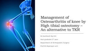
- 1. Management of Osteoarthritis of knee by High tibial osteotomy – An alternative to TKR Dr.Santhosh Raj B K Post graduate 1st year Department of Orthopaedic Surgery Prof.Dr.Rajarajan unit
- 2. Osteoarthritis of knee Osteoarthritis is a chronic degenerative disorder characterised by cartilage loss. It is extremely prevalent in society and is a major cause of disability. It is important to treat osteoarthritis effectively using a multidisciplinary approach tailored to the patient’s needs
- 3. What Happens in Osteoarthritis? In healthy joints, cartilage covers the end of each one. It provides a smooth, gliding surface for joint motion and acts as a cushion between the bones. In OA, this cartilage breaks down, leading to pain, swelling and problems using the joint. Changes also occur in the underlying bone. Bony growths called spurs develop on the edges of the joint. Bits of bone or cartilage may float loosely in the joint space. The membrane lining the joint (the synovium) often becomes inflamed, leading to joint swelling.
- 5. Risk factors The risk factors for OA can be divided into those that act at the level of individual susceptibility and those that alter the biomechanical stability of individual joints Person-level risk factors include increasing age, female sex, joint biomechanics, genetic factors and adiposity. The predominant joint-level factors are joint injury, repetitive joint use through occupation or leisure and joint malalignment.
- 6. PATHOLOGICAL FINDINGS Macroscopy • Osteoarthritic process results in cystic degeneration of the bone surrounding the joint, • Loss of cartilage • Irregular, abnormal bone formation at the edges of the joint
- 7. Pathological findings Microscopy • There is flaking and fibrillation of the articular cartilage • Destruction of the cartilage microarchitecture with formation of holes within it, as well as bony cysts
- 8. CLINICAL FEATURES Patients are usually over the age of 50 Pain and stiffness in the affected joint, which is exacerbated with activity and relieved by rest. Early morning stiffness, if present, is typically less than 30 minutes. Joint tenderness Crepitus on movement Swelling may be due to bony deformity such as osteophyte formation, or due to an effusion caused by synovial fluid accumulation. Systemic symptoms are absent, with a normal erythrocyte sedimentation rate.
- 9. INVESTIGATIONS Imaging Plain radiographs The following changes may be seen on plain radiographs • Joint space narrowing. • Osteophytes. • Bony cysts. • Subchondral sclerosis. .
- 10. Magnetic resonance imaging Used in assessing Ligament status Meniscal tears It has no place in routine clinical assessment of osteoarthritis, but may be a specific and sensitive way of quantifying cartilage loss.
- 12. Investigations Blood tests Patients would often have blood tests • Rheumatoid factor • Erythrocyte sedimentation rate (ESR) • C-reactive protein To rule out other conditions. However, these tests are not essential as a diagnosis can be made in their absence
- 14. Management The aims of management of patients with osteoarthritis are: • Patient education. • Pain control. • Improve function. • Alter the disease process. Each management plan should be individualized and patient centred, agreed on by the patient and doctor in a mutual discussion.
- 15. Treatement options Management interventions in osteoarthritis include: • Education. • Exercise. • Weight loss. • Physiotherapy. • Assisted weight bearing appliances. • Drugs. • Surgery.
- 16. Surgical options Surgery is used when medical therapy has reached its limits. •Arthroscopic debridement and lavage can improve symptoms in degenerative meniscal tears, but does not halt progression. •Autologous cartilage transplantation, where grafts of normal cartilage are taken from the edge of the diseased joint •Osteotomy in early osteoarthritis helps to relieve symptoms and slow the rate of progression. •Joint replacement surgery
- 17. High Tibial Osteotomy High tibial osteotomy (HTO) is a widely performed procedure to treat medial knee arthrosis For relatively young patients who require greater knee preservation, a surgical treatment with low operation trauma and revision rate is needed. Osteotomy around the knee, based on the notion of “knee preservation,” has been chosen as an alternative surgical treatment. Cutting and realigning the bones corrects the mechanical line of lower limb force bearing. As such, osteotomy around the knee retains normal anatomical structure and obtains good functional recovery of the knee joint.
- 18. Goals The goals of HTO are twofold: 1) To reduce knee pain by transferring weight-bearing loads to the relatively unaffected lateral compartment in varus knees 2) To delay the need for a knee replacement by slowing or stopping destruction of the medial joint compartment.
- 19. Preoperative assessment Indications Appropriate patient selection is a key to a successful HTO. Primary or secondary medial compartment degenerative arthritis is the most common indication for HTO The ideal candidate for HTO • Age 45 to 65 years • Isolated medial osteoarthritis with a varus deformity • Good range of motion (ROM) • Without ligamentous instability
- 21. Preoperative assessment Contraindications • Severe joint destruction • ≥65 years of age • Advanced patellofemoral arthritis • <90degree of ROM, • ≥15degree of flexion contracture, • Joint instability • ≥1 cm lateral tibial thrust • ≥20degree of correction • Rheumatoid arthritis
- 24. Radiographic assessment Multiple views should be obtained for preoperative radiographic assessment •Bilateral weight-bearing anterior-posterior views in full extension •Lateral views •Skyline views •Lower limb alignment can be assessed from the full length scannogram of the lower extremity that visualizes the alignment of the hip, knee, and ankle joints. •Magnetic resonance imaging can be helpful in detecting intraosseous lesions, meniscal tears, ligamentous lesions, osteochondral defects, osteonecrosis, or subchondral edema
- 25. Alignment of lower limb
- 27. Correction angle calculation Closing wedge HTO The weight-bearing line is determined by measuring from the point located at 62.5%(Fujisawa point) of the width of the tibial plateau to the center of the femoral head and the center of the ankle. The angle (α) formed at the intersection of these weight bearing lines represents the angle of correction. The wedge bone that constitutes the α angle is to be removed.
- 28. Correction angle calculation Opening wedge HTO The weight-bearing line is determined by measuring from the point located at 62.5% (Fujisawa point)of the width of the tibial plateau to the center of the femoral head and the center of the ankle. The α angle is calculated and transferred to the osteotomy site to open the proximal tibia.
- 31. Cases done in ESIC
- 35. Thank you