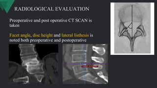
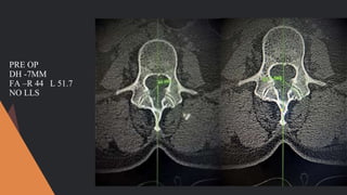
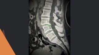
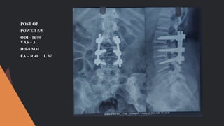
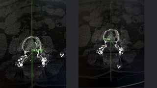
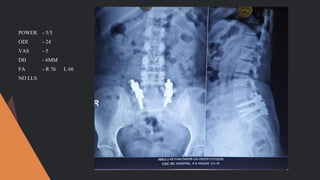
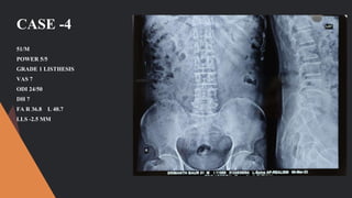
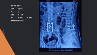
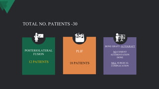
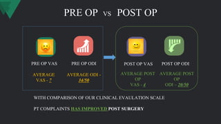
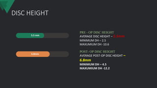
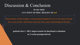
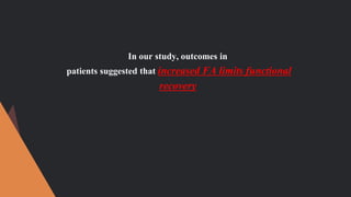
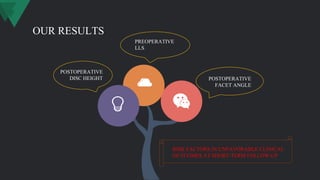
“EVALUATION OF THE PREDICTORS
FOR UNFAVORABLE CLINICAL
OUTCOMES OF LUMBAR
SPONDYLOLISTHESIS AFTER
FUSION”
DR. KARTHIK DEVARAJ,
2ND YEAR ORTHOPAEDIC RESIDENT,
PROF. DR.RAJA RAJAN UNIT,
DEPARTMENT OF OTHOPAEDIC,
ESIC KK NAGAR CHENNAI.
CONCEPTS
To prevent the negative feedback from the patients.
Our study was performed aiming to determine the
predictors that contributed to unfavourable clinical
efficacy among patients with LS after fusion
1
2
AIM
To evaluate the risk of unfavourable clinical
outcomes in short-term postoperative period in LS
after fusion
MATERIALS &METHODS
• THIS STUDY IS DONE IN
DEPARTMENT OF ORTHOPAEDIC SURGERY
ESIC MC ,KK NAGAR, CHENNAI.
• SAMPLE SIZE - 30
• STUDY DESIGN – ANALYTICAL PROSPECTIVE STUDY
CRITERIA
(1)American Society of
Anesthesiologists (ASA) rating of III
or higher
(2) Multi-level decompression and
LIF
(3) Previous lumbar surgical
intervention
(4) Revision surgery
(5) Spinal tumor, deformity, fracture,
infection or other spine diseases
(6) Rejection of implants.
(1) Patients diagnosed with
symptomatic LS
(2) Age ranged from 45 to 75 years
old
(3) Single-level spondylolisthesis
(4) With all DATA of Clinical
parameters, radiological variables,
and surgical records
INCLUSION CRITERIA: EXCLUSION CRITERIA:
CLINICAL EVALUATION
Preoperative and
Postoperative functional
evaluation, including
ODI(Oswestry Disability
Index) and VAS (Visual
Analogue Scale) will be
performed, endpoint
events will be defined as
significant relief of
symptom in the short
term (2 weeks
postoperatively)
OSWESTRY
DISABILITY
INDEX (ODI)
RADIOLOGICAL EVALUATION
X-RAY
ANTEROPOSTERIOR
AND LATERAL
VIEWS
FLEXION &
EXTENSION VIEW
CT MRI
BOTH PRE
OP
&
POST OP
2ND WEEK
CT
PREOPERATIVE
MRI
Preoperative and post operative CT SCAN is
taken
Facet angle, disc height and lateral listhesis is
noted both preoperative and postoperative
RADIOLOGICAL EVALUATION
PROCEDURE
• Surgery is done under all standard protocol
• Surgery is done by the same surgeon
• Prone position
• Standard midline incision used
• Screw method
• Pedical screw size of 6.5mm and 5.5mm is used
• Posterolateral fusion or PLIF
• Water tight closure done
• DVT prophylaxis
• Regular dressing
• Suture removal at POD - 12
POST OP REHABILITATION
• Postoperative bedside mobilization started at POD- 1
• patient made to sit in POD-2
• Made to stand using walker support in POD -3
• Made to Walk using walker support in POD -4
48/F
C/O PAIN LOWER BACK
RADIATING PAIN BOTH LOWER
LIMB
POWER 5/5
L3- L4 GRADE - I LISTHESIS
ODI - 30/50
VAS – 7
CASE 1
PRE OP
DH -7MM
FA –R 44 L 51.7
NO LLS
POST OP
POWER 5/5
ODI - 16/50
VAS – 3
DH-8 MM
FA – R 40 L 37
CASE -2
63/F
POWER 4/5 EHL LEFT
GRADE 2 LISTHESIS
VAS -8
ODI 34/50
DH 10.6
FA- R 33.5 L 51
LLS 2.5 MM
VAS -6
POWER SAME AS PRE
OP
ODI 28/50
DH 10
FA- R 31.8 L 50.4
LLS 2.5 MM
CASE -3
45/F
POWER 5/5
GRADE 2 LISTHESIS
VAS - 6
ODI - 26/50
DH -6
FA R 72 L 69.3
NO LLS
POWER - 5/5
ODI - 24
VAS - 5
DH - 6MM
FA - R 76 L 66
NO LLS
CASE -4
51/M
POWER 5/5
GRADE 1 LISTHESIS
VAS 7
ODI 24/50
DH 7
FA R 36.8 L 40.7
LLS -2.5 MM
POWER 5/5
ODI 12/50
VAS 3
DH 6 MM
FA R 34.2 L-38.6
NO LISTHESIS
POSTERIOLATERAL
FUSION
12 PATIENTS
BONE GRAFT- AUTOGRAFT
NO CEMENT
AUGMENTATION
DONE
NILL SURGICAL
COMPLICATION
PLIF
18 PATIENTS
TOTAL NO. PATIENTS -30
PRE OP VS POST OP
PRE OP VAS PRE OP ODI POST OP VAS POST OP ODI
WITH COMPARISON OF OUR CLINICAL EVAULATION SCALE
PT COMPLAINTS HAS IMPROVED POST SURGERY
AVERAGE
VAS - 7
AVERAGE ODI -
34/50
AVERAGE POST
OP
VAS - 4
AVERAGE POST
OP
ODI – 20/50
DISC HEIGHT
5.5 mm
6.8mm
AVERAGE DISC HEIGHT – 5.5mm
MINIMUM DH – 2.5
MAXUMIUM DH -10.6
PRE - OP DISC HEIGHT
AVERAGE POST-OP DISC HEIGHT –
6.8mm
MINIMUM DH – 4.5
MAXUMIUM DH -12.2
POST- OP DISC HEIGHT
FACET ANGLE
L-38.2 DEGREES
L-47.1 DEGREES
R-34.6 DEGREES
R-40 DEGREES
AVERAGE PRE OP FACET ANGLE
R-34.6 L- 38.2
MIN
R-17.8 L-14.8
MAX
R-70.8 L-78.7
PER OP FACET ANGLE
AVERAGE POST-OP FACET ANGLE
R-40.8 L- 47.1
MIN
R-30 L-32
MAX
R-66.7 L-77.2
POST OP FACET ANGLE
LATERAL LISTHESIS
LATERAL LISTHESIS WAS NOTED IN –
5 PATIENTS WITH AVERAGE OF 2.5MM
DISPLACEMENT
Discussion & Conclusion
In our study
AVG POST OP DISC HEIGHT OF 6.8
Restoration of disc height to normal physiological level or less than normal
have more better functional outcome than supra-physiological fixation
patients have > 60% improvement in functional evaluation
at 2 weeks postoperatively
In our study, outcomes in
patients suggested that increased FA limits functional
recovery
IN OUR STUDY
A limited surgical benefit
was observed in LS with LLS, including an elevated and
persistent back pain due to the
coronal displacement of the slipped segment
successful surgery is largely dependent on
acquiring a solid fusion and re-establishing normal local sagittal
and coronal balance
PURPOSEFUL SURGICAL STRATEGIES AND INSTRUMENTS ARE
RECOMMENDED TO CORRECT VERTEBRAL SLIPPAGE IN PATIENTS
WITH LATERAL SLIPPAGE
Includes
• PRESERVING LUMBAR LORDOSIS
• IMPLANTING A APPROPRIATE SIZE CAGE
• LATERALLY DISTRACTING THE DISPLACED VERTEBRAL BODY
OUR RESULTS
POSTOPERATIVE
DISC HEIGHT POSTOPERATIVE
FACET ANGLE
RISK FACTORS IN UNFAVORABLE CLINICAL
OUTCOMES AT SHORT-TERM FOLLOW-UP
PREOPERATIVE
LLS
PLIF VS PLF HAD SIMILAR OUT COME WITH EQUAL COMPLICATION RATE ,REVISON RATE AND
OPERATION TIME AND BLOOD LOSS WITH 1 YEAR FOLLOW UP
PLIF HAD HIGH FUSION RATE THAN PLF
decompression alone or decompression with interbody fusion study indicated that the patients that
attained
a <6-mm DH intraoperatively
were more likely to derive a sustained benefit from the decompression with a fusion
procedure than the patients reporting poor clinical outcomes with higher disc height
Thank You

Copy-KARTHIK MOS LISTHESIS FINAL copy.pptx

  • 1.
    “EVALUATION OF THEPREDICTORS FOR UNFAVORABLE CLINICAL OUTCOMES OF LUMBAR SPONDYLOLISTHESIS AFTER FUSION” DR. KARTHIK DEVARAJ, 2ND YEAR ORTHOPAEDIC RESIDENT, PROF. DR.RAJA RAJAN UNIT, DEPARTMENT OF OTHOPAEDIC, ESIC KK NAGAR CHENNAI.
  • 2.
    CONCEPTS To prevent thenegative feedback from the patients. Our study was performed aiming to determine the predictors that contributed to unfavourable clinical efficacy among patients with LS after fusion 1 2
  • 3.
    AIM To evaluate therisk of unfavourable clinical outcomes in short-term postoperative period in LS after fusion
  • 4.
    MATERIALS &METHODS • THISSTUDY IS DONE IN DEPARTMENT OF ORTHOPAEDIC SURGERY ESIC MC ,KK NAGAR, CHENNAI. • SAMPLE SIZE - 30 • STUDY DESIGN – ANALYTICAL PROSPECTIVE STUDY
  • 5.
    CRITERIA (1)American Society of Anesthesiologists(ASA) rating of III or higher (2) Multi-level decompression and LIF (3) Previous lumbar surgical intervention (4) Revision surgery (5) Spinal tumor, deformity, fracture, infection or other spine diseases (6) Rejection of implants. (1) Patients diagnosed with symptomatic LS (2) Age ranged from 45 to 75 years old (3) Single-level spondylolisthesis (4) With all DATA of Clinical parameters, radiological variables, and surgical records INCLUSION CRITERIA: EXCLUSION CRITERIA:
  • 6.
    CLINICAL EVALUATION Preoperative and Postoperativefunctional evaluation, including ODI(Oswestry Disability Index) and VAS (Visual Analogue Scale) will be performed, endpoint events will be defined as significant relief of symptom in the short term (2 weeks postoperatively)
  • 7.
  • 8.
    RADIOLOGICAL EVALUATION X-RAY ANTEROPOSTERIOR AND LATERAL VIEWS FLEXION& EXTENSION VIEW CT MRI BOTH PRE OP & POST OP 2ND WEEK CT PREOPERATIVE MRI
  • 9.
    Preoperative and postoperative CT SCAN is taken Facet angle, disc height and lateral listhesis is noted both preoperative and postoperative RADIOLOGICAL EVALUATION
  • 10.
    PROCEDURE • Surgery isdone under all standard protocol • Surgery is done by the same surgeon • Prone position • Standard midline incision used • Screw method • Pedical screw size of 6.5mm and 5.5mm is used • Posterolateral fusion or PLIF • Water tight closure done
  • 11.
    • DVT prophylaxis •Regular dressing • Suture removal at POD - 12
  • 12.
    POST OP REHABILITATION •Postoperative bedside mobilization started at POD- 1 • patient made to sit in POD-2 • Made to stand using walker support in POD -3 • Made to Walk using walker support in POD -4
  • 13.
    48/F C/O PAIN LOWERBACK RADIATING PAIN BOTH LOWER LIMB POWER 5/5 L3- L4 GRADE - I LISTHESIS ODI - 30/50 VAS – 7 CASE 1
  • 15.
    PRE OP DH -7MM FA–R 44 L 51.7 NO LLS
  • 17.
    POST OP POWER 5/5 ODI- 16/50 VAS – 3 DH-8 MM FA – R 40 L 37
  • 21.
    CASE -2 63/F POWER 4/5EHL LEFT GRADE 2 LISTHESIS VAS -8 ODI 34/50 DH 10.6 FA- R 33.5 L 51 LLS 2.5 MM
  • 22.
    VAS -6 POWER SAMEAS PRE OP ODI 28/50 DH 10 FA- R 31.8 L 50.4 LLS 2.5 MM
  • 23.
    CASE -3 45/F POWER 5/5 GRADE2 LISTHESIS VAS - 6 ODI - 26/50 DH -6 FA R 72 L 69.3 NO LLS
  • 24.
    POWER - 5/5 ODI- 24 VAS - 5 DH - 6MM FA - R 76 L 66 NO LLS
  • 25.
    CASE -4 51/M POWER 5/5 GRADE1 LISTHESIS VAS 7 ODI 24/50 DH 7 FA R 36.8 L 40.7 LLS -2.5 MM
  • 26.
    POWER 5/5 ODI 12/50 VAS3 DH 6 MM FA R 34.2 L-38.6 NO LISTHESIS
  • 27.
    POSTERIOLATERAL FUSION 12 PATIENTS BONE GRAFT-AUTOGRAFT NO CEMENT AUGMENTATION DONE NILL SURGICAL COMPLICATION PLIF 18 PATIENTS TOTAL NO. PATIENTS -30
  • 28.
    PRE OP VSPOST OP PRE OP VAS PRE OP ODI POST OP VAS POST OP ODI WITH COMPARISON OF OUR CLINICAL EVAULATION SCALE PT COMPLAINTS HAS IMPROVED POST SURGERY AVERAGE VAS - 7 AVERAGE ODI - 34/50 AVERAGE POST OP VAS - 4 AVERAGE POST OP ODI – 20/50
  • 29.
    DISC HEIGHT 5.5 mm 6.8mm AVERAGEDISC HEIGHT – 5.5mm MINIMUM DH – 2.5 MAXUMIUM DH -10.6 PRE - OP DISC HEIGHT AVERAGE POST-OP DISC HEIGHT – 6.8mm MINIMUM DH – 4.5 MAXUMIUM DH -12.2 POST- OP DISC HEIGHT
  • 30.
    FACET ANGLE L-38.2 DEGREES L-47.1DEGREES R-34.6 DEGREES R-40 DEGREES AVERAGE PRE OP FACET ANGLE R-34.6 L- 38.2 MIN R-17.8 L-14.8 MAX R-70.8 L-78.7 PER OP FACET ANGLE AVERAGE POST-OP FACET ANGLE R-40.8 L- 47.1 MIN R-30 L-32 MAX R-66.7 L-77.2 POST OP FACET ANGLE
  • 31.
    LATERAL LISTHESIS LATERAL LISTHESISWAS NOTED IN – 5 PATIENTS WITH AVERAGE OF 2.5MM DISPLACEMENT
  • 32.
    Discussion & Conclusion Inour study AVG POST OP DISC HEIGHT OF 6.8 Restoration of disc height to normal physiological level or less than normal have more better functional outcome than supra-physiological fixation patients have > 60% improvement in functional evaluation at 2 weeks postoperatively
  • 33.
    In our study,outcomes in patients suggested that increased FA limits functional recovery
  • 34.
    IN OUR STUDY Alimited surgical benefit was observed in LS with LLS, including an elevated and persistent back pain due to the coronal displacement of the slipped segment successful surgery is largely dependent on acquiring a solid fusion and re-establishing normal local sagittal and coronal balance
  • 35.
    PURPOSEFUL SURGICAL STRATEGIESAND INSTRUMENTS ARE RECOMMENDED TO CORRECT VERTEBRAL SLIPPAGE IN PATIENTS WITH LATERAL SLIPPAGE Includes • PRESERVING LUMBAR LORDOSIS • IMPLANTING A APPROPRIATE SIZE CAGE • LATERALLY DISTRACTING THE DISPLACED VERTEBRAL BODY
  • 36.
    OUR RESULTS POSTOPERATIVE DISC HEIGHTPOSTOPERATIVE FACET ANGLE RISK FACTORS IN UNFAVORABLE CLINICAL OUTCOMES AT SHORT-TERM FOLLOW-UP PREOPERATIVE LLS
  • 37.
    PLIF VS PLFHAD SIMILAR OUT COME WITH EQUAL COMPLICATION RATE ,REVISON RATE AND OPERATION TIME AND BLOOD LOSS WITH 1 YEAR FOLLOW UP PLIF HAD HIGH FUSION RATE THAN PLF
  • 38.
    decompression alone ordecompression with interbody fusion study indicated that the patients that attained a <6-mm DH intraoperatively were more likely to derive a sustained benefit from the decompression with a fusion procedure than the patients reporting poor clinical outcomes with higher disc height
  • 39.