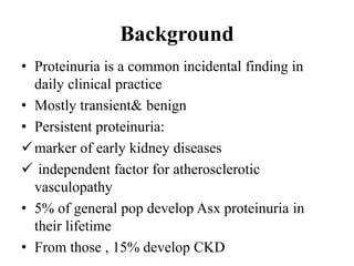
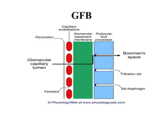
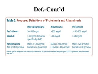
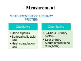
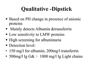
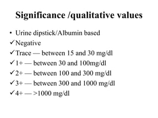
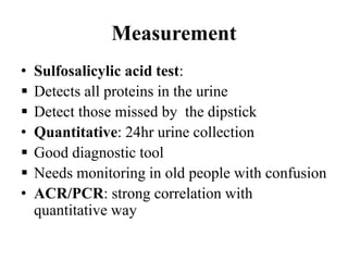
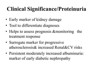
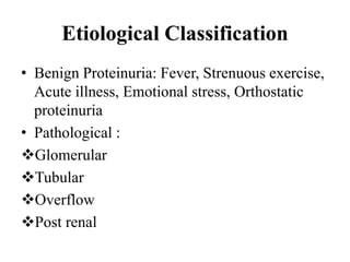
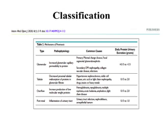
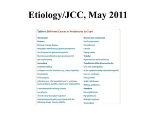
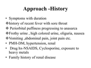
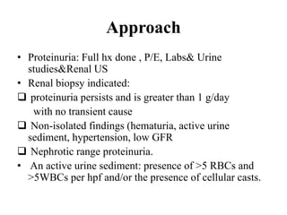
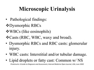
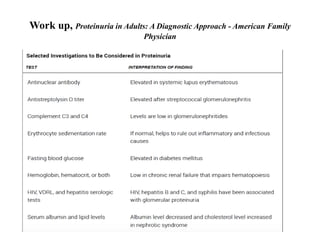
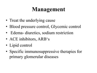
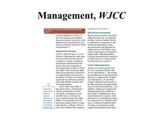
APPROACH TO
PROTEINURIA
Presenter: Dr INGABIRE Prosper
Supervisor: Dr BABANE Jean Felix
August 1st ,2022
Outline
• Background
• Basic Physiology of GFB
• Definitions
• Etiology
• Evaluation &Management
Background
• Proteinuria is a common incidental finding in
daily clinical practice
• Mostly transient& benign
• Persistent proteinuria:
marker of early kidney diseases
 independent factor for atherosclerotic
vasculopathy
• 5% of general pop develop Asx proteinuria in
their lifetime
• From those , 15% develop CKD
GFB
Physiology
• The permeability to GFB depends on size
&charge of proteins
• Small proteins <69 kDA of albumin filtered,
mostly reabsorbed in PT
• Large negatively charged molecules are retained.
• Physiological normal proteinuria: orthostatic,
febrile, exercise
• Proteinuria﹄3.5 g/day: Nephrotic range
Definitions
• Healthy individuals can excrete:
 less than 150 mg/day of Total proteins
less than 30 mg/day of albumin
• Proteinuria: Excessive amount of proteins in
urine > 150mg/24hrs
• Albuminuria: Urinary albumin excretion.
> 30mg/24hrs
Def.-Cont’d
Normal proteinuria
• 150 mg of of urinary protein/composition:
Albumin (20%)
Tamm-Horsfall proteins(40%)
Immunoglobulins
Hormones, enzymes
Mucopolysaccharides
Measurement
Qualitative -Dipstick
ļ‚§ Based on PH change in presence of anionic
proteins
ļ‚§ Mainly detects Albumin &transferrin
ļ‚§ Low sensitivity to LMW proteins
ļ‚§ High screening for albuminuria
ļ‚§ Detection level:
• 150 mg/l for albumin, 200mg/l transferrin
• 500mg/l Ig G& ﹄1000 mg/l Ig Light chains
Significance /qualitative values
• Urine dipstick/Albumin based
Negative
Trace — between 15 and 30 mg/dl
1+ — between 30 and 100mg/dl
2+ — between 100 and 300 mg/dl
3+ — between 300 and 1000 mg/dl
4+ — >1000 mg/dl
Measurement
• Sulfosalicylic acid test:
ļ‚§ Detects all proteins in the urine
ļ‚§ Detect those missed by the dipstick
• Quantitative: 24hr urine collection
ļ‚§ Good diagnostic tool
ļ‚§ Needs monitoring in old people with confusion
• ACR/PCR: strong correlation with
quantitative way
Clinical Significance/Proteinuria
• Early marker of kidney damage
• Tool to differentiate diagnoses
• Helps to assess prognosis &monitoring the
treatment response
• Surrogate marker for progressive
atherosclerosis& increased Renal&CV risks
• Persistent moderately increased albuminuria:
marker of early diabetic nephropathy
Etiological Classification
• Benign Proteinuria: Fever, Strenuous exercise,
Acute illness, Emotional stress, Orthostatic
proteinuria
• Pathological :
Glomerular
Tubular
Overflow
Post renal
Pathogenesis
Classification
Etiology/JCC, May 2011
Approach -History
• Symptoms with duration
history of recent fever with sore throat
 Periorbital puffiness progressing to anasarca
Frothy urine , high colored urine, oliguria, nausea
Vomiting ,abdominal pain, joint pain etc.
• PMH-DM, hypertension, renal
• Drug hx-NSAIDS, Cyclosporine, exposure to
heavy metals
• Family history of renal disease
Approach
• Proteinuria: Full hx done , P/E, Labs& Urine
studies&Renal US
• Renal biopsy indicated:
 proteinuria persists and is greater than 1 g/day
with no transient cause
 Non-isolated findings (hematuria, active urine
sediment, hypertension, low GFR
 Nephrotic range proteinuria.
• An active urine sediment: presence of >5 RBCs and
>5WBCs per hpf and/or the presence of cellular casts.
Microscopic Urinalysis
• Pathological findings:
Dysmorphic RBCs
WBCs (like eosinophils)
Casts (RBC, WBC, waxy and broad).
• Dysmorphic RBCs and RBC casts: glomerular
injury.
• WBC casts: Interstitial and/or tubular damage.
• Lipid droplets or fatty cast: Common w/ NS
Proteinuria: A Guide to Diagnosis and Assessment, Internal Medicine Open Journal, USA, June 2020
Work up, Proteinuria in Adults: A Diagnostic Approach - American Family
Physician
Management
• Treat the underlying cause
• Blood pressure control, Glycemic control
• Edema- diuretics, sodium restriction
• ACE inhibitors, ARBā€˜s
• Lipid control
• Specific immunosuppressive therapies for
primary glomerular diseases
Management, WJCC
Take Home Message
• Proteinuria is not a disease, it’s a marker of
Kidney damage
• Albuminuria is associated with bad
outcomes(CVD, Mortality, ESRD)
• Screen risk patients regularly
• Focus on CV risk factor reduction as patients
with early stage CKD die from CV diseases
References
1. Michael F.(2000).Proteinuria in Adults: A
Diagnostic Approach, Madison accessed on
April 18,2021
2. Sasha et.al (2020), ā€˜Proteinuria: A Guide to
Diagnosis and Assessment’ INTERNAL
MEDICINE Open Journal. Doi:
I0.17140/IMOJ-4-112
3. Uptodate
4. KDIGO Clinical practice 2021 for
management of diabetes

PROTEINURIA .pptx

  • 1.
    APPROACH TO PROTEINURIA Presenter: DrINGABIRE Prosper Supervisor: Dr BABANE Jean Felix August 1st ,2022
  • 2.
    Outline • Background • BasicPhysiology of GFB • Definitions • Etiology • Evaluation &Management
  • 3.
    Background • Proteinuria isa common incidental finding in daily clinical practice • Mostly transient& benign • Persistent proteinuria: marker of early kidney diseases  independent factor for atherosclerotic vasculopathy • 5% of general pop develop Asx proteinuria in their lifetime • From those , 15% develop CKD
  • 4.
  • 5.
    Physiology • The permeabilityto GFB depends on size &charge of proteins • Small proteins <69 kDA of albumin filtered, mostly reabsorbed in PT • Large negatively charged molecules are retained. • Physiological normal proteinuria: orthostatic, febrile, exercise • Proteinuria﹄3.5 g/day: Nephrotic range
  • 6.
    Definitions • Healthy individualscan excrete:  less than 150 mg/day of Total proteins less than 30 mg/day of albumin • Proteinuria: Excessive amount of proteins in urine > 150mg/24hrs • Albuminuria: Urinary albumin excretion. > 30mg/24hrs
  • 7.
  • 8.
    Normal proteinuria • 150mg of of urinary protein/composition: Albumin (20%) Tamm-Horsfall proteins(40%) Immunoglobulins Hormones, enzymes Mucopolysaccharides
  • 9.
  • 10.
    Qualitative -Dipstick ļ‚§ Basedon PH change in presence of anionic proteins ļ‚§ Mainly detects Albumin &transferrin ļ‚§ Low sensitivity to LMW proteins ļ‚§ High screening for albuminuria ļ‚§ Detection level: • 150 mg/l for albumin, 200mg/l transferrin • 500mg/l Ig G& ﹄1000 mg/l Ig Light chains
  • 11.
    Significance /qualitative values •Urine dipstick/Albumin based Negative Trace — between 15 and 30 mg/dl 1+ — between 30 and 100mg/dl 2+ — between 100 and 300 mg/dl 3+ — between 300 and 1000 mg/dl 4+ — >1000 mg/dl
  • 12.
    Measurement • Sulfosalicylic acidtest: ļ‚§ Detects all proteins in the urine ļ‚§ Detect those missed by the dipstick • Quantitative: 24hr urine collection ļ‚§ Good diagnostic tool ļ‚§ Needs monitoring in old people with confusion • ACR/PCR: strong correlation with quantitative way
  • 13.
    Clinical Significance/Proteinuria • Earlymarker of kidney damage • Tool to differentiate diagnoses • Helps to assess prognosis &monitoring the treatment response • Surrogate marker for progressive atherosclerosis& increased Renal&CV risks • Persistent moderately increased albuminuria: marker of early diabetic nephropathy
  • 14.
    Etiological Classification • BenignProteinuria: Fever, Strenuous exercise, Acute illness, Emotional stress, Orthostatic proteinuria • Pathological : Glomerular Tubular Overflow Post renal
  • 15.
  • 16.
  • 17.
  • 18.
    Approach -History • Symptomswith duration history of recent fever with sore throat  Periorbital puffiness progressing to anasarca Frothy urine , high colored urine, oliguria, nausea Vomiting ,abdominal pain, joint pain etc. • PMH-DM, hypertension, renal • Drug hx-NSAIDS, Cyclosporine, exposure to heavy metals • Family history of renal disease
  • 19.
    Approach • Proteinuria: Fullhx done , P/E, Labs& Urine studies&Renal US • Renal biopsy indicated:  proteinuria persists and is greater than 1 g/day with no transient cause  Non-isolated findings (hematuria, active urine sediment, hypertension, low GFR  Nephrotic range proteinuria. • An active urine sediment: presence of >5 RBCs and >5WBCs per hpf and/or the presence of cellular casts.
  • 20.
    Microscopic Urinalysis • Pathologicalfindings: Dysmorphic RBCs WBCs (like eosinophils) Casts (RBC, WBC, waxy and broad). • Dysmorphic RBCs and RBC casts: glomerular injury. • WBC casts: Interstitial and/or tubular damage. • Lipid droplets or fatty cast: Common w/ NS Proteinuria: A Guide to Diagnosis and Assessment, Internal Medicine Open Journal, USA, June 2020
  • 21.
    Work up, Proteinuriain Adults: A Diagnostic Approach - American Family Physician
  • 22.
    Management • Treat theunderlying cause • Blood pressure control, Glycemic control • Edema- diuretics, sodium restriction • ACE inhibitors, ARBā€˜s • Lipid control • Specific immunosuppressive therapies for primary glomerular diseases
  • 23.
  • 24.
    Take Home Message •Proteinuria is not a disease, it’s a marker of Kidney damage • Albuminuria is associated with bad outcomes(CVD, Mortality, ESRD) • Screen risk patients regularly • Focus on CV risk factor reduction as patients with early stage CKD die from CV diseases
  • 25.
    References 1. Michael F.(2000).Proteinuriain Adults: A Diagnostic Approach, Madison accessed on April 18,2021 2. Sasha et.al (2020), ā€˜Proteinuria: A Guide to Diagnosis and Assessment’ INTERNAL MEDICINE Open Journal. Doi: I0.17140/IMOJ-4-112 3. Uptodate 4. KDIGO Clinical practice 2021 for management of diabetes