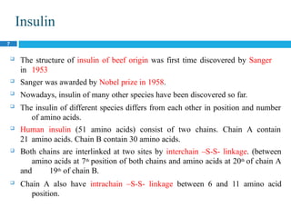
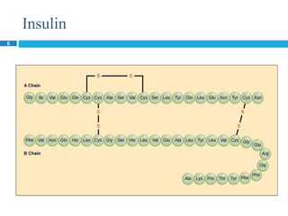
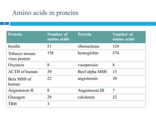
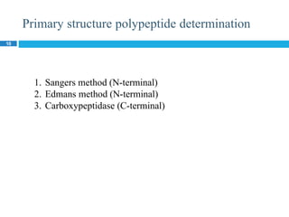
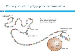
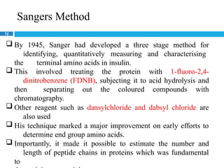
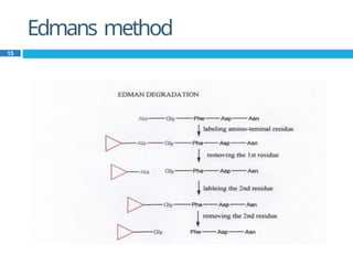
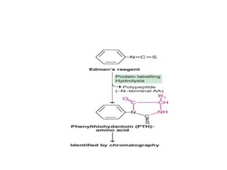
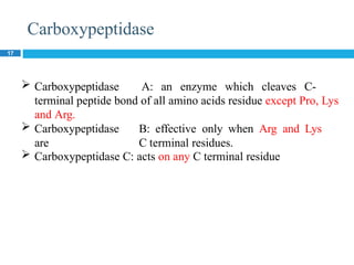
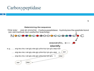
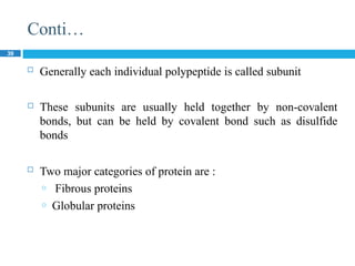
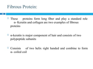
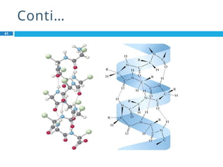
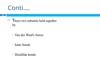
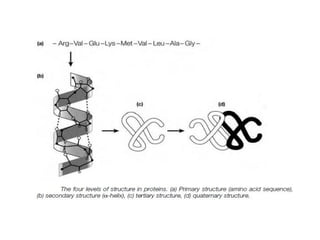
Biochemistry
Organization level of protein
Lecturer ARCP
1
Organizational Level of Protein:
2
 About thousands of proteins are present in human body.
 Basic building block of all these proteins are amino acids.
 Standard amino acids are 20.
 All these protein perform different functions.
 The difference in all of these proteins is due to
certain factors:
 Quantity of amino acids
 Quality of amino acids
 Sequence of amino acids
 Shape of protein
Organizational Level of Protein:
3
 Every protein has a unique three dimensional structure
that is responsible for specific function.
1. Primary structure
2. Secondary structure
3. Tertiary structure
4. Quaternary structure
Primary Structure:
4
 The specific linear sequence of amino acids residue in a
poly peptide chain is called Primary structure of Protein
 The number of amino acids varies from few to many that
are linked together by peptide bonds (amide Bond).
 The numbering of amino acids residue in peptide chain start
from amino acid residue containing free NH2 group.
 The number and position of amino acids is so unique that
any change result in abnormal functioning of protein.
C C
N N
Conti…
5
R
H O
R
N
C
C
H C N C
H
H H O
H O R H O
R
C
R
O
H
C N
H
C C
H H O H
Sickle cell anemia
6
 Hemoglobin S (Hb S) differs from normal hemoglobin in that
valine is present in place of glutamic acid at position six of
beta chain.
 Normal hemoglobin has four chain (574 amino acids). Two
alpha and two beta chains. 141 amino acids are present in each
alpha chain and 146 amino acids are present in each beta chain.
Insulin
7
 The structure of insulin of beef origin was first time discovered by Sanger
in 1953
 Sanger was awarded by Nobel prize in 1958.
 Nowadays, insulin of many other species have been discovered so far.
 The insulin of different species differs from each other in position and number
of amino acids.
 Human insulin (51 amino acids) consist of two chains. Chain A contain
21 amino acids. Chain B contain 30 amino acids.
 Both chains are interlinked at two sites by interchain –S-S- linkage. (between
amino acids at 7th position of both chains and amino acids at 20th of chain A
and 19th of chain B.
 Chain A also have intrachain –S-S- linkage between 6 and 11 amino acid
position.
Insulin
8
Amino acids in proteins
9
Protein Number of
amino acids
Protein Number of
amino acids
Insulin 51 ribonuclease 124
Tobacco mosaic
virus protein
158 hemoglobin 574
Oxytocin 8 vasopressin 8
ACTH of human 39 Beef alpha MSH 13
Beta MSH of
human
22 angiotensin 10
Angiotensin II 8 Angiotensin III 7
Glucagon 29 calcitonin 32
TRH 3
Primary structure polypeptide determination
10
1. Sangers method (N-terminal)
2. Edmans method (N-terminal)
3. Carboxypeptidase (C-terminal)
Primary structure polypeptide determination
11
Sangers Method
12
 By 1945, Sanger had developed a three stage method for
identifying, quantitatively measuring and characterising
the terminal amino acids in insulin.
 This involved treating the protein with 1-fluoro-2,4-
dinitrobenzene (FDNB), subjecting it to acid hydrolysis and
then separating out the coloured compounds with
chromatography.
 Other reagent such as dansylchloride and dabsyl chloride are
also used
 His technique marked a major improvement on early efforts to
determine end group amino acids.
 Importantly, it made it possible to estimate the number and
length of peptide chains in proteins which was fundamental
to
Sangers Method
13
Edmans method
14
 By the action of enzyme trypsin or cyanogen bromide, peptide
hydrolysed into smaller fragments.
 These smaller peptides are then treated with phenylisothiocyanate
(Edmans reagent). This labelled amino group of terminal amino
acid broken down from remaining peptide and is treated with
ninhydrin reagent and thus identified.
 The remaining amino acids are treated with edmans reagent and
same process is carried out until all amino acids in peptide chain
can be identified.
 Sequentially removes one residue at a time from amino end of a
peptide.
 This process become automated and amino acids analyser
determine A.A sequence. Sequenator machine is used for this
process.
Edmans method
15
Carboxypeptidase
17
 Carboxypeptidase A: an enzyme which cleaves C-
terminal peptide bond of all amino acids residue except Pro, Lys
and Arg.
 Carboxypeptidase B: effective only when Arg and Lys
are C terminal residues.
 Carboxypeptidase C: acts on any C terminal residue
Carboxypeptidase
18
Secondary structure:
19
 Once the primary structure of the polypeptide is formed, it
begins to twist into regular patterns that makes the secondary
structures
 Following are the major types of protein secondary structure
are known;
o alpha helix
o beta pleated sheet
a-Helix:
20
 Clockwise, rodlike spiral and form by intrachain
hydrogen bonding between O for carboxylic
group of one amino acid and NH2 of second
amino acid.
 In an a-helix (alpha helix), the polypeptide
backbone follows a helical path.
 There are 3.6 amino acid residues per turn of the
helix and successive turn is 0.54nm above the
turn below it
 The amino acid Proline tends to interrupt an a-
helix
Structure of α-
Helix:
21
 These twists are formed as a result of the
regular pattern of hydrogen bonding between
N-H and C=O gaps on the polypeptide chain
Conti…
22
 The α-Helix is a rod like structure that contains
the backbone on the inner portion of the helix
and side chain contains outer portion
 Each amino acid uses its N-H group to form
hydrogen bond and the C=O group of the
amino acid that is four units ahead of it
Conti…
23
 The hydrogen bonds are individually weak but
collectively , they are strong enough to stabilize
the helix. Helical structure imparts stability to the
peptide molecule because each peptide bond
participates in the hydrogen bonding.
 Proteins with α-helix show great strength,
elasticity and can be easily stretched as they
are in the form of tight coil. A complete turn of
the helix contains an average of 3.6 amino acid
residues.
Pleated β
Sheet:
25
 Two main forms of  pleated sheets
o Antiparallel  pleated sheet
o Parallel  pleated sheet
 Antiparallel  pleated sheet in which the
neighboring hydrogen bonded polypeptide
chains run in opposite directions
 Parallel  pleated sheet in which the
hydrogen bonded chains extend in the same
direction
Structure:
26
Beta Sheet Features:
28
 Proteins containing these structures are
inelastic because the hydrogen bonds are at
right angle to the direction of stretching and
simply hold the bundles of peptide chains
together. The β-pleated sheets cannot further
extend as it is almost fully extended. It should
be noted that hydrogen bonding in α- helix is
intra-chain while hydrogen bonding in β-pleated
sheet is inter-chain.
Various proteins have α-helix and β-pleated
sheets structures to a highly variable extent but
one of these structures may be predominate.
This is due to the presence of certain amino
acids in protein molecules which may interfere
with the formation of certain structure e.g.
proline whose peptide N cannot form the
hydrogen bond prevent alpha helix.
Different proteins show different structure e.g.
Chymotrypsin virtually devoid of α-helix but
Myoglobin and Hemoglobin predominantly
show α-helix structure. Collagen show β-
pleated sheet structure.
Tertiary Structure:
32
 The molecules are folded and refolded in such a way that
globular structure is formed that is called tertiary structure.
 A three dimensional shape or conformation is formed.
 If myoglobin protein is extended then it could be expected that
its length may be 20 time more than its width. But X-
ray diffraction analysis showed that its structure is
just like football.
Factors maintaining tertiary structure
33
 There are several
important maintaining tertiary
structure.
 Hydrophobic Interactions :
factors that are involved in
 Most protein exist in aqueous solution we know that when non-
polar molecules are placed into water, They will
aggregate together because that will create
thermodynamically more stable systems.
 These amino acid with hydrophobic side chain will tend to be
inside
 These amino acid with hydrophillic side chain will tend to be
found on the outside of the protein
Conti…
34
 Disulfide
Bridge
Conti…
35
 In some proteins usually ones cysteine to be extra-cellular the
polypeptide chain can be cross linked via disulfide bridge
between system, and form Cystines
 Hydrogen Bonding:
 The polar and hydrophilic side chains on the surface interact
with water molecules via hydrogen bonding
 Ionic Interaction:
 Two oppositely charged side chains can interact via ionic
bonds
 For example lysine form ionic bond with aspartate
Conti…
36
 Vander Waals Interaction :
 The non polar amino acid of the protein
interaction with each other via dipole
moments
 These Van der Waals forces are very weak.
 Ester linkage between a –COOH group and –
OH group on two different amino acids
X-ray diffraction analysis
37
 Determine the three dimensional structure of protein.
 When X-rays strike an atom, these rays are diffracted in proportion
to the number of electrons present outside the nucleus of atom.
 Heavier atom that contain more electrons will produce
more diffraction than lighter atoms.
 X-rays are bombarded on protein molecule, a series of
electron
density photographic pattern are obtained from different
planes. Through which three dimensional structure is formed.
Quaternary Structure:
38
When a protein molecule is made up of more than one polypeptide
chain subunits, each of which has its own primary, secondary and
tertiary structure, then the number as well as the arrangement of
each of these subunits is referred to as quaternary structure of
protein
 In some cases large protein consists of two or more
polypeptide chains
 Quaternary structure refers in which two or more
polypeptide interact with one another
 A dimmer is a simplest case of quaternary structure
 In a dimmer there are two polypeptide that constitute
protein
Conti…
39
 Generally each individual polypeptide is called subunit
 These subunits are usually held together by non-covalent
bonds, but can be held by covalent bond such as disulfide
bonds
 Two major categories of protein are :
o Fibrous proteins
o Globular proteins
Fibrous Protein:
40
 These proteins form long fiber and play a standard role
α- Keratin and collagen are two examples of fibrous
proteins
 α-keratin is major component of hair and consists of two
polypeptide subunits
 Consists of two helix right handed and combine to form
α- coiled coil
Conti…
41
Conti….
42
 These two subunits held together
by
o Van der Waal's forces
o Ionic bonds
o Disulfide bonds
Globular Proteins:
43
 These proteins have wide range of function and are special
in shape
 Example is Hemoglobin
Conti…
44
 Hemoglobin is a tetramer that consists of four
individual subunits
 Each group is equipped with haem group that is capable
of binding an oxygen molecule
Conti…
45
Functions of protein
 Enzymes are biological proteins
 Each one gram of protein give 4.1 kilocalories
 Haemoglobin act as carrier of oxygen.
 Proteins have role in contraction of muscle such as myosin.
 Protein contribute to the structure of tissue such as collagen,
elastins and keratin.
 Antibodies against infectious diseases are also proteins.
 In plasma membrane protein act as a carrier molecule.
 Protein is integral part of viruses.
 Protein is essential part of protoplasm.
46

Organization Level (Protein Structure)1 (1) (2).pptx

  • 1.
    Biochemistry Organization level ofprotein Lecturer ARCP 1
  • 2.
    Organizational Level ofProtein: 2  About thousands of proteins are present in human body.  Basic building block of all these proteins are amino acids.  Standard amino acids are 20.  All these protein perform different functions.  The difference in all of these proteins is due to certain factors:  Quantity of amino acids  Quality of amino acids  Sequence of amino acids  Shape of protein
  • 3.
    Organizational Level ofProtein: 3  Every protein has a unique three dimensional structure that is responsible for specific function. 1. Primary structure 2. Secondary structure 3. Tertiary structure 4. Quaternary structure
  • 4.
    Primary Structure: 4  Thespecific linear sequence of amino acids residue in a poly peptide chain is called Primary structure of Protein  The number of amino acids varies from few to many that are linked together by peptide bonds (amide Bond).  The numbering of amino acids residue in peptide chain start from amino acid residue containing free NH2 group.  The number and position of amino acids is so unique that any change result in abnormal functioning of protein.
  • 5.
    C C N N Conti… 5 R HO R N C C H C N C H H H O H O R H O R C R O H C N H C C H H O H
  • 6.
    Sickle cell anemia 6 Hemoglobin S (Hb S) differs from normal hemoglobin in that valine is present in place of glutamic acid at position six of beta chain.  Normal hemoglobin has four chain (574 amino acids). Two alpha and two beta chains. 141 amino acids are present in each alpha chain and 146 amino acids are present in each beta chain.
  • 7.
    Insulin 7  The structureof insulin of beef origin was first time discovered by Sanger in 1953  Sanger was awarded by Nobel prize in 1958.  Nowadays, insulin of many other species have been discovered so far.  The insulin of different species differs from each other in position and number of amino acids.  Human insulin (51 amino acids) consist of two chains. Chain A contain 21 amino acids. Chain B contain 30 amino acids.  Both chains are interlinked at two sites by interchain –S-S- linkage. (between amino acids at 7th position of both chains and amino acids at 20th of chain A and 19th of chain B.  Chain A also have intrachain –S-S- linkage between 6 and 11 amino acid position.
  • 8.
  • 9.
    Amino acids inproteins 9 Protein Number of amino acids Protein Number of amino acids Insulin 51 ribonuclease 124 Tobacco mosaic virus protein 158 hemoglobin 574 Oxytocin 8 vasopressin 8 ACTH of human 39 Beef alpha MSH 13 Beta MSH of human 22 angiotensin 10 Angiotensin II 8 Angiotensin III 7 Glucagon 29 calcitonin 32 TRH 3
  • 10.
    Primary structure polypeptidedetermination 10 1. Sangers method (N-terminal) 2. Edmans method (N-terminal) 3. Carboxypeptidase (C-terminal)
  • 11.
  • 12.
    Sangers Method 12  By1945, Sanger had developed a three stage method for identifying, quantitatively measuring and characterising the terminal amino acids in insulin.  This involved treating the protein with 1-fluoro-2,4- dinitrobenzene (FDNB), subjecting it to acid hydrolysis and then separating out the coloured compounds with chromatography.  Other reagent such as dansylchloride and dabsyl chloride are also used  His technique marked a major improvement on early efforts to determine end group amino acids.  Importantly, it made it possible to estimate the number and length of peptide chains in proteins which was fundamental to
  • 13.
  • 14.
    Edmans method 14  Bythe action of enzyme trypsin or cyanogen bromide, peptide hydrolysed into smaller fragments.  These smaller peptides are then treated with phenylisothiocyanate (Edmans reagent). This labelled amino group of terminal amino acid broken down from remaining peptide and is treated with ninhydrin reagent and thus identified.  The remaining amino acids are treated with edmans reagent and same process is carried out until all amino acids in peptide chain can be identified.  Sequentially removes one residue at a time from amino end of a peptide.  This process become automated and amino acids analyser determine A.A sequence. Sequenator machine is used for this process.
  • 15.
  • 17.
    Carboxypeptidase 17  Carboxypeptidase A:an enzyme which cleaves C- terminal peptide bond of all amino acids residue except Pro, Lys and Arg.  Carboxypeptidase B: effective only when Arg and Lys are C terminal residues.  Carboxypeptidase C: acts on any C terminal residue
  • 18.
  • 19.
    Secondary structure: 19  Oncethe primary structure of the polypeptide is formed, it begins to twist into regular patterns that makes the secondary structures  Following are the major types of protein secondary structure are known; o alpha helix o beta pleated sheet
  • 20.
    a-Helix: 20  Clockwise, rodlikespiral and form by intrachain hydrogen bonding between O for carboxylic group of one amino acid and NH2 of second amino acid.  In an a-helix (alpha helix), the polypeptide backbone follows a helical path.  There are 3.6 amino acid residues per turn of the helix and successive turn is 0.54nm above the turn below it  The amino acid Proline tends to interrupt an a- helix
  • 21.
    Structure of α- Helix: 21 These twists are formed as a result of the regular pattern of hydrogen bonding between N-H and C=O gaps on the polypeptide chain
  • 23.
    Conti… 22  The α-Helixis a rod like structure that contains the backbone on the inner portion of the helix and side chain contains outer portion  Each amino acid uses its N-H group to form hydrogen bond and the C=O group of the amino acid that is four units ahead of it
  • 24.
    Conti… 23  The hydrogenbonds are individually weak but collectively , they are strong enough to stabilize the helix. Helical structure imparts stability to the peptide molecule because each peptide bond participates in the hydrogen bonding.  Proteins with α-helix show great strength, elasticity and can be easily stretched as they are in the form of tight coil. A complete turn of the helix contains an average of 3.6 amino acid residues.
  • 25.
    Pleated β Sheet: 25  Twomain forms of  pleated sheets o Antiparallel  pleated sheet o Parallel  pleated sheet  Antiparallel  pleated sheet in which the neighboring hydrogen bonded polypeptide chains run in opposite directions  Parallel  pleated sheet in which the hydrogen bonded chains extend in the same direction
  • 26.
  • 27.
    Beta Sheet Features: 28 Proteins containing these structures are inelastic because the hydrogen bonds are at right angle to the direction of stretching and simply hold the bundles of peptide chains together. The β-pleated sheets cannot further extend as it is almost fully extended. It should be noted that hydrogen bonding in α- helix is intra-chain while hydrogen bonding in β-pleated sheet is inter-chain.
  • 28.
    Various proteins haveα-helix and β-pleated sheets structures to a highly variable extent but one of these structures may be predominate. This is due to the presence of certain amino acids in protein molecules which may interfere with the formation of certain structure e.g. proline whose peptide N cannot form the hydrogen bond prevent alpha helix. Different proteins show different structure e.g. Chymotrypsin virtually devoid of α-helix but Myoglobin and Hemoglobin predominantly show α-helix structure. Collagen show β- pleated sheet structure.
  • 29.
    Tertiary Structure: 32  Themolecules are folded and refolded in such a way that globular structure is formed that is called tertiary structure.  A three dimensional shape or conformation is formed.  If myoglobin protein is extended then it could be expected that its length may be 20 time more than its width. But X- ray diffraction analysis showed that its structure is just like football.
  • 30.
    Factors maintaining tertiarystructure 33  There are several important maintaining tertiary structure.  Hydrophobic Interactions : factors that are involved in  Most protein exist in aqueous solution we know that when non- polar molecules are placed into water, They will aggregate together because that will create thermodynamically more stable systems.  These amino acid with hydrophobic side chain will tend to be inside  These amino acid with hydrophillic side chain will tend to be found on the outside of the protein
  • 31.
  • 32.
    Conti… 35  In someproteins usually ones cysteine to be extra-cellular the polypeptide chain can be cross linked via disulfide bridge between system, and form Cystines  Hydrogen Bonding:  The polar and hydrophilic side chains on the surface interact with water molecules via hydrogen bonding  Ionic Interaction:  Two oppositely charged side chains can interact via ionic bonds  For example lysine form ionic bond with aspartate
  • 33.
    Conti… 36  Vander WaalsInteraction :  The non polar amino acid of the protein interaction with each other via dipole moments  These Van der Waals forces are very weak.  Ester linkage between a –COOH group and – OH group on two different amino acids
  • 34.
    X-ray diffraction analysis 37 Determine the three dimensional structure of protein.  When X-rays strike an atom, these rays are diffracted in proportion to the number of electrons present outside the nucleus of atom.  Heavier atom that contain more electrons will produce more diffraction than lighter atoms.  X-rays are bombarded on protein molecule, a series of electron density photographic pattern are obtained from different planes. Through which three dimensional structure is formed.
  • 35.
    Quaternary Structure: 38 When aprotein molecule is made up of more than one polypeptide chain subunits, each of which has its own primary, secondary and tertiary structure, then the number as well as the arrangement of each of these subunits is referred to as quaternary structure of protein  In some cases large protein consists of two or more polypeptide chains  Quaternary structure refers in which two or more polypeptide interact with one another  A dimmer is a simplest case of quaternary structure  In a dimmer there are two polypeptide that constitute protein
  • 36.
    Conti… 39  Generally eachindividual polypeptide is called subunit  These subunits are usually held together by non-covalent bonds, but can be held by covalent bond such as disulfide bonds  Two major categories of protein are : o Fibrous proteins o Globular proteins
  • 37.
    Fibrous Protein: 40  Theseproteins form long fiber and play a standard role α- Keratin and collagen are two examples of fibrous proteins  α-keratin is major component of hair and consists of two polypeptide subunits  Consists of two helix right handed and combine to form α- coiled coil
  • 38.
  • 39.
    Conti…. 42  These twosubunits held together by o Van der Waal's forces o Ionic bonds o Disulfide bonds
  • 40.
    Globular Proteins: 43  Theseproteins have wide range of function and are special in shape  Example is Hemoglobin
  • 41.
    Conti… 44  Hemoglobin isa tetramer that consists of four individual subunits  Each group is equipped with haem group that is capable of binding an oxygen molecule
  • 42.
  • 44.
    Functions of protein Enzymes are biological proteins  Each one gram of protein give 4.1 kilocalories  Haemoglobin act as carrier of oxygen.  Proteins have role in contraction of muscle such as myosin.  Protein contribute to the structure of tissue such as collagen, elastins and keratin.  Antibodies against infectious diseases are also proteins.
  • 45.
     In plasmamembrane protein act as a carrier molecule.  Protein is integral part of viruses.  Protein is essential part of protoplasm.
  • 46.