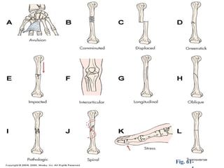
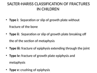
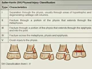
FRACTURES IN CHILDREN
BY
VARENCE
Fractures
• is partial or complete break in the continiuty of the bone
bone
• A fracture is the medical term for a broken bone.
• Fractures can be closed/ simple (where the skin is intact)
or open/compound (where there is a wound of the skin).
Open fractures may lead to serious bone infection.
• Suspect an open fracture if there is an associated
wound. A child’s bones are different from adults; instead
of breaking they often bend like a stick
General types of fractures
• Complete when the fragments are separated
• Incomplete / when the fragments are attached together
• Transverse crosswise fracture at the right to the long axis [of the bone
• Oblique slanting but straight between horizontal and perpendicular
direction
• Spiral slanting and circular twisting around the bone shaft
• Comminuted when the fragments of the fractured bone lie in the
surrounding the tissue (crashed bone)
• Proximal fragments are closer to the midline
• Distal fragments are farther to the midline
Fig. 61-
Common types of fractures seen in children
• Plastic deformation occurs when the bone is bent
but not broken . A child flexible bone can be bent at
45°or more before breaking
• Buckle or torus fracture produced by compression of
the porous bone appears as raised of the fracture
site. These fracture occur near the metaphysis
(portion of bone shaft adjacent to the epiphysis)
Contn
• Green stick occurs when the bone is angulated
beyond the limits of bending. The compressed side
bends and the tension side fails causing incomplete
fracture similar to when a green stick is broken
• Complete. Divides the fragment. These fragments
often remain attached by periosteal hinge which can
aid or hinder reduction
Causes
• Fractures happen when more force is applied
to the bone can absorb. Bones are weakest
when they are twisted
• Breaks in bones can happen from over use
injuries , falls, trauma, or direct hit to the body
SALTER-HARISS CLASSIFICATION OF FRACTURES
IN CHILDREN
• Type i: Separation or slip of growth plate without
fracture of the bone
• Type ii: Separation or slip of growth plate breaking off
the of the section of metaphysis
• Type iii: fracture of epiphysis extending through the joint
• Type iv: fracture of growth plate epiphysis and
metaphysis
• Type v: crushing of epiphysis
Investigations
• History taking & physical Examination
• X-tray
• CT scan
• MRI
• Bone scan
• Blood tests CBC, HCT
Clinical manifestation
• Generalized swelling
• Pain and tender
• Deformity
• Diminished functional use of limb
• Bruising
• Severe muscular rigidity
• Crepitus ( granting sensation at fracture site)
5Ps TO R/O COMPATMENT SYNDROME
• Parasthesia; tingling and burning sensations
• Pulselessness; inability to palpate pulse distal to the
fracture/ compartment
• Pressure; The limbs feels tense and warm skin is
tight and shin. Pressure within the compartment
• Pain; Elevation of the limb or movement the pain
• Paralysis; Inability to move the extremity or digits
Fracture Healing
• bone reparative process of self-healing (union) occurs in the
following stages:
1. Fracture hematoma ( bleeding, edema)
2. Granulation tissue → osteoid (3 – 14 days post injury)
3. Callus formation (minerals deposited in osteoid)
4. Ossification (3 wks – 6 months)
5. Consolidation (distance between fragments decreases → closes).
6. Remodeling (union completed; remodels to original shape,
strength)
Bone Healing
Fig.
MANAGEMENT
• AIMS :
• To regain alignment and length of the bony
fragment (reduction)
• To retain alignment and bony length
(Immobilization)
• To restore function to the injured party
• To prevent further injury or deformity
First aid mgt
• ABCD
• Determine the mechanism of an injury
• Move the injured part as little as possible
• Assess 5 Ps
• Cover the open wound with clean or sterile dressing
• Arrest any bleeding
• Use soft splits when immobilizing the injured part by aiming at securing joint above and
below of the fracture
• In extreme cases particularly lower limbs uninjured part can serve as splint
• Elevate the injured part
• Apply the cold compress to injured area
• Assess neurovascular status
HOSPITAL MGT
• Open reduction like internal fixation
• Cast application spica cast
• Calcium tablets
• Drugs analgesics
• Closed reduction internal fixation
Internal fixation
Fracture Immobilization
–Pins, plates, intramedullary
wires , and screws
–Surgically inserted at the time of
realignment
Application of casts
• Casts are temporary circumferential immobilization
device commonly used following the closed
reduction
• Spica cast are suitable for age of 6m-6 yrs can be
applied covering mid torso (trunk) and continues
down to the ankle on the affected side and to the
knee on the affected side immobile the hip joint. The
is an opening left in perineal area for toileting
pavlik harness spica
External fixation
Metallic device composed of pins that are
inserted into the bone and attached to
external rods
`

FRACTURES IN CHILDREN POWER POINT IN NURSING

  • 1.
  • 2.
    Fractures • is partialor complete break in the continiuty of the bone bone • A fracture is the medical term for a broken bone. • Fractures can be closed/ simple (where the skin is intact) or open/compound (where there is a wound of the skin). Open fractures may lead to serious bone infection. • Suspect an open fracture if there is an associated wound. A child’s bones are different from adults; instead of breaking they often bend like a stick
  • 3.
    General types offractures • Complete when the fragments are separated • Incomplete / when the fragments are attached together • Transverse crosswise fracture at the right to the long axis [of the bone • Oblique slanting but straight between horizontal and perpendicular direction • Spiral slanting and circular twisting around the bone shaft • Comminuted when the fragments of the fractured bone lie in the surrounding the tissue (crashed bone) • Proximal fragments are closer to the midline • Distal fragments are farther to the midline
  • 5.
  • 6.
    Common types offractures seen in children • Plastic deformation occurs when the bone is bent but not broken . A child flexible bone can be bent at 45°or more before breaking • Buckle or torus fracture produced by compression of the porous bone appears as raised of the fracture site. These fracture occur near the metaphysis (portion of bone shaft adjacent to the epiphysis)
  • 7.
    Contn • Green stickoccurs when the bone is angulated beyond the limits of bending. The compressed side bends and the tension side fails causing incomplete fracture similar to when a green stick is broken • Complete. Divides the fragment. These fragments often remain attached by periosteal hinge which can aid or hinder reduction
  • 8.
    Causes • Fractures happenwhen more force is applied to the bone can absorb. Bones are weakest when they are twisted • Breaks in bones can happen from over use injuries , falls, trauma, or direct hit to the body
  • 9.
    SALTER-HARISS CLASSIFICATION OFFRACTURES IN CHILDREN • Type i: Separation or slip of growth plate without fracture of the bone • Type ii: Separation or slip of growth plate breaking off the of the section of metaphysis • Type iii: fracture of epiphysis extending through the joint • Type iv: fracture of growth plate epiphysis and metaphysis • Type v: crushing of epiphysis
  • 11.
    Investigations • History taking& physical Examination • X-tray • CT scan • MRI • Bone scan • Blood tests CBC, HCT
  • 12.
    Clinical manifestation • Generalizedswelling • Pain and tender • Deformity • Diminished functional use of limb • Bruising • Severe muscular rigidity • Crepitus ( granting sensation at fracture site)
  • 13.
    5Ps TO R/OCOMPATMENT SYNDROME • Parasthesia; tingling and burning sensations • Pulselessness; inability to palpate pulse distal to the fracture/ compartment • Pressure; The limbs feels tense and warm skin is tight and shin. Pressure within the compartment • Pain; Elevation of the limb or movement the pain • Paralysis; Inability to move the extremity or digits
  • 14.
    Fracture Healing • bonereparative process of self-healing (union) occurs in the following stages: 1. Fracture hematoma ( bleeding, edema) 2. Granulation tissue → osteoid (3 – 14 days post injury) 3. Callus formation (minerals deposited in osteoid) 4. Ossification (3 wks – 6 months) 5. Consolidation (distance between fragments decreases → closes). 6. Remodeling (union completed; remodels to original shape, strength)
  • 15.
  • 16.
    MANAGEMENT • AIMS : •To regain alignment and length of the bony fragment (reduction) • To retain alignment and bony length (Immobilization) • To restore function to the injured party • To prevent further injury or deformity
  • 17.
    First aid mgt •ABCD • Determine the mechanism of an injury • Move the injured part as little as possible • Assess 5 Ps • Cover the open wound with clean or sterile dressing • Arrest any bleeding • Use soft splits when immobilizing the injured part by aiming at securing joint above and below of the fracture • In extreme cases particularly lower limbs uninjured part can serve as splint • Elevate the injured part • Apply the cold compress to injured area • Assess neurovascular status
  • 18.
    HOSPITAL MGT • Openreduction like internal fixation • Cast application spica cast • Calcium tablets • Drugs analgesics • Closed reduction internal fixation
  • 19.
    Internal fixation Fracture Immobilization –Pins,plates, intramedullary wires , and screws –Surgically inserted at the time of realignment
  • 20.
    Application of casts •Casts are temporary circumferential immobilization device commonly used following the closed reduction • Spica cast are suitable for age of 6m-6 yrs can be applied covering mid torso (trunk) and continues down to the ankle on the affected side and to the knee on the affected side immobile the hip joint. The is an opening left in perineal area for toileting
  • 22.
  • 23.
    External fixation Metallic devicecomposed of pins that are inserted into the bone and attached to external rods
  • 24.