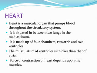
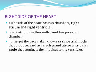
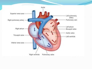
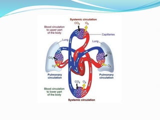
Introduction to Cardiovascular
System
 Cardiovascular system includes heart and blood
vessels.
 Heart pumps blood into the blood vessels.
 Blood vessels circulate the blood throughout the
body.
 Blood transports nutrients and oxygen to the
tissues and removes carbon dioxide and waste
products from the tissues.
HEART
 Heart is a muscular organ that pumps blood
throughout the circulatory system.
 It is situated in between two lungs in the
mediastinum.
 It is made up of four chambers, two atria and two
ventricles.
 The musculature of ventricles is thicker than that of
atria.
 Force of contraction of heart depends upon the
muscles.
RIGHT SIDE OF THE HEART
 Right side of the heart has two chambers, right
atrium and right ventricle.
 Right atrium is a thin walled and low pressure
chamber.
 It has got the pacemaker known as sinoatrial node
that produces cardiac impulses and atrioventricular
node that conducts the impulses to the ventricles.
RIGHT SIDE OF THE HEART
 Right atrium receives venous (deoxygenated) blood via
two large veins:
 1. Superior vena cava that returns venous blood from
the head, neck and upper limbs.
 2. Inferior vena cava that returns venous blood from
lower parts of the body.
 Right atrium communicates with right ventricle
through tricuspid valve. Wall of right ventricle is thick.
Venous blood from the right atrium enters the right
ventricle through this valve.
RIGHT SIDE OF THE HEART
 From the right ventricle, pulmonary artery arises.
 It carries the venous blood from right ventricle to
lungs.
 In the lungs, the deoxygenated blood is oxygenated.
LEFT SIDE OF THE HEART
 Left side of the heart has two chambers, left atrium
and left ventricle.
 Left atrium is a thin walled and low pressure chamber.
It receives oxygenated blood from the lungs through
pulmonary veins. This is the only exception in the
body, where an artery carries venous blood and vein
carries the arterial blood.
 Blood from left atrium enters the left ventricle through
mitral valve (bicuspid valve). Wall of the left ventricle
is very thick. Left ventricle pumps the arterial blood to
different parts of the body through systemic aorta.
SEPTA OF THE HEART
 Right and left atria are separated from one another by
a fibrous septum called interatrial septum.
 Right and left ventricles are separated from one
another by interventricular septum.
 The upper part of this septum is a membranous
structure, whereas the lower part of it is muscular in
nature.
LAYERS OF WALL OF THE HEART
 Heart is made up of three layers of tissues:
 1. Outer pericardium
 2. Middle myocardium
 3. Inner endocardium.
 All three have an important functions.
ACTIONS OF THE HEART
 Actions of the heart are classified into four types:
 1. Chronotropic action
 2. Inotropic action
 3. Dromotropic action
 4. Bathmotropic action
 CHRONOTROPIC ACTION
Chronotropic action is the frequency of heartbeat
or heart rate. It is of two types:
i. Tachycardia or increase in heart rate
ii. Bradycardia or decrease in heart rate
INOTROPIC ACTION
 Force of contraction of heart is called inotropic action.
 It is of two types:
 i. Positive inotropic action or increase in the force of contraction
 ii. Negative inotropic action or decrease in the force of contraction
DROMOTROPIC ACTION
Dromotropic action is the conduction of impulse through heart. It is of
two types:
i. Positive dromotropic action or increase in the velocity of conduction
ii. Negative dromotropic action or decrease in the velocity of conduction
„BATHMOTROPIC ACTION
Bathmotropic action is the excitability of cardiac muscles
It is also of two types:
i. Positive bathmotropic action or increase in the excitability of cardiac muscle
ii. Negative bathmotropic action or decrease in the excitability of cardiac muscle.
Regulation of Actions of Heart
 All the actions of heart are continuously regulated. It is
essential for the heart to cope up with the needs of the
body.
 All the actions are altered by stimulation of nerves
supplying the heart or some hormones or hormonal
substances secreted in the body.
BLOOD VESSELS
 Vessels of circulatory system are the
 aorta,
 arteries,
 arterioles,
 capillaries,
 venules,
 veins
 venae cavae.
ARTERIAL SYSTEM
 Arterial system comprises the aorta, arteries and
arterioles.
 Walls of the aorta and arteries are formed by three
layers:
1. Outer tunica adventitia
2. Middle tunica media
3. Inner tunica intima
VENOUS SYSTEM
 From the capillaries, venous system starts and it
includes venules, veins and venae cavae. Capillaries
end in venules, which are the smaller vessels with thin
muscular wall than the arterioles.
 Diameter of the venules is about 20 µ. At a time, a
large quantity of blood is held in venules and hence
the venules are called capacitance vessels. Venules
are continued as veins, which have the diameter of 5
mm. Veins form superior and inferior venae cavae,
which have a diameter of about 30 mm.
COMPLICATIONS IN BLOOD
VESSELS
 Arteriosclerosis is the disease of the arteries, associated
with hardening, thickening and loss of elasticity in the wall
of the vessels.
 Atherosclerosis is the disease marked by the narrowing of
lumen of arterial vessel due to deposition of cholesterol.
 Arterioles When the tone of the smooth muscles in the
arterioles increases, hypertension occurs.
 Capillaries Permeability of the capillary membrane may
increase resulting in shock or edema due to leakage of
fluid, proteins and other substances from blood.
COMPLICATIONS IN BLOOD
VESSELS
 Veins Inflammation of the wall of veins leads to the
formation of intravascular clot called thrombosis.
 The clot gets dislodged, as thrombus.
 The thrombus travels through blood and causes
embolism. Embolism obstructs the blood flow to vital
organs such as brain, heart and lungs, leading to many
complications.
DIVISIONS OF CIRCULATION
 Blood flows through two divisions of circulatory
system:
 1. Systemic circulation
 2. Pulmonary circulation
 Systemic circulation
 Also known as greater circulation. Blood pumped
from left ventricle passes through a series of blood
vessels, arterial system and reaches the tissues.
Exchange of various substances between blood and the
tissues occurs at the capillaries.
 After exchange of materials, blood enters the venous
system and returns to right atrium of the heart. From
right atrium, blood enters the right ventricle.
 Thus, through systemic circulation, oxygenated blood
is supplied from heart to the tissues and venous blood
returns to the heart from tissues.
 Pulmonary Circulation
Pulmonary circulation is otherwise called lesser
circulation. Blood is pumped from right ventricle to
lungs through pulmonary artery. Exchange of gases
occurs between blood and alveoli of the lungs at
pulmonary capillaries. Oxygenated blood returns to
left atrium through the pulmonary veins.
Thus, left side of the heart contains oxygenated or
arterial blood and the right side of the heart contains
deoxygenated or venous blood

Introduction to Cardiovascular System.pptx

  • 1.
    Introduction to Cardiovascular System Cardiovascular system includes heart and blood vessels.  Heart pumps blood into the blood vessels.  Blood vessels circulate the blood throughout the body.  Blood transports nutrients and oxygen to the tissues and removes carbon dioxide and waste products from the tissues.
  • 2.
    HEART  Heart isa muscular organ that pumps blood throughout the circulatory system.  It is situated in between two lungs in the mediastinum.  It is made up of four chambers, two atria and two ventricles.  The musculature of ventricles is thicker than that of atria.  Force of contraction of heart depends upon the muscles.
  • 3.
    RIGHT SIDE OFTHE HEART  Right side of the heart has two chambers, right atrium and right ventricle.  Right atrium is a thin walled and low pressure chamber.  It has got the pacemaker known as sinoatrial node that produces cardiac impulses and atrioventricular node that conducts the impulses to the ventricles.
  • 4.
    RIGHT SIDE OFTHE HEART  Right atrium receives venous (deoxygenated) blood via two large veins:  1. Superior vena cava that returns venous blood from the head, neck and upper limbs.  2. Inferior vena cava that returns venous blood from lower parts of the body.  Right atrium communicates with right ventricle through tricuspid valve. Wall of right ventricle is thick. Venous blood from the right atrium enters the right ventricle through this valve.
  • 5.
    RIGHT SIDE OFTHE HEART  From the right ventricle, pulmonary artery arises.  It carries the venous blood from right ventricle to lungs.  In the lungs, the deoxygenated blood is oxygenated.
  • 6.
    LEFT SIDE OFTHE HEART  Left side of the heart has two chambers, left atrium and left ventricle.  Left atrium is a thin walled and low pressure chamber. It receives oxygenated blood from the lungs through pulmonary veins. This is the only exception in the body, where an artery carries venous blood and vein carries the arterial blood.  Blood from left atrium enters the left ventricle through mitral valve (bicuspid valve). Wall of the left ventricle is very thick. Left ventricle pumps the arterial blood to different parts of the body through systemic aorta.
  • 8.
    SEPTA OF THEHEART  Right and left atria are separated from one another by a fibrous septum called interatrial septum.  Right and left ventricles are separated from one another by interventricular septum.  The upper part of this septum is a membranous structure, whereas the lower part of it is muscular in nature.
  • 9.
    LAYERS OF WALLOF THE HEART  Heart is made up of three layers of tissues:  1. Outer pericardium  2. Middle myocardium  3. Inner endocardium.  All three have an important functions.
  • 10.
    ACTIONS OF THEHEART  Actions of the heart are classified into four types:  1. Chronotropic action  2. Inotropic action  3. Dromotropic action  4. Bathmotropic action  CHRONOTROPIC ACTION Chronotropic action is the frequency of heartbeat or heart rate. It is of two types: i. Tachycardia or increase in heart rate ii. Bradycardia or decrease in heart rate
  • 11.
    INOTROPIC ACTION  Forceof contraction of heart is called inotropic action.  It is of two types:  i. Positive inotropic action or increase in the force of contraction  ii. Negative inotropic action or decrease in the force of contraction DROMOTROPIC ACTION Dromotropic action is the conduction of impulse through heart. It is of two types: i. Positive dromotropic action or increase in the velocity of conduction ii. Negative dromotropic action or decrease in the velocity of conduction „BATHMOTROPIC ACTION Bathmotropic action is the excitability of cardiac muscles It is also of two types: i. Positive bathmotropic action or increase in the excitability of cardiac muscle ii. Negative bathmotropic action or decrease in the excitability of cardiac muscle.
  • 12.
    Regulation of Actionsof Heart  All the actions of heart are continuously regulated. It is essential for the heart to cope up with the needs of the body.  All the actions are altered by stimulation of nerves supplying the heart or some hormones or hormonal substances secreted in the body.
  • 13.
    BLOOD VESSELS  Vesselsof circulatory system are the  aorta,  arteries,  arterioles,  capillaries,  venules,  veins  venae cavae.
  • 14.
    ARTERIAL SYSTEM  Arterialsystem comprises the aorta, arteries and arterioles.  Walls of the aorta and arteries are formed by three layers: 1. Outer tunica adventitia 2. Middle tunica media 3. Inner tunica intima
  • 15.
    VENOUS SYSTEM  Fromthe capillaries, venous system starts and it includes venules, veins and venae cavae. Capillaries end in venules, which are the smaller vessels with thin muscular wall than the arterioles.  Diameter of the venules is about 20 µ. At a time, a large quantity of blood is held in venules and hence the venules are called capacitance vessels. Venules are continued as veins, which have the diameter of 5 mm. Veins form superior and inferior venae cavae, which have a diameter of about 30 mm.
  • 16.
    COMPLICATIONS IN BLOOD VESSELS Arteriosclerosis is the disease of the arteries, associated with hardening, thickening and loss of elasticity in the wall of the vessels.  Atherosclerosis is the disease marked by the narrowing of lumen of arterial vessel due to deposition of cholesterol.  Arterioles When the tone of the smooth muscles in the arterioles increases, hypertension occurs.  Capillaries Permeability of the capillary membrane may increase resulting in shock or edema due to leakage of fluid, proteins and other substances from blood.
  • 17.
    COMPLICATIONS IN BLOOD VESSELS Veins Inflammation of the wall of veins leads to the formation of intravascular clot called thrombosis.  The clot gets dislodged, as thrombus.  The thrombus travels through blood and causes embolism. Embolism obstructs the blood flow to vital organs such as brain, heart and lungs, leading to many complications.
  • 18.
    DIVISIONS OF CIRCULATION Blood flows through two divisions of circulatory system:  1. Systemic circulation  2. Pulmonary circulation  Systemic circulation  Also known as greater circulation. Blood pumped from left ventricle passes through a series of blood vessels, arterial system and reaches the tissues. Exchange of various substances between blood and the tissues occurs at the capillaries.
  • 19.
     After exchangeof materials, blood enters the venous system and returns to right atrium of the heart. From right atrium, blood enters the right ventricle.  Thus, through systemic circulation, oxygenated blood is supplied from heart to the tissues and venous blood returns to the heart from tissues.
  • 21.
     Pulmonary Circulation Pulmonarycirculation is otherwise called lesser circulation. Blood is pumped from right ventricle to lungs through pulmonary artery. Exchange of gases occurs between blood and alveoli of the lungs at pulmonary capillaries. Oxygenated blood returns to left atrium through the pulmonary veins. Thus, left side of the heart contains oxygenated or arterial blood and the right side of the heart contains deoxygenated or venous blood