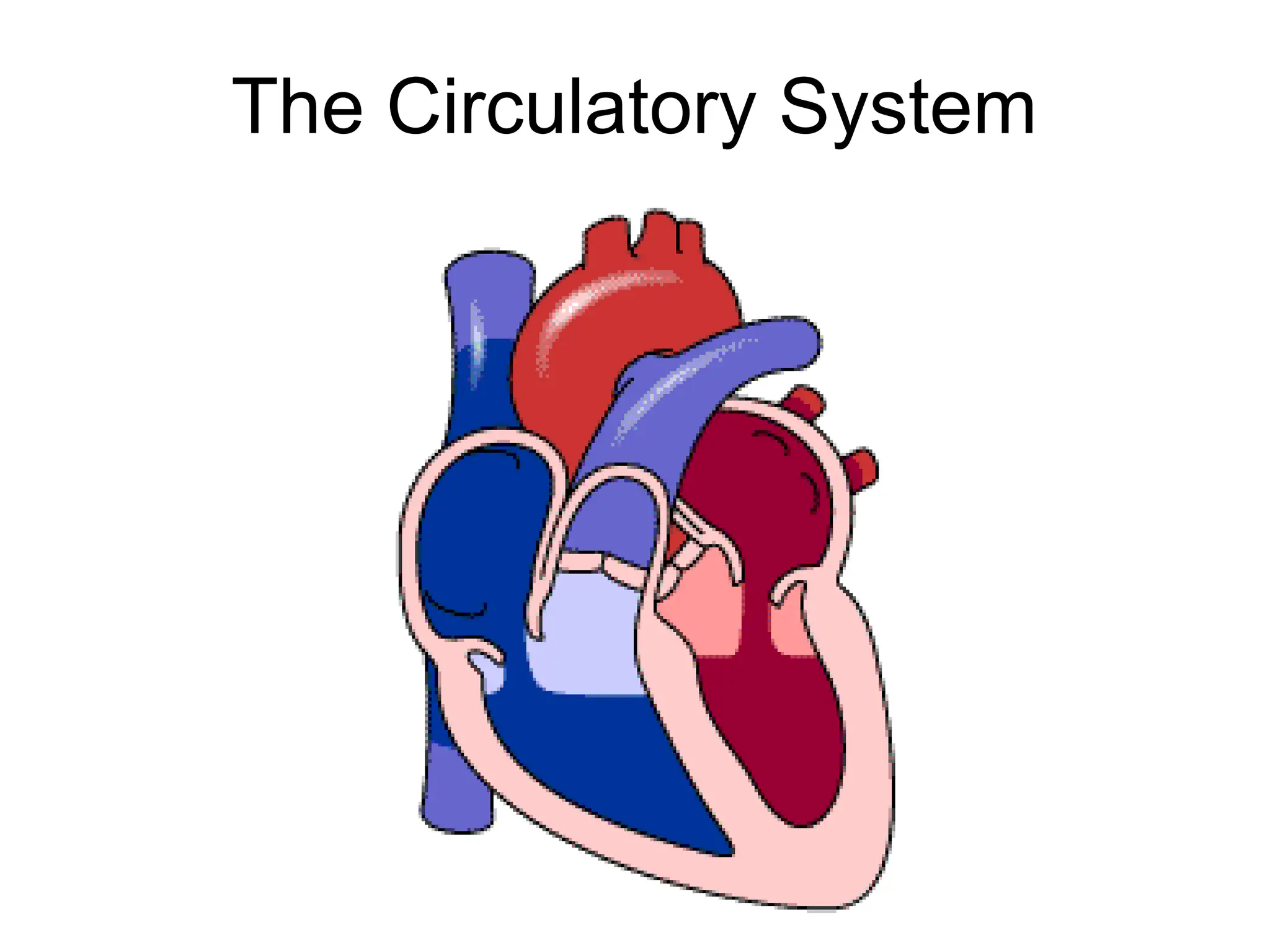
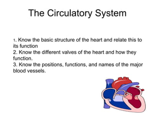
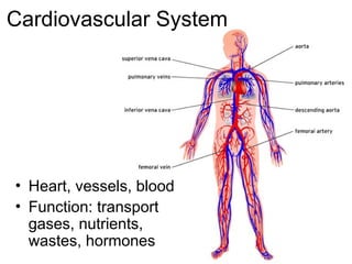
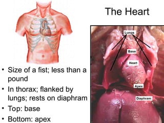
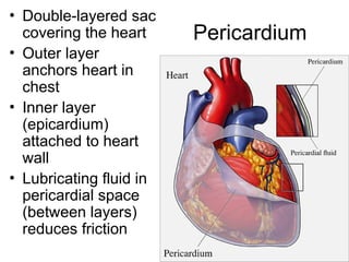
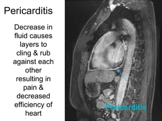
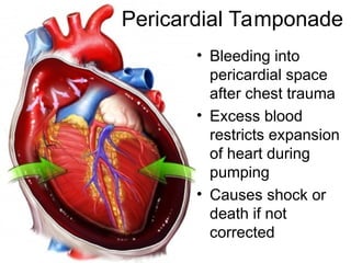
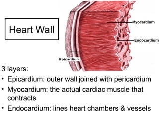
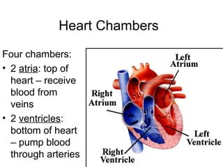
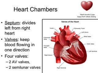
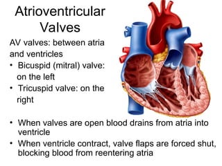
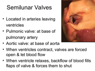
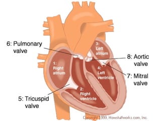
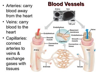
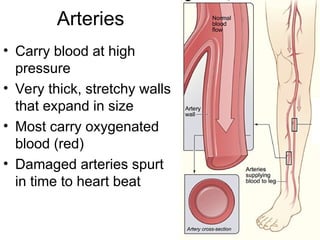
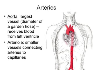
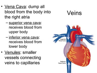
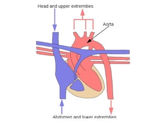
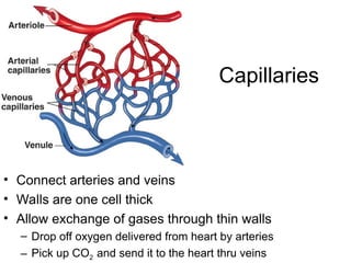
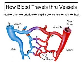
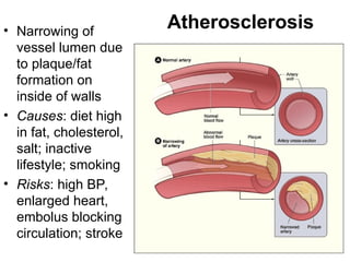
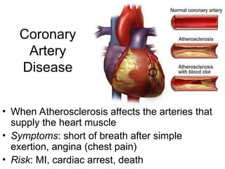
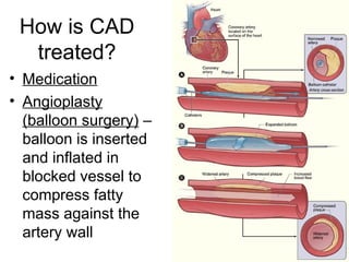
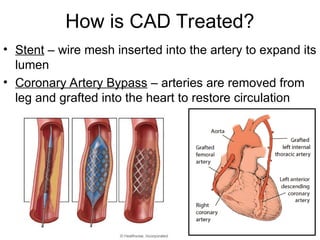
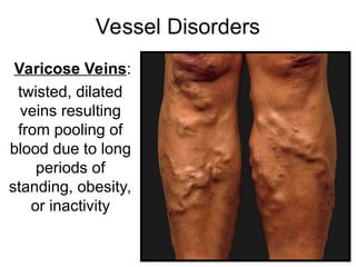
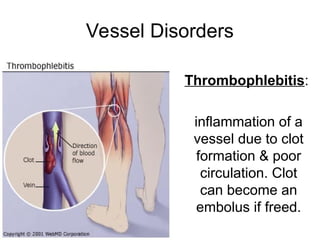
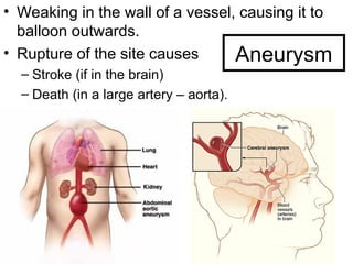
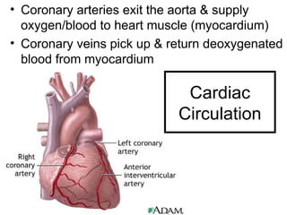
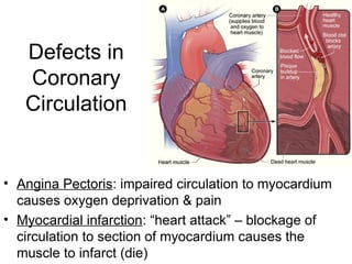
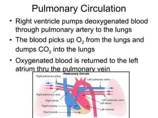
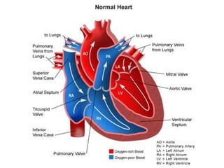
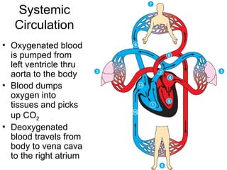
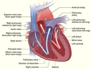
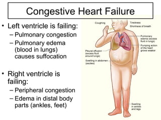
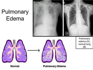
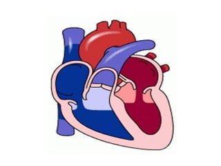
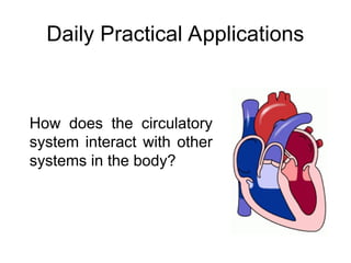
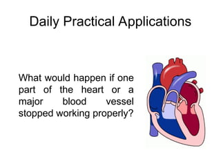
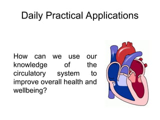
The Circulatory System
1. Know the basic structure of the heart and relate this to
its function
2. Know the different valves of the heart and how they
function.
3. Know the positions, functions, and names of the major
blood vessels.
Cardiovascular System
• Heart, vessels, blood
• Function: transport
gases, nutrients,
wastes, hormones
The Heart
• Size of a fist; less than a
pound
• In thorax; flanked by
lungs; rests on diaphram
• Top: base
• Bottom: apex
Pericardium
• Double-layered sac
covering the heart
• Outer layer
anchors heart in
chest
• Inner layer
(epicardium)
attached to heart
wall
• Lubricating fluid in
pericardial space
(between layers)
reduces friction
Pericarditis
Decrease in
fluid causes
layers to
cling & rub
against each
other
resulting in
pain &
decreased
efficiency of
heart
Pericardial Tamponade
• Bleeding into
pericardial space
after chest trauma
• Excess blood
restricts expansion
of heart during
pumping
• Causes shock or
death if not
corrected
Heart Wall
3 layers:
• Epicardium: outer wall joined with pericardium
• Myocardium: the actual cardiac muscle that
contracts
• Endocardium: lines heart chambers & vessels
Heart Chambers
Four chambers:
• 2 atria: top of
heart – receive
blood from
veins
• 2 ventricles:
bottom of heart
– pump blood
through arteries
Heart Chambers
• Septum: divides
left from right
heart
• Valves: keep
blood flowing in
one direction
• Four valves:
– 2 AV valves,
– 2 semilunar valves
Heart sounds (Lupp-
dupp) from valves closing
Atrioventricular
Valves
AV valves: between atria
and ventricles
• Bicuspid (mitral) valve:
on the left
• Tricuspid valve: on the
right
• When valves are open blood drains from atria into
ventricle
• When ventricle contract, valve flaps are forced shut,
blocking blood from reentering atria
Semilunar Valves
• Located in arteries leaving
ventricles
• Pulmonic valve: at base of
pulmonary artery
• Aortic valve: at base of aorta
• When ventricles contract, valves are forced
open & let blood flow
• When ventricle relaxes, backflow of blood fills
flaps of valve & forces them to shut
Blood Vessels
• Arteries: carry
blood away
from the heart
• Veins: carry
blood to the
heart
• Capillaries:
connect
arteries to
veins &
exchange
gases with
tissues
Arteries
• Carry blood at high
pressure
• Very thick, stretchy walls
that expand in size
• Most carry oxygenated
blood (red)
• Damaged arteries spurt
in time to heart beat
Arteries
• Aorta: largest
vessel (diameter of
a garden hose) –
receives blood
from left ventricle
• Arteriole: smaller
vessels connecting
arteries to
capillaries
Veins
• Carry blood at low
pressure
• Have valves to
prevent backflow of
blood against gravity
• Most carry de-
oxygenated blood
(purple)
• Damaged veins ooze
blood
Veins
• Vena Cava: dump all
blood from the body into
the right atria
– superior vena cava:
receives blood from
upper body
– inferior vena cava:
receives blood from
lower body
• Venules: smaller
vessels connecting
veins to capillaries
Capillaries
• Connect arteries and veins
• Walls are one cell thick
• Allow exchange of gases through thin walls
– Drop off oxygen delivered from heart by arteries
– Pick up CO2 and send it to the heart thru veins
How Blood Travels thru Vessels
heart artery arteriole capillary venule vein heart
• Narrowing of
vessel lumen due
to plaque/fat
formation on
inside of walls
• Causes: diet high
in fat, cholesterol,
salt; inactive
lifestyle; smoking
• Risks: high BP,
enlarged heart,
embolus blocking
circulation; stroke
Atherosclerosis
Coronary
Artery
Disease
• When Atherosclerosis affects the arteries that
supply the heart muscle
• Symptoms: short of breath after simple
exertion, angina (chest pain)
• Risk: MI, cardiac arrest, death
How is CAD
treated?
• Medication
• Angioplasty
(balloon surgery) –
balloon is inserted
and inflated in
blocked vessel to
compress fatty
mass against the
artery wall
How is CAD Treated?
• Stent – wire mesh inserted into the artery to expand its
lumen
• Coronary Artery Bypass – arteries are removed from
leg and grafted into the heart to restore circulation
Vessel Disorders
Varicose Veins:
twisted, dilated
veins resulting
from pooling of
blood due to long
periods of
standing, obesity,
or inactivity
Vessel Disorders
Thrombophlebitis:
inflammation of a
vessel due to clot
formation & poor
circulation. Clot
can become an
embolus if freed.
Aneurysm
• Weaking in the wall of a vessel, causing it to
balloon outwards.
• Rupture of the site causes
– Stroke (if in the brain)
– Death (in a large artery – aorta).
Cardiac
Circulation
• Coronary arteries exit the aorta & supply
oxygen/blood to heart muscle (myocardium)
• Coronary veins pick up & return deoxygenated
blood from myocardium
Defects in
Coronary
Circulation
• Angina Pectoris: impaired circulation to myocardium
causes oxygen deprivation & pain
• Myocardial infarction: “heart attack” – blockage of
circulation to section of myocardium causes the
muscle to infarct (die)
Pulmonary Circulation
• Right ventricle pumps deoxygenated blood
through pulmonary artery to the lungs
• The blood picks up O2 from the lungs and
dumps CO2 into the lungs
• Oxygenated blood is returned to the left
atrium thru the pulmonary vein
Systemic
Circulation
• Oxygenated blood
is pumped from
left ventricle thru
aorta to the body
• Blood dumps
oxygen into
tissues and picks
up CO2
• Deoxygenated
blood travels from
body to vena cava
to the right atrium
Congestive Heart Failure
• Heart is ‘worn out’ from hypertension,
multiple MI, atherosclerosis, or age
• Heart pumps too weakly to meet tissue
needs
• If one side is weaker than the other, blood
will back up in system
Congestive Heart Failure
• Left ventricle is failing:
– Pulmonary congestion
– Pulmonary edema
(blood in lungs)
causes suffocation
• Right ventricle is
failing:
– Peripheral congestion
– Edema in distal body
parts (ankles, feet)
Pulmonary
Edema
Pulmonary
edema (A);
normal lung
(B)
Group Activity
Instructions:
1.Group by 4
2.Discuss and answer the given questions
3.10 mins will be given for each group to
finished the task
4.One representative will discuss about the
given questions
Questions
1.Can you name the four chambers of the
heart and describe their locations?
2.What are the four main valves of the heart?
3.Describe the pathway of blood through the
heart starting from the vena cava.
4.How does oxygenated blood differ from
deoxygenated blood in terms of their
pathways in the heart?
Daily Practical Applications
How does the circulatory
system interact with other
systems in the body?
Daily Practical Applications
What would happen if one
part of the heart or a
major blood vessel
stopped working properly?
Daily Practical Applications
How can we use our
knowledge of the
circulatory system to
improve overall health and
wellbeing?

circulatory-system-for academics purposes

  • 1.
  • 2.
    The Circulatory System 1.Know the basic structure of the heart and relate this to its function 2. Know the different valves of the heart and how they function. 3. Know the positions, functions, and names of the major blood vessels.
  • 4.
    Cardiovascular System • Heart,vessels, blood • Function: transport gases, nutrients, wastes, hormones
  • 5.
    The Heart • Sizeof a fist; less than a pound • In thorax; flanked by lungs; rests on diaphram • Top: base • Bottom: apex
  • 6.
    Pericardium • Double-layered sac coveringthe heart • Outer layer anchors heart in chest • Inner layer (epicardium) attached to heart wall • Lubricating fluid in pericardial space (between layers) reduces friction
  • 7.
    Pericarditis Decrease in fluid causes layersto cling & rub against each other resulting in pain & decreased efficiency of heart
  • 8.
    Pericardial Tamponade • Bleedinginto pericardial space after chest trauma • Excess blood restricts expansion of heart during pumping • Causes shock or death if not corrected
  • 9.
    Heart Wall 3 layers: •Epicardium: outer wall joined with pericardium • Myocardium: the actual cardiac muscle that contracts • Endocardium: lines heart chambers & vessels
  • 10.
    Heart Chambers Four chambers: •2 atria: top of heart – receive blood from veins • 2 ventricles: bottom of heart – pump blood through arteries
  • 11.
    Heart Chambers • Septum:divides left from right heart • Valves: keep blood flowing in one direction • Four valves: – 2 AV valves, – 2 semilunar valves Heart sounds (Lupp- dupp) from valves closing
  • 12.
    Atrioventricular Valves AV valves: betweenatria and ventricles • Bicuspid (mitral) valve: on the left • Tricuspid valve: on the right • When valves are open blood drains from atria into ventricle • When ventricle contract, valve flaps are forced shut, blocking blood from reentering atria
  • 13.
    Semilunar Valves • Locatedin arteries leaving ventricles • Pulmonic valve: at base of pulmonary artery • Aortic valve: at base of aorta • When ventricles contract, valves are forced open & let blood flow • When ventricle relaxes, backflow of blood fills flaps of valve & forces them to shut
  • 15.
    Blood Vessels • Arteries:carry blood away from the heart • Veins: carry blood to the heart • Capillaries: connect arteries to veins & exchange gases with tissues
  • 16.
    Arteries • Carry bloodat high pressure • Very thick, stretchy walls that expand in size • Most carry oxygenated blood (red) • Damaged arteries spurt in time to heart beat
  • 17.
    Arteries • Aorta: largest vessel(diameter of a garden hose) – receives blood from left ventricle • Arteriole: smaller vessels connecting arteries to capillaries
  • 18.
    Veins • Carry bloodat low pressure • Have valves to prevent backflow of blood against gravity • Most carry de- oxygenated blood (purple) • Damaged veins ooze blood
  • 19.
    Veins • Vena Cava:dump all blood from the body into the right atria – superior vena cava: receives blood from upper body – inferior vena cava: receives blood from lower body • Venules: smaller vessels connecting veins to capillaries
  • 21.
    Capillaries • Connect arteriesand veins • Walls are one cell thick • Allow exchange of gases through thin walls – Drop off oxygen delivered from heart by arteries – Pick up CO2 and send it to the heart thru veins
  • 22.
    How Blood Travelsthru Vessels heart artery arteriole capillary venule vein heart
  • 23.
    • Narrowing of vessellumen due to plaque/fat formation on inside of walls • Causes: diet high in fat, cholesterol, salt; inactive lifestyle; smoking • Risks: high BP, enlarged heart, embolus blocking circulation; stroke Atherosclerosis
  • 24.
    Coronary Artery Disease • When Atherosclerosisaffects the arteries that supply the heart muscle • Symptoms: short of breath after simple exertion, angina (chest pain) • Risk: MI, cardiac arrest, death
  • 25.
    How is CAD treated? •Medication • Angioplasty (balloon surgery) – balloon is inserted and inflated in blocked vessel to compress fatty mass against the artery wall
  • 26.
    How is CADTreated? • Stent – wire mesh inserted into the artery to expand its lumen • Coronary Artery Bypass – arteries are removed from leg and grafted into the heart to restore circulation
  • 27.
    Vessel Disorders Varicose Veins: twisted,dilated veins resulting from pooling of blood due to long periods of standing, obesity, or inactivity
  • 28.
    Vessel Disorders Thrombophlebitis: inflammation ofa vessel due to clot formation & poor circulation. Clot can become an embolus if freed.
  • 29.
    Aneurysm • Weaking inthe wall of a vessel, causing it to balloon outwards. • Rupture of the site causes – Stroke (if in the brain) – Death (in a large artery – aorta).
  • 30.
    Cardiac Circulation • Coronary arteriesexit the aorta & supply oxygen/blood to heart muscle (myocardium) • Coronary veins pick up & return deoxygenated blood from myocardium
  • 31.
    Defects in Coronary Circulation • AnginaPectoris: impaired circulation to myocardium causes oxygen deprivation & pain • Myocardial infarction: “heart attack” – blockage of circulation to section of myocardium causes the muscle to infarct (die)
  • 32.
    Pulmonary Circulation • Rightventricle pumps deoxygenated blood through pulmonary artery to the lungs • The blood picks up O2 from the lungs and dumps CO2 into the lungs • Oxygenated blood is returned to the left atrium thru the pulmonary vein
  • 34.
    Systemic Circulation • Oxygenated blood ispumped from left ventricle thru aorta to the body • Blood dumps oxygen into tissues and picks up CO2 • Deoxygenated blood travels from body to vena cava to the right atrium
  • 36.
    Congestive Heart Failure •Heart is ‘worn out’ from hypertension, multiple MI, atherosclerosis, or age • Heart pumps too weakly to meet tissue needs • If one side is weaker than the other, blood will back up in system
  • 37.
    Congestive Heart Failure •Left ventricle is failing: – Pulmonary congestion – Pulmonary edema (blood in lungs) causes suffocation • Right ventricle is failing: – Peripheral congestion – Edema in distal body parts (ankles, feet)
  • 38.
  • 40.
    Group Activity Instructions: 1.Group by4 2.Discuss and answer the given questions 3.10 mins will be given for each group to finished the task 4.One representative will discuss about the given questions
  • 41.
    Questions 1.Can you namethe four chambers of the heart and describe their locations? 2.What are the four main valves of the heart? 3.Describe the pathway of blood through the heart starting from the vena cava. 4.How does oxygenated blood differ from deoxygenated blood in terms of their pathways in the heart?
  • 42.
    Daily Practical Applications Howdoes the circulatory system interact with other systems in the body?
  • 43.
    Daily Practical Applications Whatwould happen if one part of the heart or a major blood vessel stopped working properly?
  • 44.
    Daily Practical Applications Howcan we use our knowledge of the circulatory system to improve overall health and wellbeing?