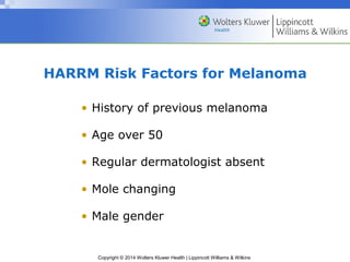
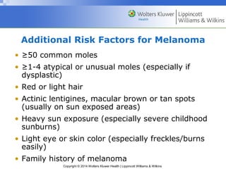
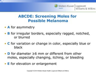
Chapter 6 
The Skin, Hair, and Nails 
Copyright © 2014 Wolters Kluwer Health | Lippincott Williams & Wilkins
Anatomy and Physiology 
• Major function of skin is to keep the body in 
homeostasis 
– Provides boundaries for body fluid 
– Protects underlying tissues from microorganisms, 
harmful substances, and radiation 
– Modulates body temperature 
– Synthesizes vitamin D 
• Heaviest single organ in body 
– 16% of body weight 
Copyright © 2014 Wolters Kluwer Health | Lippincott Williams & Wilkins
Anatomy and Physiology (cont.) 
• Three layers 
– Epidermis 
– Dermis 
– Subcutaneous tissue 
• Hair, nails, and sebaceous and sweat glands 
are appendages of skin 
Copyright © 2014 Wolters Kluwer Health | Lippincott Williams & Wilkins
Anatomy and Physiology (cont.) 
Copyright © 2014 Wolters Kluwer Health | Lippincott Williams & Wilkins 
• Hair 
– Vellus hair – short, fine, less pigmentation 
– Terminal hair – coarser, pigmented (scalp/eyebrows) 
• Nails - protect distal ends of fingers/toes 
• Sebaceous glands – present all surfaces except palms/soles; 
produce a fatty substance secreted onto skin surface through 
hair follicles 
• Sweat glands 
– Eccrine glands – widely distributed, open directly onto 
skin surface, help control body temperature 
– Apocrine glands – found in axilla and groin, stimulated by 
emotional stress
The Health History 
• Common or concerning symptoms 
Copyright © 2014 Wolters Kluwer Health | Lippincott Williams & Wilkins 
– Hair loss 
– Rash 
– Moles 
• Ask the patient 
– “Have you noticed any changes in your skin or your 
hair?” 
– “Have you noticed any moles that have changed size, 
shape, color, or sensation?” 
– “Have you noticed any new moles?”
Health Promotion and Counseling 
• Clinicians play an important role in educating 
patients 
– Early detection of suspicious moles 
– Protective measures for skin care 
– Hazards of excessive sun exposure 
• Skin cancers are most common cancers in the U.S. 
– Most prevalent on hands, neck, and head 
Copyright © 2014 Wolters Kluwer Health | Lippincott Williams & Wilkins
Skin Cancers 
• Basal cell carcinoma 
– Comprises 80% of skin cancers 
– Shiny and translucent, they grow slowly and rarely 
metastasize 
• Squamous cell carcinoma 
– Comprises 16% of skin cancers 
– Crusted, scaly, and ulcerated, they can metastasize 
Copyright © 2014 Wolters Kluwer Health | Lippincott Williams & Wilkins 
• Melanoma 
– Comprises 4% of skin cancers 
– Rapidly increasing in frequency, they spread rapidly
HARRM Risk Factors for Melanoma 
• History of previous melanoma 
• Age over 50 
• Regular dermatologist absent 
• Mole changing 
• Male gender 
Copyright © 2014 Wolters Kluwer Health | Lippincott Williams & Wilkins
Additional Risk Factors for Melanoma 
• ≥50 common moles 
• ≥1-4 atypical or unusual moles (especially if 
dysplastic) 
• Red or light hair 
• Actinic lentigines, macular brown or tan spots 
(usually on sun exposed areas) 
• Heavy sun exposure (especially severe childhood 
sunburns) 
• Light eye or skin color (especially freckles/burns 
easily) 
• Family history of melanoma 
Copyright © 2014 Wolters Kluwer Health | Lippincott Williams & Wilkins
ABCDE: Screening Moles for 
Possible Melanoma 
• A for asymmetry 
• B for irregular borders, especially ragged, notched, 
or blurred 
• C for variation or change in color, especially blue or 
Copyright © 2014 Wolters Kluwer Health | Lippincott Williams & Wilkins 
black 
• D for diameter ≥6 mm or different from other 
moles, especially changing, itching, or bleeding 
• E for elevation or enlargement
Question 
Which of the following is not considered a risk 
factor for the development of melanoma? 
a. Light hair and eyes 
b. Female gender 
c. Severe sunburns in childhood 
d. 1-4 dysplastic moles 
Copyright © 2014 Wolters Kluwer Health | Lippincott Williams & Wilkins
Answer 
b. Female gender 
Males have an increased risk of melanoma (1.4). 
Copyright © 2014 Wolters Kluwer Health | Lippincott Williams & Wilkins
Techniques of Examination 
• Examination of the skin, hair, and nails begins with the 
general survey of the patient 
• Make sure the patient wears a gown 
– Drape appropriately to facilitate close inspection of 
hair, anterior and posterior surfaces of body, palms 
and soles, and webspaces 
• Inspect entire skin surface in good light 
– Preferably in natural light (or artificial light that 
resembles natural) 
o Artificial light often distorts colors 
Copyright © 2014 Wolters Kluwer Health | Lippincott Williams & Wilkins
Techniques of Examination (cont.) 
• Inspect and palpate skin 
• Note characteristics of: 
– Color 
– Moisture 
– Temperature 
– Texture 
– Mobility and turgor 
– Lesions 
Copyright © 2014 Wolters Kluwer Health | Lippincott Williams & Wilkins
Techniques of Examination (cont.) 
Copyright © 2014 Wolters Kluwer Health | Lippincott Williams & Wilkins 
• Color 
– Patients often notice change in color before physician 
– Look for increased pigmentation, loss of pigmentation 
– Look for redness, pallor, cyanosis, and yellowing 
o Red color of oxyhemoglobin best assessed at 
fingertips, lips, and mucous membranes 
 In dark-skinned people, palms and soles 
o For central cyanosis, look in lips, oral mucosa, 
and tongue 
o Jaundice - sclera
Techniques of Examination (cont.) 
• Moisture 
– Dryness, sweating, and oiliness 
• Temperature 
– Use back of fingertips 
– Identify warmth or coolness of skin 
Copyright © 2014 Wolters Kluwer Health | Lippincott Williams & Wilkins 
• Texture 
– Roughness or smoothness 
• Mobility and turgor 
– Lift fold of skin 
– Note ease with which it lifts up (mobility) and 
speed with which it returns to place (turgor)
Techniques of Examination (cont.) 
Copyright © 2014 Wolters Kluwer Health | Lippincott Williams & Wilkins 
• Lesions 
– Note characteristics 
o Anatomic location and distribution 
o Patterns and shapes 
o Type of lesion (macules, papules, nevi, 
vesicles) 
o Color
Techniques of Examination (cont.) 
• Skin lesions in context 
– Whenever you see a skin lesion, look it up 
in a well-illustrated textbook of dermatology 
– To arrive at a dermatologic diagnosis, consider 
the type of lesions, location, and distribution, 
along with the patient’s history and physical 
Copyright © 2014 Wolters Kluwer Health | Lippincott Williams & Wilkins
Techniques of Examination (cont.) 
• Hair 
– Inspect and palpate 
– Note quantity, distribution, and texture 
Copyright © 2014 Wolters Kluwer Health | Lippincott Williams & Wilkins 
• Nails 
– Inspect and palpate fingernails/toenails 
– Note color and shape 
– Note lesions 
o Longitudinal bands of pigment may be a 
normal finding in people with darker skin
Evaluating the Bedbound Patient 
• People confined to bed are particularly susceptible 
to skin damage and ulceration 
– Pressure sores result when sustained 
compression obliterates arteriolar and capillary 
blood flow to the skin 
• Assess these patients by carefully inspecting the 
skin that overlies the sacrum, buttocks, greater 
trochanters, knees, and heels 
• Roll patient onto one side to see sacrum and 
buttocks 
Copyright © 2014 Wolters Kluwer Health | Lippincott Williams & Wilkins
Recording the Physical Examination 
• Initially you may use sentences to describe findings; 
later you will use phrases 
• Examples: 
– “Color good. Skin warm and moist. Nails without 
clubbing or cyanosis. No suspicious nevi. No rash, 
petechiae, or ecchymoses.” 
– “Marked facial pallor, with circumoral cyanosis. Palms 
cold and moist. Cyanosis in nailbeds of fingers and 
toes. One raised blue-black nevus, 1x2 cm, with 
irregular border on right forearm. No rash.” 
Copyright © 2014 Wolters Kluwer Health | Lippincott Williams & Wilkins

Ppt06

  • 1.
    Chapter 6 TheSkin, Hair, and Nails Copyright © 2014 Wolters Kluwer Health | Lippincott Williams & Wilkins
  • 2.
    Anatomy and Physiology • Major function of skin is to keep the body in homeostasis – Provides boundaries for body fluid – Protects underlying tissues from microorganisms, harmful substances, and radiation – Modulates body temperature – Synthesizes vitamin D • Heaviest single organ in body – 16% of body weight Copyright © 2014 Wolters Kluwer Health | Lippincott Williams & Wilkins
  • 3.
    Anatomy and Physiology(cont.) • Three layers – Epidermis – Dermis – Subcutaneous tissue • Hair, nails, and sebaceous and sweat glands are appendages of skin Copyright © 2014 Wolters Kluwer Health | Lippincott Williams & Wilkins
  • 4.
    Anatomy and Physiology(cont.) Copyright © 2014 Wolters Kluwer Health | Lippincott Williams & Wilkins • Hair – Vellus hair – short, fine, less pigmentation – Terminal hair – coarser, pigmented (scalp/eyebrows) • Nails - protect distal ends of fingers/toes • Sebaceous glands – present all surfaces except palms/soles; produce a fatty substance secreted onto skin surface through hair follicles • Sweat glands – Eccrine glands – widely distributed, open directly onto skin surface, help control body temperature – Apocrine glands – found in axilla and groin, stimulated by emotional stress
  • 5.
    The Health History • Common or concerning symptoms Copyright © 2014 Wolters Kluwer Health | Lippincott Williams & Wilkins – Hair loss – Rash – Moles • Ask the patient – “Have you noticed any changes in your skin or your hair?” – “Have you noticed any moles that have changed size, shape, color, or sensation?” – “Have you noticed any new moles?”
  • 6.
    Health Promotion andCounseling • Clinicians play an important role in educating patients – Early detection of suspicious moles – Protective measures for skin care – Hazards of excessive sun exposure • Skin cancers are most common cancers in the U.S. – Most prevalent on hands, neck, and head Copyright © 2014 Wolters Kluwer Health | Lippincott Williams & Wilkins
  • 7.
    Skin Cancers •Basal cell carcinoma – Comprises 80% of skin cancers – Shiny and translucent, they grow slowly and rarely metastasize • Squamous cell carcinoma – Comprises 16% of skin cancers – Crusted, scaly, and ulcerated, they can metastasize Copyright © 2014 Wolters Kluwer Health | Lippincott Williams & Wilkins • Melanoma – Comprises 4% of skin cancers – Rapidly increasing in frequency, they spread rapidly
  • 8.
    HARRM Risk Factorsfor Melanoma • History of previous melanoma • Age over 50 • Regular dermatologist absent • Mole changing • Male gender Copyright © 2014 Wolters Kluwer Health | Lippincott Williams & Wilkins
  • 9.
    Additional Risk Factorsfor Melanoma • ≥50 common moles • ≥1-4 atypical or unusual moles (especially if dysplastic) • Red or light hair • Actinic lentigines, macular brown or tan spots (usually on sun exposed areas) • Heavy sun exposure (especially severe childhood sunburns) • Light eye or skin color (especially freckles/burns easily) • Family history of melanoma Copyright © 2014 Wolters Kluwer Health | Lippincott Williams & Wilkins
  • 10.
    ABCDE: Screening Molesfor Possible Melanoma • A for asymmetry • B for irregular borders, especially ragged, notched, or blurred • C for variation or change in color, especially blue or Copyright © 2014 Wolters Kluwer Health | Lippincott Williams & Wilkins black • D for diameter ≥6 mm or different from other moles, especially changing, itching, or bleeding • E for elevation or enlargement
  • 11.
    Question Which ofthe following is not considered a risk factor for the development of melanoma? a. Light hair and eyes b. Female gender c. Severe sunburns in childhood d. 1-4 dysplastic moles Copyright © 2014 Wolters Kluwer Health | Lippincott Williams & Wilkins
  • 12.
    Answer b. Femalegender Males have an increased risk of melanoma (1.4). Copyright © 2014 Wolters Kluwer Health | Lippincott Williams & Wilkins
  • 13.
    Techniques of Examination • Examination of the skin, hair, and nails begins with the general survey of the patient • Make sure the patient wears a gown – Drape appropriately to facilitate close inspection of hair, anterior and posterior surfaces of body, palms and soles, and webspaces • Inspect entire skin surface in good light – Preferably in natural light (or artificial light that resembles natural) o Artificial light often distorts colors Copyright © 2014 Wolters Kluwer Health | Lippincott Williams & Wilkins
  • 14.
    Techniques of Examination(cont.) • Inspect and palpate skin • Note characteristics of: – Color – Moisture – Temperature – Texture – Mobility and turgor – Lesions Copyright © 2014 Wolters Kluwer Health | Lippincott Williams & Wilkins
  • 15.
    Techniques of Examination(cont.) Copyright © 2014 Wolters Kluwer Health | Lippincott Williams & Wilkins • Color – Patients often notice change in color before physician – Look for increased pigmentation, loss of pigmentation – Look for redness, pallor, cyanosis, and yellowing o Red color of oxyhemoglobin best assessed at fingertips, lips, and mucous membranes  In dark-skinned people, palms and soles o For central cyanosis, look in lips, oral mucosa, and tongue o Jaundice - sclera
  • 16.
    Techniques of Examination(cont.) • Moisture – Dryness, sweating, and oiliness • Temperature – Use back of fingertips – Identify warmth or coolness of skin Copyright © 2014 Wolters Kluwer Health | Lippincott Williams & Wilkins • Texture – Roughness or smoothness • Mobility and turgor – Lift fold of skin – Note ease with which it lifts up (mobility) and speed with which it returns to place (turgor)
  • 17.
    Techniques of Examination(cont.) Copyright © 2014 Wolters Kluwer Health | Lippincott Williams & Wilkins • Lesions – Note characteristics o Anatomic location and distribution o Patterns and shapes o Type of lesion (macules, papules, nevi, vesicles) o Color
  • 18.
    Techniques of Examination(cont.) • Skin lesions in context – Whenever you see a skin lesion, look it up in a well-illustrated textbook of dermatology – To arrive at a dermatologic diagnosis, consider the type of lesions, location, and distribution, along with the patient’s history and physical Copyright © 2014 Wolters Kluwer Health | Lippincott Williams & Wilkins
  • 19.
    Techniques of Examination(cont.) • Hair – Inspect and palpate – Note quantity, distribution, and texture Copyright © 2014 Wolters Kluwer Health | Lippincott Williams & Wilkins • Nails – Inspect and palpate fingernails/toenails – Note color and shape – Note lesions o Longitudinal bands of pigment may be a normal finding in people with darker skin
  • 20.
    Evaluating the BedboundPatient • People confined to bed are particularly susceptible to skin damage and ulceration – Pressure sores result when sustained compression obliterates arteriolar and capillary blood flow to the skin • Assess these patients by carefully inspecting the skin that overlies the sacrum, buttocks, greater trochanters, knees, and heels • Roll patient onto one side to see sacrum and buttocks Copyright © 2014 Wolters Kluwer Health | Lippincott Williams & Wilkins
  • 21.
    Recording the PhysicalExamination • Initially you may use sentences to describe findings; later you will use phrases • Examples: – “Color good. Skin warm and moist. Nails without clubbing or cyanosis. No suspicious nevi. No rash, petechiae, or ecchymoses.” – “Marked facial pallor, with circumoral cyanosis. Palms cold and moist. Cyanosis in nailbeds of fingers and toes. One raised blue-black nevus, 1x2 cm, with irregular border on right forearm. No rash.” Copyright © 2014 Wolters Kluwer Health | Lippincott Williams & Wilkins