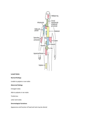
Download to read offline

The document provides an overview of assessing the skin, head, and neck by describing the anatomy and physiology of these systems, outlining the subjective and objective components of the physical exam including inspection, palpation, and documentation of normal and abnormal findings, and noting age-related variations and differences to consider in the assessment. The assessment approach is systematic and includes evaluating characteristics like color, temperature, texture, and integrity while inspecting for lesions, rashes, lumps or other abnormalities.


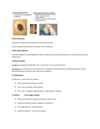
![APPROACH TO FEVER IN PEDIATRICS[1].pptTT](https://cdn.slidesharecdn.com/ss_thumbnails/approachtofeverinpediatrics1-260125081456-d559e079-thumbnail.jpg?width=640&height=640&fit=bounds)


