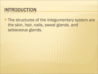
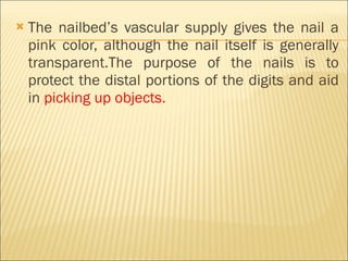
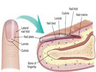
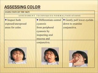
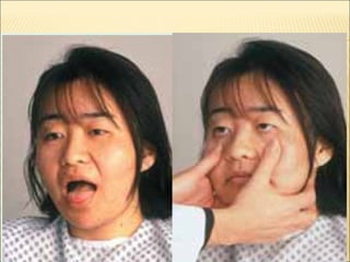
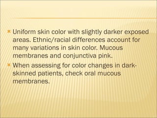
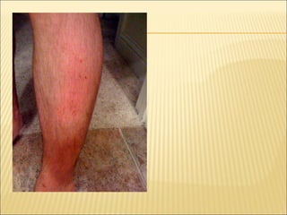
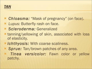
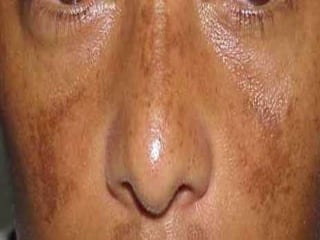
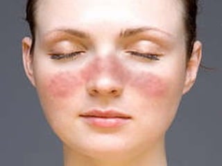
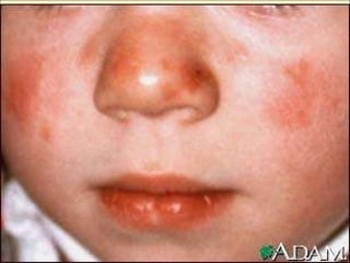
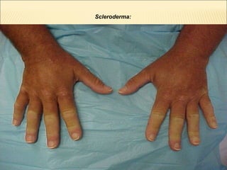
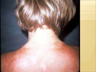
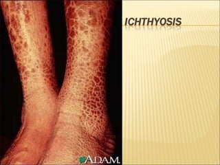
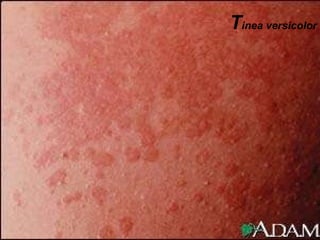
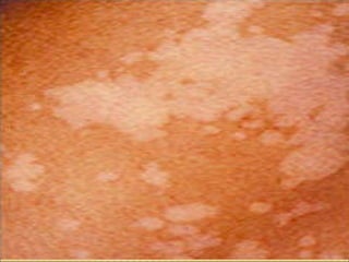
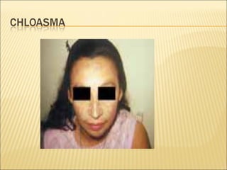
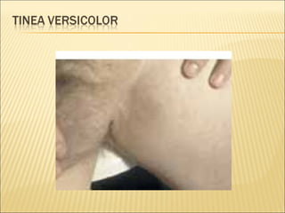
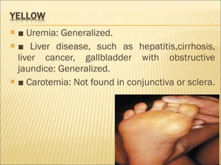
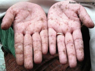
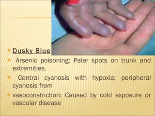
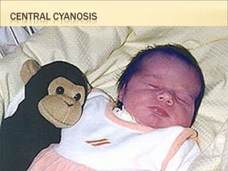
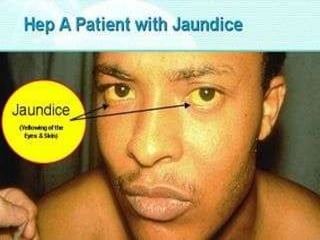
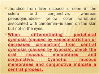
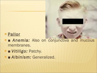
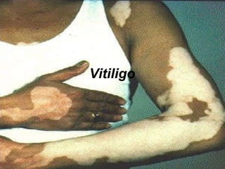
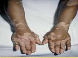
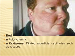
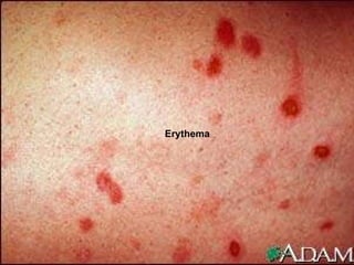
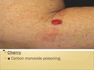
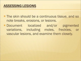
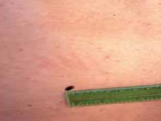
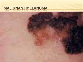
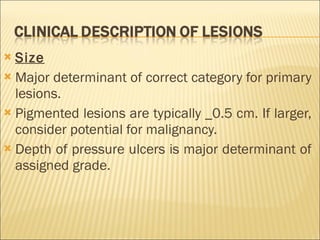
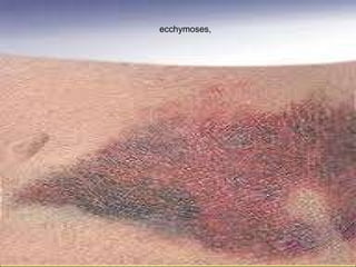
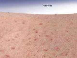
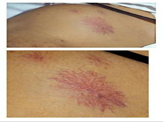
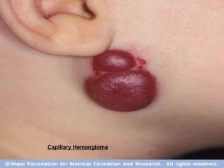
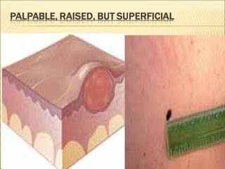
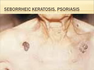
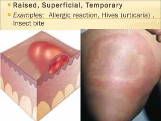
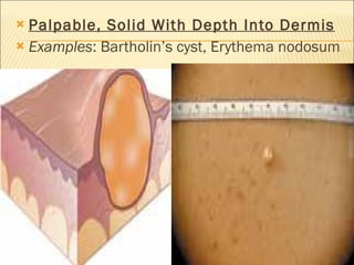
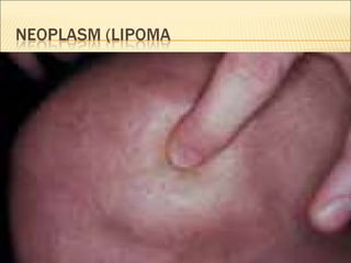
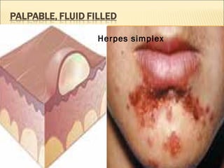
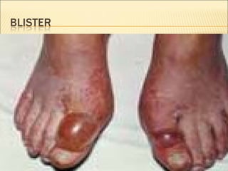
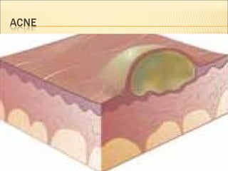
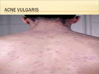
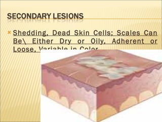
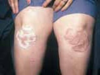
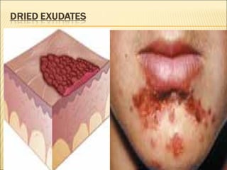
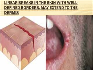
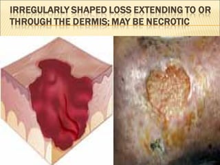
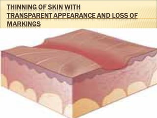
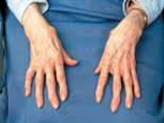
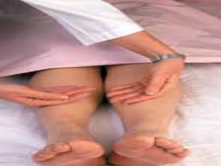
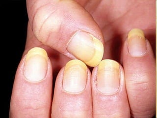
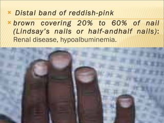
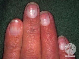
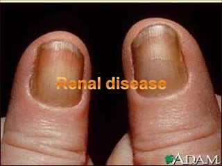
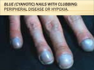
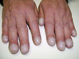
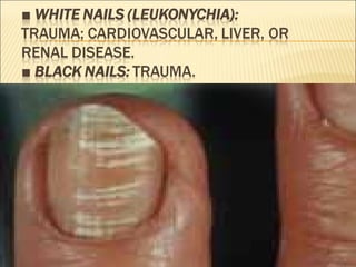
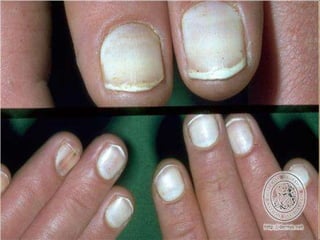
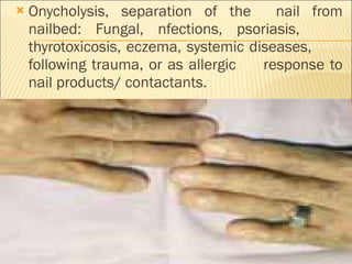
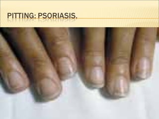
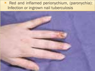
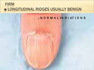
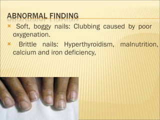
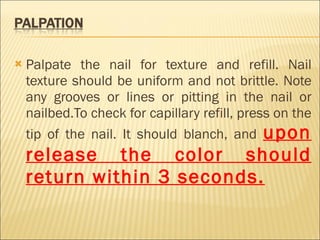
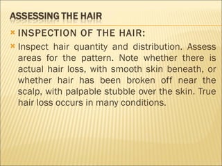
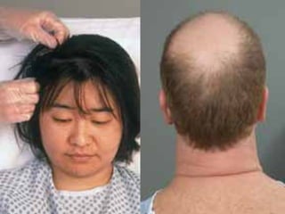
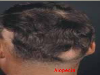
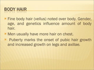
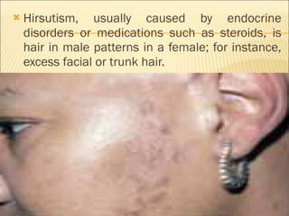
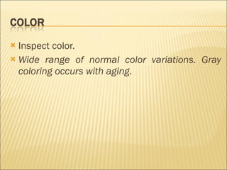
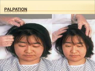
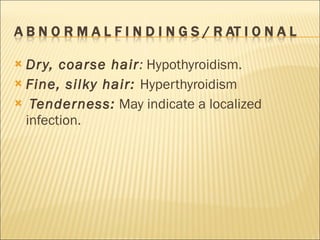
Assessing the Integumentary System
The structures of the integumentary system are the skin, hair, nails, sweat glands, and sebaceous glands.
 
The  skin  is a layer of tissue that covers all exposed body surfaces. Although similar to the mucous membranes, the skin also includes appendages such as hair follicles and sebaceous glands.
the outer visible layer, contains keratin, an extremely tough, protective protein substance that can cause tissue to become hard or horny.
The deeper  dermis  is made up of proteins and  muco-poly-saccharides , thick, gelatinous material that provides a supporting matrix for nerve tissue, blood vessels, sweat and sebum glands, and hair follicles.
made up of fatty connective tissue.  Together,  the layers of the skin protect underlying structures from physical trauma and ultraviolet (UV) radiation. The skin is essential to  maintaining body temperature, fluid balance, and sensation . It is involved in absorption and excretion, immunity, and the synthesis of vitamin D from the sun.
is also made up of keratinized cells. Hair is found over most of the body. It grows from hair follicles supplied by blood vessels located in the dermis.  Vellus   ,which is short, pale, and fine hair, is located over all of the body.  Terminal hairs ,  which are dark and coarse, are found on the scalp, brows, and, after puberty, on the legs, axillae , and perineum. The texture and color of hair are highly variable.  Hair provides protection by covering the scalp and filtering dust and debris away from the nose, ears, and eyes .
Nails  are made up of hard, keratinized cells and grow from a nail root under the cuticle. Other nail structures include the free edge, which overhangs the tip of the finger or toe; the nailbed, or epithelial layer of skin; and the  lunula , the proximal part of the nail.
The nailbed’s vascular supply gives the nail a pink color, although the nail itself is generally transparent.The purpose of the nails is to protect the distal portions of the digits and aid in  picking up objects.
 
 
Other appendages to the integument include the  sweat glands  and  sebaceous glands . There are two types of sweat glands:  eccrine glands ,  which are distributed over much of the body, and  apocrine glands ,  which are limited to the genitalia, axillae, and areolae.  Sebaceous glands are located near hair follicles, over  most of the body.  They secrete  sebum , which lubricates the hair shaft.
ENDOCRINE Thyroid affects growth and texture of skin, hair and nails. Hormones stimulate sebaceous glands.  Sex hormones affect hair growth and distribution, fat and subcutaneous tissue distribution and activity of apocrine sweat glands.  Adrenal hormones affect dermal blood supply and mobilize lipids from adipocytes.
URINARY Kidneys remove waste and maintain normal pH. Skin helps eliminate water and waste. Skin prevents excess fluid loss. RESPIRATORY Provides oxygen to and removes carbon dioxide from integumentary system. Color of skin and nails can reflect changes in respiratory system
CARDIOVASCULAR   Mast cell stimulation produces localized changes in blood flow and capillary permeability.  CV system provides nutrients and removes wastes. Delivers hormones and lymphocytes. Provides heat for skin temperature.
DIGESTIVE Skin synthesizes vitamin D for calcium and phosphorous absorption. Supplies nutrients while skin stores lipids. SKELETAL Skin synthesizes vitamin D needed for calcium and phosphorus absorption. Skeletal system provides a framework for skin.
MUSCULAR   Skin synthesizes vitamin D needed for calcium absorption for muscle contraction. Gives shape to and supports skin. Contraction of facial muscles allows communication through expressions. REPRODUCTIVE   Provides sensory receptors for sexual stimulation.
NEUROLOGICAL Sensory receptors in dermis to touch, temperature, pressure, vibration and pain. Provides communication with external environment. Controls blood flow and sweating through thermoregulation.
Once you have taken the history, proceed to collect objective data through your physical examination. Even though the skin,hair,and nails are easily accessible and we look at them every day, you still need to be very objective and attentive to details that could easily be overlooked.
The techniques used in the examination of the integument are inspection and palpation.
Assessing the Skin   Inspection Examine the patient’s skin,noting color,odor,and the presence of lesions.  Once you have determined the patient’s overall skin coloring,  take a moment to decide if the coloring suggests something other than a normal variation.
INSPECTION OF THE SKIN A S S E S S M E N T  T E C H N I Q U E S / N O R M A L VA R I AT I O N S ■ Inspect both exposed/unexposed areas for color. ■ Differentiate central cyanosis from peripheral cyanosis by inspecting oral mucosa and conjunctiva. ■ Gently pull lower eyelids down to examine conjunctiva.
 
 
Uniform skin color with slightly darker exposed areas. Ethnic/racial differences account for many variations in skin color. Mucous membranes and conjunctiva pink. When assessing for color changes in dark-skinned patients, check oral mucous membranes.
No unusual odor
Unusual body odor:   Poor hygiene or underlying disease. If from poor hygiene, may be related to self-care deficit that warrants nursing intervention. Odors from excessive sweating (hyperhydrosis):   Possible thyrotoxicosis. Odors from night sweats :  Possible tuberculosis. Urine odor:   Incontinence problem. Stale urine odor may be associated with uremia. Mousy odor:   Liver disease.
Differentiate primary and secondary lesions. Identify vascular lesions. If vascular lesions present, gently palpate and note blanching and pulsation. Describe ABCD of suspicious lesions.  A  is for asymmetry,  B  is for border irregularity,  C  is for color variations, and  D  is for diameter _ 0.5 cm . No skin lesions. No vascular lesions.
Primary lesion is an initial alteration in the skin. Secondary lesion arises from a change in a primary lesion. A thorough description of lesions should include: Morphological (clinical) description: ■  Size. ■  Shape. ■  Color.
■  Texture. ■  Surface relationship. ■  Exudate. ■  Tenderness. ■  Configuration. ■  Location and distribution. ■  For vascular lesions, also note: ■  Pulsations. ■  Blanching.
Bronzing/Tanning   ■  Addison’s disease/adrenal insufficiency :  Generalized, most   evident over exposed areas. ■  Hemo-chroma-tosis:   Generalized, may be gray-brown coloring.
 
H emo-chroma-tosis
Addison's disease
 
Chloasma:   “Mask of pregnancy” (on face). Lupus:  Butterfly rash on face. Scleroderma:   Generalized tanning/yellowing of skin, associated with loss of elasticity. Ichthyosis:   With coarse scaliness. Sprue:  Tan/brown patches of any area. Tinea versicolor:   Fawn color or yellow patchy.


Scleroderma:
 
 
T inea versicolor


■  Uremia: Generalized. ■  Liver disease, such as hepatitis,cirrhosis, liver cancer, gallbladder with obstructive jaundice: Generalized. ■  Carotemia: Not found in conjunctiva or sclera.
 
Arsenic poisoning
Dusky Blue Arsenic poisoning: Paler spots on trunk and extremities. Central cyanosis with hypoxia; peripheral cyanosis from vasoconstriction: Caused by cold exposure or vascular disease


Jaundice from liver disease is seen in the sclera and conjunctiva, whereas pseudojaundice— yellow color variations associated with carotemia—is seen on the skin but not in the eyes. When differentiating peripheral cyanosis (caused by vasoconstriction or decreased circulation) from central cyanosis (caused by hypoxia), check the oral mucous membranes and conjunctiva. Cyanotic mucous membranes and conjunctiva indicate a central process.
Pallor ■  Anemia:   Also on conjunctiva and mucous membranes. ■  Vitiligo:   Patchy. ■  Albinism:   Generalized.
Vitiligo
 
Albinism
Red ■  Polycythemia. ■  Erythema:   Dilated superficial capillaries, such as rosacea.
Erythema
Cherry   ■  Carbon monoxide poisoning.
The skin should be a continuous tissue, and so note breaks, erosions, or lesions. Document localized and/or pigmented variations, including moles, freckles, or vascular lesions, and examine them closely.
lesion is solid or fluid filled Fluid-filled lesions have a yellow or pink glow, whereas solid lesions do not. Don’t forget to Clean the ruler after each use Press the ruler or slide gently against the lesion, noting whether it blanches or pales with the pressure. Vascular lesions are red to purple in color. They may be caused by an  extravasation  of blood into the skin tissue or by visible superficial vascular irregularities
Pressure ulcers are a type of secondary lesion caused by unrelieved pressure.  Moles are generally uniformly tan or brown, round or oval in shape, and have well-defined borders. A person many normally have 10 to 40 scattered moles, which generally appear above the waist. When a mole changes in appearance, it needs evaluation, including biopsy, to rule out malignant melanoma.
 
Pressure ulcers often develop over bony prominences, such as the sacrum and heels, so inspect these areas carefully.
 
Size Major determinant of correct category for primary lesions. Pigmented lesions are typically _0.5 cm. If larger, consider potential for malignancy. Depth of pressure ulcers is major determinant of assigned grade.
Macules, wheals, and vesicles are circumscribed. Fissures are linear. Irregular borders are associated with melanoma
Varies widely, and many changes are diagnostic of specific skin diseases. Variegated-colored lesions may signal melanoma. Pustules are usually yellow-white. New scars are red and raised; old scars, white or silver. Petechiae are red. Purpura are red to purplish. Vitiligo is white.
Macules are smooth. Warts are rough. Psoriasis is scaly.
Surface characteristics help differentiate potential causes of a change and between various primary and secondary lesions: Flat (nonpalpable):  Macules, patches, purpura, ecchymoses, spider angioma, venous spider. Raised (palpable) solid:  Papules, plaques, nodules, tumors, wheals, scale, crust. Raised (palpable) cystic:  Vesicles, pustules, bullae, cysts. Depressed:  Atrophy, erosion, ulcer, fissures. Pedunculated:  Skin tags, cutaneous horns.
ecchymoses,
Pattechiae
 
Capillary Hemangioma
Port-Wine Stain
Clear or pale, straw-yellow exudate:  Serous oozing/weeping from noninfected lesion. Thicker, purulent discharge:  Infected lesion. Clear serous exudates:  Vesicles, as seen with herpes simplex; or bullae, larger than vesicles, as seen with second-degree burns. Yellow pus exudates:  Pustules, as seen with impetigo or acne
Tenderness or pain associated with a lesion depends on the underlying cause. May be associated with bullae from a burn or ecchymoses (bruise
Ecchymosis   Extravasation of blood into skin layer. Caused by trauma/injury. Does not blanch.
Petechiae or Purpura Extravasations of blood into skin. Caused by steroids, vasculitis, systemic diseases. Does not blanch.
Venous Star Blue color. Irregular-shaped, linear, spider. Does not blanch. Caused by increased pressure on superficial veins.
Telangiectasia Red color. Very fine and irregular vessels. Blanches. Seen with dilation of capillaries.
Spider Angioma Red color, type of telangiectasis. Looks like a spider, with central body and fine radiating legs. Blanches; seen in liver disease, vitamin B deficiencies, idiopathic origin.
Capillary Hemangioma ■  Red color. ■  Irregular-shaped macular patch.
Port-Wine Stain Red color. Does not blanch. Seen with dilation of dermal capillaries.
Flat, Nonpalpable


Raised, Superficial, Temporary Examples :  Allergic reaction, Hives (urticaria) ,  Insect bite
Palpable, Solid With Depth Into Dermis Examples : Bartholin’s cyst, Erythema nodosum
 
Herpes simplex


Shedding, Dead Skin Cells; Scales Can Be\ Either Dry or Oily, Adherent or Loose, Variable in Color


Palpation is used to determine the skin’s temperature, moisture, texture, and turgor.
Compare side to side using the dorsal aspect of your hand. Skin warm. Temperature varies depending on area being assessed; for example, exposed areas may be cooler than unexposed areas
 
Local area with increased temperature: Inflammatory process, infection, or burn, caused by increased circulation to area. Generalized increase in temperature: Fever. Local area with decreased temperature: Decreased circulation to area, as with arterial occlusion. Generalized decrease in skin temperature: Exposure or shock.
 
Use light palpation to assess skin moisture. Depends on environmental conditions and patient’s age. Elderly people have drier skin because of decreased sweat production. Exposed areas are usually drier than unexposed areas. Also, moisture varies according to body area; for example , the axillae are usually more moist than other areas.
Increased moisture :  Fever, thyrotoxicosis. Decreased moisture:   Dehydration, myxedema, chronic nephritis
Use light palpation to assess texture. Varies from soft and fine to coarse and thick, depending on area assessed and patient’s age. Exposed skin usually not as soft as unexposed. Extensor surfaces, such as elbows, have coarser skin. Usually, the younger the patient, the softer the skin, so infants have very soft skin.
Coarse, thick, dry skin: Hypothyroidism. Skin that becomes more fine-textured: Hyperthyroidism. Smooth, thin, shiny skin: Arterial insufficiency. Thick, rough skin: Venous insufficiency.
Test turgor by gently pinching a fold of skin on an unexposed area (such as below the clavicle) and note any “tenting.” Elasticity decreases with age . Exposed areas may have less turgor . Turgor
 
Decreased turgor or tenting: Dehydration or normal aging. With scleroderma, the turgor is actually increased and the tension does not allow the skin to be pinched upward. This may also be seen with edema.
Inspect the color and shape of the nails. Nail texture should be uniform and not brittle.  Note any grooves or lines in the nail or nailbed Also assess for  clubbing , or loss of the normal angle (Lovibond’s angle) between the nail base and the finger.
Inspect nails for color. Normal nails vary from pink in light-skinned patients to light brown in darker-skinned patients.
Color changes in nails may indicate a local or systemic problem. Very distal band of reddish-pink or brown covering _20% of nail (Terry’s nails): Cirrhosis, disorders causing hypo-albuminemia.


Distal band of reddish-pink brown covering 20% to 60% of nail (Lindsay’s nails or half-andhalf nails) :  Renal disease, hypoalbuminemia.
Lindsay’s nails


Inspect nail shape. Have patient place fingers together and note space  (opening) between nails. Angle of nail attachment 160 degrees; nails convex Clubbing is present if nails meet and angle of  attachment is 180 degrees or greater.
 
Splinter hemorrhages: Bacterial endocarditis or trauma. Angle of nail attachment 180 degrees or more: Clubbing associated with diseases that affect level of oxygenation, such as congenital heart disorders, cystic fibrosis, and chronic pulmonary diseases . Spooning or concave nail (koilonychia): Severe iron deficiency anemia, hemochromatosis, thyroid and circulatory diseases, in response to some skin diseases and local trauma.
 
 
Onycholysis, separation of the  nail from nailbed: Fungal, nfections, psoriasis,  thyrotoxicosis, eczema, systemic diseases,  following trauma, or as allergic  response to nail products/ contactants.
 
Red and inflamed perionychium, (paronychia): Infection or ingrown nail tuberculosis
 
/  N O R M A L VA R I AT I O N S
Soft, boggy nails: Clubbing caused by poor  oxygenation. Brittle nails: Hyperthyroidism, malnutrition, calcium and iron deficiency,
 
Gently press on nail and note blanching, then release and note speed of refill (color return).
Palpate the nail for texture and refill. Nail texture should be uniform and not brittle. Note any grooves or lines or pitting in the nail or nailbed.To check for capillary refill, press on the tip of the nail. It should blanch, and  upon release the color should return within 3 seconds.
INSPECTION OF THE HAIR: Inspect hair quantity and distribution. Assess areas for the pattern. Note whether there is actual hair loss, with smooth skin beneath, or whether hair has been broken off near the scalp, with palpable stubble over the skin. True hair loss occurs in many conditions.
 
Gender, age, and genetics affect hair distribution. Hair should be evenly distributed; exceptions are normal balding patterns common to men or persons of advanced age. Hair thins with age.  
Generalized hair loss :  Nutritional deficiencies, hypothyroidism, lupus, thyroid disease, and in response to disorders or situations that stress the integumentary system, such as serious illnesses or side effects of medications. Patchy alopecia   associated with alopecia areata, and fungal infections such as tinea capitis.
Alopecia
Tinea capitis
Alopecia areata
Fine body hair (vellus) noted over body. Gender, age, and genetics influence amount of body hair. Men usually have more hair on chest. Puberty marks the onset of pubic hair growth and increased growth on legs and axillae .
Hirsutism, usually caused by endocrine disorders or medications such as steroids, is hair in male patterns in a female; for instance, excess facial or trunk hair.
Inspect color. Wide range of normal color variations. Gray coloring occurs with aging.
Localized areas of white or gray hair: In patients recovering from alopecia areata and in those with  vitiligo. Diffuse white hair: Albinism. Green hair: Copper exposure and anemia
Inspect scalp. Scalp intact and free of lesions and pediculosis A B N O R M A L F I N D I N G S / R AT I O N A L E  Scaling of scalp:  Dandruff, seborrhea, psoriasis, certain tineas, and eczema (atopic dermatitis).
 
Use light palpation to assess hair Use light palpation to assess scalp.  Scalp mobile, nontender
Dry, coarse hair :  Hypothyroidism. Fine, silky hair:  Hyperthyroidism Tenderness:   May indicate a localized infection.

Assessing the integumentary system new

  • 1.
  • 2.
    The structures ofthe integumentary system are the skin, hair, nails, sweat glands, and sebaceous glands.
  • 3.
  • 4.
    The skin is a layer of tissue that covers all exposed body surfaces. Although similar to the mucous membranes, the skin also includes appendages such as hair follicles and sebaceous glands.
  • 5.
    the outer visiblelayer, contains keratin, an extremely tough, protective protein substance that can cause tissue to become hard or horny.
  • 6.
    The deeper dermis is made up of proteins and muco-poly-saccharides , thick, gelatinous material that provides a supporting matrix for nerve tissue, blood vessels, sweat and sebum glands, and hair follicles.
  • 7.
    made up offatty connective tissue. Together, the layers of the skin protect underlying structures from physical trauma and ultraviolet (UV) radiation. The skin is essential to maintaining body temperature, fluid balance, and sensation . It is involved in absorption and excretion, immunity, and the synthesis of vitamin D from the sun.
  • 8.
    is also madeup of keratinized cells. Hair is found over most of the body. It grows from hair follicles supplied by blood vessels located in the dermis. Vellus ,which is short, pale, and fine hair, is located over all of the body. Terminal hairs , which are dark and coarse, are found on the scalp, brows, and, after puberty, on the legs, axillae , and perineum. The texture and color of hair are highly variable. Hair provides protection by covering the scalp and filtering dust and debris away from the nose, ears, and eyes .
  • 9.
    Nails aremade up of hard, keratinized cells and grow from a nail root under the cuticle. Other nail structures include the free edge, which overhangs the tip of the finger or toe; the nailbed, or epithelial layer of skin; and the lunula , the proximal part of the nail.
  • 10.
    The nailbed’s vascularsupply gives the nail a pink color, although the nail itself is generally transparent.The purpose of the nails is to protect the distal portions of the digits and aid in picking up objects.
  • 11.
  • 12.
  • 13.
    Other appendages tothe integument include the sweat glands and sebaceous glands . There are two types of sweat glands: eccrine glands , which are distributed over much of the body, and apocrine glands , which are limited to the genitalia, axillae, and areolae. Sebaceous glands are located near hair follicles, over most of the body. They secrete sebum , which lubricates the hair shaft.
  • 14.
    ENDOCRINE Thyroid affectsgrowth and texture of skin, hair and nails. Hormones stimulate sebaceous glands. Sex hormones affect hair growth and distribution, fat and subcutaneous tissue distribution and activity of apocrine sweat glands. Adrenal hormones affect dermal blood supply and mobilize lipids from adipocytes.
  • 15.
    URINARY Kidneys removewaste and maintain normal pH. Skin helps eliminate water and waste. Skin prevents excess fluid loss. RESPIRATORY Provides oxygen to and removes carbon dioxide from integumentary system. Color of skin and nails can reflect changes in respiratory system
  • 16.
    CARDIOVASCULAR   Mastcell stimulation produces localized changes in blood flow and capillary permeability. CV system provides nutrients and removes wastes. Delivers hormones and lymphocytes. Provides heat for skin temperature.
  • 17.
    DIGESTIVE Skin synthesizesvitamin D for calcium and phosphorous absorption. Supplies nutrients while skin stores lipids. SKELETAL Skin synthesizes vitamin D needed for calcium and phosphorus absorption. Skeletal system provides a framework for skin.
  • 18.
    MUSCULAR   Skinsynthesizes vitamin D needed for calcium absorption for muscle contraction. Gives shape to and supports skin. Contraction of facial muscles allows communication through expressions. REPRODUCTIVE   Provides sensory receptors for sexual stimulation.
  • 19.
    NEUROLOGICAL Sensory receptorsin dermis to touch, temperature, pressure, vibration and pain. Provides communication with external environment. Controls blood flow and sweating through thermoregulation.
  • 20.
    Once you havetaken the history, proceed to collect objective data through your physical examination. Even though the skin,hair,and nails are easily accessible and we look at them every day, you still need to be very objective and attentive to details that could easily be overlooked.
  • 21.
    The techniques usedin the examination of the integument are inspection and palpation.
  • 22.
    Assessing the Skin  Inspection Examine the patient’s skin,noting color,odor,and the presence of lesions. Once you have determined the patient’s overall skin coloring, take a moment to decide if the coloring suggests something other than a normal variation.
  • 23.
    INSPECTION OF THESKIN A S S E S S M E N T T E C H N I Q U E S / N O R M A L VA R I AT I O N S ■ Inspect both exposed/unexposed areas for color. ■ Differentiate central cyanosis from peripheral cyanosis by inspecting oral mucosa and conjunctiva. ■ Gently pull lower eyelids down to examine conjunctiva.
  • 24.
  • 25.
  • 26.
    Uniform skin colorwith slightly darker exposed areas. Ethnic/racial differences account for many variations in skin color. Mucous membranes and conjunctiva pink. When assessing for color changes in dark-skinned patients, check oral mucous membranes.
  • 27.
  • 28.
    Unusual body odor: Poor hygiene or underlying disease. If from poor hygiene, may be related to self-care deficit that warrants nursing intervention. Odors from excessive sweating (hyperhydrosis): Possible thyrotoxicosis. Odors from night sweats : Possible tuberculosis. Urine odor: Incontinence problem. Stale urine odor may be associated with uremia. Mousy odor: Liver disease.
  • 29.
    Differentiate primary andsecondary lesions. Identify vascular lesions. If vascular lesions present, gently palpate and note blanching and pulsation. Describe ABCD of suspicious lesions. A is for asymmetry, B is for border irregularity, C is for color variations, and D is for diameter _ 0.5 cm . No skin lesions. No vascular lesions.
  • 30.
    Primary lesion isan initial alteration in the skin. Secondary lesion arises from a change in a primary lesion. A thorough description of lesions should include: Morphological (clinical) description: ■ Size. ■ Shape. ■ Color.
  • 31.
    ■ Texture.■ Surface relationship. ■ Exudate. ■ Tenderness. ■ Configuration. ■ Location and distribution. ■ For vascular lesions, also note: ■ Pulsations. ■ Blanching.
  • 32.
    Bronzing/Tanning   ■ Addison’s disease/adrenal insufficiency : Generalized, most evident over exposed areas. ■ Hemo-chroma-tosis: Generalized, may be gray-brown coloring.
  • 33.
  • 34.
  • 35.
  • 36.
  • 37.
    Chloasma: “Mask of pregnancy” (on face). Lupus: Butterfly rash on face. Scleroderma: Generalized tanning/yellowing of skin, associated with loss of elasticity. Ichthyosis: With coarse scaliness. Sprue: Tan/brown patches of any area. Tinea versicolor: Fawn color or yellow patchy.
  • 38.
  • 39.
  • 40.
  • 41.
  • 42.
  • 43.
  • 44.
  • 45.
  • 46.
  • 47.
  • 48.
  • 49.
    ■ Uremia:Generalized. ■ Liver disease, such as hepatitis,cirrhosis, liver cancer, gallbladder with obstructive jaundice: Generalized. ■ Carotemia: Not found in conjunctiva or sclera.
  • 50.
  • 51.
  • 52.
    Dusky Blue Arsenicpoisoning: Paler spots on trunk and extremities. Central cyanosis with hypoxia; peripheral cyanosis from vasoconstriction: Caused by cold exposure or vascular disease
  • 53.
  • 54.
  • 55.
  • 56.
    Jaundice from liverdisease is seen in the sclera and conjunctiva, whereas pseudojaundice— yellow color variations associated with carotemia—is seen on the skin but not in the eyes. When differentiating peripheral cyanosis (caused by vasoconstriction or decreased circulation) from central cyanosis (caused by hypoxia), check the oral mucous membranes and conjunctiva. Cyanotic mucous membranes and conjunctiva indicate a central process.
  • 57.
    Pallor ■ Anemia: Also on conjunctiva and mucous membranes. ■ Vitiligo: Patchy. ■ Albinism: Generalized.
  • 58.
  • 59.
  • 60.
  • 61.
    Red ■ Polycythemia. ■ Erythema: Dilated superficial capillaries, such as rosacea.
  • 62.
  • 63.
    Cherry   ■ Carbon monoxide poisoning.
  • 64.
    The skin shouldbe a continuous tissue, and so note breaks, erosions, or lesions. Document localized and/or pigmented variations, including moles, freckles, or vascular lesions, and examine them closely.
  • 65.
    lesion is solidor fluid filled Fluid-filled lesions have a yellow or pink glow, whereas solid lesions do not. Don’t forget to Clean the ruler after each use Press the ruler or slide gently against the lesion, noting whether it blanches or pales with the pressure. Vascular lesions are red to purple in color. They may be caused by an extravasation of blood into the skin tissue or by visible superficial vascular irregularities
  • 66.
    Pressure ulcers area type of secondary lesion caused by unrelieved pressure. Moles are generally uniformly tan or brown, round or oval in shape, and have well-defined borders. A person many normally have 10 to 40 scattered moles, which generally appear above the waist. When a mole changes in appearance, it needs evaluation, including biopsy, to rule out malignant melanoma.
  • 67.
  • 68.
    Pressure ulcers oftendevelop over bony prominences, such as the sacrum and heels, so inspect these areas carefully.
  • 69.
  • 70.
    Size Major determinantof correct category for primary lesions. Pigmented lesions are typically _0.5 cm. If larger, consider potential for malignancy. Depth of pressure ulcers is major determinant of assigned grade.
  • 71.
    Macules, wheals, andvesicles are circumscribed. Fissures are linear. Irregular borders are associated with melanoma
  • 72.
    Varies widely, andmany changes are diagnostic of specific skin diseases. Variegated-colored lesions may signal melanoma. Pustules are usually yellow-white. New scars are red and raised; old scars, white or silver. Petechiae are red. Purpura are red to purplish. Vitiligo is white.
  • 73.
    Macules are smooth.Warts are rough. Psoriasis is scaly.
  • 74.
    Surface characteristics helpdifferentiate potential causes of a change and between various primary and secondary lesions: Flat (nonpalpable): Macules, patches, purpura, ecchymoses, spider angioma, venous spider. Raised (palpable) solid: Papules, plaques, nodules, tumors, wheals, scale, crust. Raised (palpable) cystic: Vesicles, pustules, bullae, cysts. Depressed: Atrophy, erosion, ulcer, fissures. Pedunculated: Skin tags, cutaneous horns.
  • 75.
  • 76.
  • 77.
  • 78.
  • 79.
  • 80.
    Clear or pale,straw-yellow exudate: Serous oozing/weeping from noninfected lesion. Thicker, purulent discharge: Infected lesion. Clear serous exudates: Vesicles, as seen with herpes simplex; or bullae, larger than vesicles, as seen with second-degree burns. Yellow pus exudates: Pustules, as seen with impetigo or acne
  • 81.
    Tenderness or painassociated with a lesion depends on the underlying cause. May be associated with bullae from a burn or ecchymoses (bruise
  • 82.
    Ecchymosis   Extravasationof blood into skin layer. Caused by trauma/injury. Does not blanch.
  • 83.
    Petechiae or PurpuraExtravasations of blood into skin. Caused by steroids, vasculitis, systemic diseases. Does not blanch.
  • 84.
    Venous Star Bluecolor. Irregular-shaped, linear, spider. Does not blanch. Caused by increased pressure on superficial veins.
  • 85.
    Telangiectasia Red color.Very fine and irregular vessels. Blanches. Seen with dilation of capillaries.
  • 86.
    Spider Angioma Redcolor, type of telangiectasis. Looks like a spider, with central body and fine radiating legs. Blanches; seen in liver disease, vitamin B deficiencies, idiopathic origin.
  • 87.
    Capillary Hemangioma ■ Red color. ■ Irregular-shaped macular patch.
  • 88.
    Port-Wine Stain Redcolor. Does not blanch. Seen with dilation of dermal capillaries.
  • 89.
  • 90.
  • 91.
  • 92.
  • 93.
    Raised, Superficial, TemporaryExamples : Allergic reaction, Hives (urticaria) , Insect bite
  • 94.
    Palpable, Solid WithDepth Into Dermis Examples : Bartholin’s cyst, Erythema nodosum
  • 95.
  • 96.
  • 97.
  • 98.
  • 99.
  • 100.
    Shedding, Dead SkinCells; Scales Can Be\ Either Dry or Oily, Adherent or Loose, Variable in Color
  • 101.
  • 102.
  • 103.
  • 104.
  • 105.
  • 106.
  • 107.
  • 108.
  • 109.
  • 110.
  • 111.
    Palpation is usedto determine the skin’s temperature, moisture, texture, and turgor.
  • 112.
    Compare side toside using the dorsal aspect of your hand. Skin warm. Temperature varies depending on area being assessed; for example, exposed areas may be cooler than unexposed areas
  • 113.
  • 114.
    Local area withincreased temperature: Inflammatory process, infection, or burn, caused by increased circulation to area. Generalized increase in temperature: Fever. Local area with decreased temperature: Decreased circulation to area, as with arterial occlusion. Generalized decrease in skin temperature: Exposure or shock.
  • 115.
  • 116.
    Use light palpationto assess skin moisture. Depends on environmental conditions and patient’s age. Elderly people have drier skin because of decreased sweat production. Exposed areas are usually drier than unexposed areas. Also, moisture varies according to body area; for example , the axillae are usually more moist than other areas.
  • 117.
    Increased moisture : Fever, thyrotoxicosis. Decreased moisture: Dehydration, myxedema, chronic nephritis
  • 118.
    Use light palpationto assess texture. Varies from soft and fine to coarse and thick, depending on area assessed and patient’s age. Exposed skin usually not as soft as unexposed. Extensor surfaces, such as elbows, have coarser skin. Usually, the younger the patient, the softer the skin, so infants have very soft skin.
  • 119.
    Coarse, thick, dryskin: Hypothyroidism. Skin that becomes more fine-textured: Hyperthyroidism. Smooth, thin, shiny skin: Arterial insufficiency. Thick, rough skin: Venous insufficiency.
  • 120.
    Test turgor bygently pinching a fold of skin on an unexposed area (such as below the clavicle) and note any “tenting.” Elasticity decreases with age . Exposed areas may have less turgor . Turgor
  • 121.
  • 122.
    Decreased turgor ortenting: Dehydration or normal aging. With scleroderma, the turgor is actually increased and the tension does not allow the skin to be pinched upward. This may also be seen with edema.
  • 123.
    Inspect the colorand shape of the nails. Nail texture should be uniform and not brittle. Note any grooves or lines in the nail or nailbed Also assess for clubbing , or loss of the normal angle (Lovibond’s angle) between the nail base and the finger.
  • 124.
    Inspect nails forcolor. Normal nails vary from pink in light-skinned patients to light brown in darker-skinned patients.
  • 125.
    Color changes innails may indicate a local or systemic problem. Very distal band of reddish-pink or brown covering _20% of nail (Terry’s nails): Cirrhosis, disorders causing hypo-albuminemia.
  • 126.
  • 127.
  • 128.
  • 129.
    Distal band ofreddish-pink brown covering 20% to 60% of nail (Lindsay’s nails or half-andhalf nails) : Renal disease, hypoalbuminemia.
  • 130.
  • 131.
  • 132.
  • 133.
  • 134.
  • 135.
  • 136.
  • 137.
  • 138.
    Inspect nail shape.Have patient place fingers together and note space (opening) between nails. Angle of nail attachment 160 degrees; nails convex Clubbing is present if nails meet and angle of attachment is 180 degrees or greater.
  • 139.
  • 140.
    Splinter hemorrhages: Bacterialendocarditis or trauma. Angle of nail attachment 180 degrees or more: Clubbing associated with diseases that affect level of oxygenation, such as congenital heart disorders, cystic fibrosis, and chronic pulmonary diseases . Spooning or concave nail (koilonychia): Severe iron deficiency anemia, hemochromatosis, thyroid and circulatory diseases, in response to some skin diseases and local trauma.
  • 141.
  • 142.
  • 143.
    Onycholysis, separation ofthe nail from nailbed: Fungal, nfections, psoriasis, thyrotoxicosis, eczema, systemic diseases, following trauma, or as allergic response to nail products/ contactants.
  • 144.
  • 145.
    Red and inflamedperionychium, (paronychia): Infection or ingrown nail tuberculosis
  • 146.
  • 147.
    / NO R M A L VA R I AT I O N S
  • 148.
    Soft, boggy nails:Clubbing caused by poor oxygenation. Brittle nails: Hyperthyroidism, malnutrition, calcium and iron deficiency,
  • 149.
  • 150.
    Gently press onnail and note blanching, then release and note speed of refill (color return).
  • 151.
    Palpate the nailfor texture and refill. Nail texture should be uniform and not brittle. Note any grooves or lines or pitting in the nail or nailbed.To check for capillary refill, press on the tip of the nail. It should blanch, and upon release the color should return within 3 seconds.
  • 152.
    INSPECTION OF THEHAIR: Inspect hair quantity and distribution. Assess areas for the pattern. Note whether there is actual hair loss, with smooth skin beneath, or whether hair has been broken off near the scalp, with palpable stubble over the skin. True hair loss occurs in many conditions.
  • 153.
  • 154.
    Gender, age, andgenetics affect hair distribution. Hair should be evenly distributed; exceptions are normal balding patterns common to men or persons of advanced age. Hair thins with age.  
  • 155.
    Generalized hair loss: Nutritional deficiencies, hypothyroidism, lupus, thyroid disease, and in response to disorders or situations that stress the integumentary system, such as serious illnesses or side effects of medications. Patchy alopecia associated with alopecia areata, and fungal infections such as tinea capitis.
  • 156.
  • 157.
  • 158.
  • 159.
    Fine body hair(vellus) noted over body. Gender, age, and genetics influence amount of body hair. Men usually have more hair on chest. Puberty marks the onset of pubic hair growth and increased growth on legs and axillae .
  • 160.
    Hirsutism, usually causedby endocrine disorders or medications such as steroids, is hair in male patterns in a female; for instance, excess facial or trunk hair.
  • 161.
    Inspect color. Widerange of normal color variations. Gray coloring occurs with aging.
  • 162.
    Localized areas ofwhite or gray hair: In patients recovering from alopecia areata and in those with vitiligo. Diffuse white hair: Albinism. Green hair: Copper exposure and anemia
  • 163.
    Inspect scalp. Scalpintact and free of lesions and pediculosis A B N O R M A L F I N D I N G S / R AT I O N A L E  Scaling of scalp: Dandruff, seborrhea, psoriasis, certain tineas, and eczema (atopic dermatitis).
  • 164.
  • 165.
    Use light palpationto assess hair Use light palpation to assess scalp. Scalp mobile, nontender
  • 166.
    Dry, coarse hair: Hypothyroidism. Fine, silky hair: Hyperthyroidism Tenderness: May indicate a localized infection.