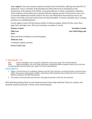
Skin, Hair, Nails               Ch (6) P. (163-195)
Cognitive Objectives            Upon completion of this lesson the student should be able to:
1. List and describe the 3 layers of the skin. (163)
   Epidermis: most superficial layer, no blood vessels, divided into
   1 - outer horny layer of dead keratinized cells
   2 - inner cellular layer where (melanin and keratin) are formed. Migration from inner to top layer takes approximately
   a month. It depends on the dermis for
   nutrition.

   Dermis: well supplied with blood, it contains connective tissue, sebaceous glands, sweat glands, and hair follicles.
   Subcutaneous (adipose) tissues: just under the dermis layer. Also know as fat.
2. 5 functions of the skin. (163)
       1) Keep body in homeostasis.
       2) Provide boundaries for body fluids.
       3) Protecting underlying tissues from microorganisms, harmful substances, and radiations.
       4) Modulate body temperature.
       5) Synthesizes vitamin D.
           Hair, nails, sebaceous and sweat glands are considered appendages of the skin. Color depend on 4 pigments
           (melanin (brown), carotene (golden yellow), oxyhemoglobin (red), deoxyhemoglobin (blue)).
3. sebaceous, eccrine and apocrine glands. (165)
       Sebaceous glands: produce a fatty substance secreted onto the skin surface through the hair follicles, they are
       present on all skin surfaces except (palms and soles).
       Sweat glands: two types
       1) Eccrine: widely distributed, open directly onto skin surface, by the sweat production help control body
       temperature.
       2) Apocrine: found chiefly in the axillary and genitals regions, open onto hair follicles and are stimulated by
       emotional stress. (Bacteria decomposition of apocrine glands responsible for adult body odor.
4. Discuss the cause of central cyanosis and peripheral cyanosis. (165)
       If Oxygen is low in arterial blood, cyanosis is central best identified in the lips, oral mucosa, & tongue.
       If Oxygen is normal in arterial blood normal cyanosis is peripheral. Peripheral occurs when blood flow slows and
       decreases and tissues extract more oxygen than usual from the blood, it maybe a normal response to anxiety or a
       cold environment. Central or peripheral could be identified in the nails, hands, & feet.
5. Identify the changes in the skin that occur with aging. 895
       With age, the skin wrinkles, becomes lax, and loses turgor. The vascularity of the dermis decreases, causing
       lighter skin to look paler and more opaque. Skin on the backs of the hands and forearms appears thin, fragile,
       loose, and transparent. There may be purple patches or macules, termed actinic purpura, that fade over time.
       These spots and patches come from blood that has leaked through poorly supported capillaries and spread within
       the dermis.
6. Identify risk factors and preventive measures related to skin. (166-167)
       Risk factors (melanoma):
       1) having 50 or more common moles,
       2) 1-4 atypical or unusual moles (dysplastic), red or light hair, actinic lentigenes.
       3) macular brown spots usually on sun exposed areas.
       4) Ultraviolet radiation exposure, sunlamps, tanning booths.
       5) Male gender.

       Preventative measures: total body skin examination by clinician. Routine cancer related check every 3 years
       (age 20-40), yearly (older than 40). Reducing direct sun light exposure, using sunscreens.
       Mole Evaluation:
       A=asymmetry
       B=Border
C=Color=
        Size= <6mm
7. 6 elements noted in the examination of the skin. (169-170)
    Skin: 1) color: of lips, oral mucosa, tongue, nails, palms, & feet. Look for yellow color in the sclera
    (jaundice).
        2) moisture: dryness in hypothyroidism, sweating, oily in
            acne.
        3) 3) temperature: use back of figures, note temp of any red
            area.
        4) 4) texture: roughness in hypothyrodism and
            smoothness
        5) 5) mobility and turgor: lift folds of the skin note the ease of lifting and turgor (return to
            place).
        6) 6) lesions: note their characteristics. (4 types discussed next question).
8. List 4 features to note regarding skin lesions. (169)
        A) Location & Distribution: any exposed surfaces. This is the most important type.
        B) B) Pattern & shape: linear, clustered, arciform, Geographic, Serpigious. (pics 177)
        C) C) Types: (e.g., macules, papules, vesicles, nevi) . (pics
       178-180)
        D) D) Color: they didn’t write anything under color. Evaluation the bedbound patient: if they are emaciated (thin,
       wasted), elderly, they are susceptible to skin damage & ulceration. Pressure sores results when sustained
       compression demolishes arterial capillary blood flow to the skin. Also might occur from dragged rather than lifted
       up of bed. (170)
9. primary skin lesions: (178-180 check the pictures in the book please)
        Non-palpable lesion with changes in skin color.        (there are more types but she asked just for
        these)
        1) Macule: small flat spot (up to 1cm).         Ex. Hemangioma, Vitiligo
        Palpable elevation: Solid masses
        1) Papule: (up to 1cm).                           Ex. Psoriasis
        2) Wheal: somewhat irregular, relatively transient, superficial area of localized skin edema. Ex. Urticaria
        3) Nodule: marble-like lesion (larger than 0.5cm) often deeper and firmer than a papule. Ex. Dermatofibroma
        4) Plaque: superficial (larger than 1cm) often found by coalescence of papules.            Ex. Psoriasis
        Palpable elevation: with fluid-filled cavities
        1) Pustule: filled with pus.                             Ex. Acne
        2) Vesicle: (up to 1cm) filled with serous fluid. Ex. Herpes (Simplex, Zoster)
        Tumor: 185               TABLE 6-9 Skin Tumors
        Actinic Keratosis                                          Seborrheic Keratosis
        Superficial, flattened papules covered by a dry scale.     Common, benign, yellowish to brown raised lesions that
        Often multiple; can be round or irregular; pink, tan, or   feel slightly greasy and velvety or warty and have a
        grayish. Appear on sun-exposed skin of older, fair-        “stuck on” appearance. Typically multiple and
        skinned people. Though benign, 1 of every 1000 per         symmetrically distributed on the trunk of older people,
        year develop into squamous cell carcinoma (suggested       but may also appear on the face and elsewhere. In black
        by rapid growth, induration, redness at the base, and      people, often younger women, may appear as small,
        ulceration). Keratoses on face and hand, typical           deeply pigmented papules on the cheeks and temples
        locations, are shown.                                      (dermatosis papulosa nigra).
Basal Cell Carcinoma                                       Squamous Cell Carcinoma
       A basal cell carcinoma, though malignant, grows slowly Usually appears on sun-exposed skin of fair-skinned
       and seldom metastasizes. It is most common in fair-        adults older than 60 years. May develop in an actinic
       skinned adults 40 years or older, and usually appears on keratosis. Usually grows more quickly than a basal cell
       the face. An initial translucent nodule spreads, leaving a carcinoma, is firmer, and looks redder. The face and the
       depressed center and a firm, elevated border.              back of the hand are often affected, as shown here.
       Telangiectatic vessels are often visible.


       Vascular lesion:
                                                                                                              1)
       Cherry angioma: (P. 184) A) color and size: bright or ruby red may become brownish by age (1-3mm). B)
       Shape: round, flat and sometimes raised, maybe surrounded with a pale halo. C) Pulsatility and Effect of
       Pressure: not pulsating, D) Distribution: trunk and extremities, E) Significance: none, increases in size and
       numbers by age.
       2) Telangectasias: or dilated small vessels that look red or bluish, they are 2 types, listed in table below
       (A&B).

                              A) Spider Angioma*                     B) Spider Vein*             Cherry Angioma


  Color and Size      Fiery red. From very small to 2 cm       Bluish. Size variable, from Bright or ruby red; may
                                                               very small to several inches become brownish with age.
                                                                                             1-3 mm
     Shape              Central body, sometimes raised,          Variable. May resemble a      Round, flat or sometimes
                        surrounded by erythema and radiating spider or be linear, irregular, raised, may be surrounded
                        legs                                     cascading                     by a pale halo
     Pulsatility and    Often seen in center of the spider, when Absent. Pressure over the     Absent. May show partial
     Effect of Pressure pressure with a glass slide is applied. center does not cause          blanching, especially if
                        Pressure on the body causes blanching blanching, but diffuse           pressure applied with edge
                        of the spider.                           pressure blanches the veins. of a pinpoint
     Distribution       Face, neck, arms, and upper trunk;       Most often on the legs, near Trunk; also extremities
                        almost never below the waist             veins; also on the anterior
                                                                 chest
     Significance       Liver disease, pregnancy, vitamin B      Often accompanies             None; increases in size and
                        deficiency; also occurs normally in      increased pressure in the     numbers with aging
                        some people                              superficial veins, as in
                                                                 varicose veins
10. secondary skin lesions: (table page 181-182)
   Scar: connective tissue that arises from injury or disease. Pic: of Hypertrophic scar from steroids injections
   Scale: a thin flake of dead exfoliated epidermis.              Ex.Ichthyosis Vulgaris & Dry Skin
   Fissure: linear crack in the skin, resulting from excessive dryness. Ex.Athlete’s foot
   Crust: dried residue of skin exudates such a serum, pus or blood.       Ex.Impetigo
   Erosion: non-scaring loss of the superficial epidermis, surface is moist but does not bleed. Ex.Aphthous stomatitis
   Excoriation: Linear or punctate erosions caused by scratching. Ex.Cat scratches
   Ulcer: deeper loss of epidermis and dermis may bleed and scar.          Ex. Stasis ulcer of
   venous                                                                                                 Ex. insufficiency,
   Syphilitic chancre             Keloid: Hypertrophic scaring that extends beyond the borders of the initiating injury.
   Ex.Keloid-ear lobe           Lichenification: visible and palpable thickening of the epidermis and roughening of the
skin with increase visibility of the normal skin furrows (often from chronic rubbing).
                Ex.Neurodermatitis                                   Atrophic scar:        can’t find it
11. vascular skin lesions. (184)         Same answer as table in Question 9
12. abnormalities for the nails: (193-194) (there are more types but she asked just for these)
                                                 Clubbing of the Fingers
                                                 Clinically a bulbous swelling of the soft tissue at the nail base, with loss
                                                 of the normal angle between the nail and the proximal nail fold. The
                                                 angle increases to 180° or more, and the nail bed feels spongy or floating.
                                                 The mechanism is still unknown but involves vasodilatation with
                                                 increased blood flow to the distal portion of the digits and changes in
                                                 connective tissue, possibly from hypoxia, changes in innervation,
                                                 genetics, or a platelet-derived growth factor from fragments of platelet
                                                 clumps. Seen in congenital heart disease, interstitial lung disease and lung
                                                 cancer, inflammatory bowel diseases, and malignancies.
                                                 Terry's Nails
                                                 Nail plate turns white with a ground-glass appearance, a distal band of
                                                 reddish brown, and obliteration of the lunula. Commonly affects all
                                                 fingers, although may appear in only one finger. Seen in liver disease,
                                                 usually cirrhosis, congestive heart failure, and diabetes. May arise from
                                                 decreased vascularity and increased connective tissue in nail bed.


                                                 Transverse White Bands (Mees' Lines)
                                                 Curving transverse white bands that cross the nail parallel to the lunula.
                                                 Arising from the disrupted matrix of the proximal nail, they vary in width
                                                 and move distally as the nail grows out. Seen in arsenic poisoning, heart
                                                 failure, Hodgkin's disease, chemotherapy, carbon monoxide poisoning,
                                                 and leprosy.

                                                 Transverse Linear Depressions (Beau's Lines)
                                                 Transverse depressions of the nail plates, usually bilateral, resulting
                                                 from temporary disruption of proximal nail growth from systemic illness.
                                                 As with Mees' lines, timing of the illness may be estimated by measuring
                                                 the distance from the line to the nail bed (nails grow approximately 1 mm
                                                 every 6 to 10 days). Seen in severe illness, trauma, and cold exposure if
                                                 Raynaud's disease is present.


13. Compare and contrast primary and secondary skin lesions. (183)
    Primary lesions are present at the onset of the disease. Secondary result from changes over time caused by disease
    progression, manipulation or treatment. (scratching, secondary infection…)

                              TABLE 6-7 Acne Vulgaris—Primary and Secondary Lesions
Acne vulgaris is the most common cutaneous disorder in the United States, affecting more than 85% of
        adolescents. Acne is a disorder of the pilosebaceous follicle that involves proliferation of the
        keratinocytes at the opening of the follicle; increased production of sebum, stimulated by androgens,
        which combines with keratinocytes to plug the follicular opening; growth of Propionibacterium acnes,
        an anaerobic diphtheroid normally found on the skin; and inflammation from bacterial activity and
        release of free fatty acids and enzymes from activated neutrophils. Cosmetics, humidity, heavy sweating,
        and stress are contributing factors.
        Lesions appear in areas with the greatest number of sebaceous glands, namely the face, neck, chest,
        upper back, and upper arms. They may be primary, secondary, or mixed.
        Primary Lesions                                                                                 Secondary Lesions
        Mild Acne                                                                                  Acne With Pitting and
        Scars
        Open and closed comedones, occasional papules
        Moderate Acne
        Comedones, papules, pustules
        Severe Cystic Acne


14. hair and nails. (170)
    Hair:       inspect and palpate, note its quantity, distribution, and texture. (page 192 for abnormalities)
    Nails:      inspect and palpate, note color shape and lesions, longitudinal bands of pigment maybe seen in the nails
    of normal people who have darker skin. (page 193-194 for abnormalities)
Clinical Objectives
    1. Obtain a relevant history for complaints relating to the skin, hair, and nails to include the history of present illness
       (HPI), relevant past medical history (PMH), social history (SH) and family history (FH) and review of system(s)
       (ROS) as outlined in the H&P Plus Booklet.
    2. The student will demonstrate and document a thorough examination of the skin, hair and nails.


Skill: blanching (pushing blood out with topical pressure) lesions helps determine if they are systemic, and
should be treated systemically or if they can be treated topically.

Lucas french haap-skin

  • 1.
    Skin, Hair, Nails Ch (6) P. (163-195) Cognitive Objectives Upon completion of this lesson the student should be able to: 1. List and describe the 3 layers of the skin. (163) Epidermis: most superficial layer, no blood vessels, divided into 1 - outer horny layer of dead keratinized cells 2 - inner cellular layer where (melanin and keratin) are formed. Migration from inner to top layer takes approximately a month. It depends on the dermis for nutrition. Dermis: well supplied with blood, it contains connective tissue, sebaceous glands, sweat glands, and hair follicles. Subcutaneous (adipose) tissues: just under the dermis layer. Also know as fat. 2. 5 functions of the skin. (163) 1) Keep body in homeostasis. 2) Provide boundaries for body fluids. 3) Protecting underlying tissues from microorganisms, harmful substances, and radiations. 4) Modulate body temperature. 5) Synthesizes vitamin D. Hair, nails, sebaceous and sweat glands are considered appendages of the skin. Color depend on 4 pigments (melanin (brown), carotene (golden yellow), oxyhemoglobin (red), deoxyhemoglobin (blue)). 3. sebaceous, eccrine and apocrine glands. (165) Sebaceous glands: produce a fatty substance secreted onto the skin surface through the hair follicles, they are present on all skin surfaces except (palms and soles). Sweat glands: two types 1) Eccrine: widely distributed, open directly onto skin surface, by the sweat production help control body temperature. 2) Apocrine: found chiefly in the axillary and genitals regions, open onto hair follicles and are stimulated by emotional stress. (Bacteria decomposition of apocrine glands responsible for adult body odor. 4. Discuss the cause of central cyanosis and peripheral cyanosis. (165) If Oxygen is low in arterial blood, cyanosis is central best identified in the lips, oral mucosa, & tongue. If Oxygen is normal in arterial blood normal cyanosis is peripheral. Peripheral occurs when blood flow slows and decreases and tissues extract more oxygen than usual from the blood, it maybe a normal response to anxiety or a cold environment. Central or peripheral could be identified in the nails, hands, & feet. 5. Identify the changes in the skin that occur with aging. 895 With age, the skin wrinkles, becomes lax, and loses turgor. The vascularity of the dermis decreases, causing lighter skin to look paler and more opaque. Skin on the backs of the hands and forearms appears thin, fragile, loose, and transparent. There may be purple patches or macules, termed actinic purpura, that fade over time. These spots and patches come from blood that has leaked through poorly supported capillaries and spread within the dermis. 6. Identify risk factors and preventive measures related to skin. (166-167) Risk factors (melanoma): 1) having 50 or more common moles, 2) 1-4 atypical or unusual moles (dysplastic), red or light hair, actinic lentigenes. 3) macular brown spots usually on sun exposed areas. 4) Ultraviolet radiation exposure, sunlamps, tanning booths. 5) Male gender. Preventative measures: total body skin examination by clinician. Routine cancer related check every 3 years (age 20-40), yearly (older than 40). Reducing direct sun light exposure, using sunscreens. Mole Evaluation: A=asymmetry B=Border
  • 2.
    C=Color= Size= <6mm 7. 6 elements noted in the examination of the skin. (169-170) Skin: 1) color: of lips, oral mucosa, tongue, nails, palms, & feet. Look for yellow color in the sclera (jaundice). 2) moisture: dryness in hypothyroidism, sweating, oily in acne. 3) 3) temperature: use back of figures, note temp of any red area. 4) 4) texture: roughness in hypothyrodism and smoothness 5) 5) mobility and turgor: lift folds of the skin note the ease of lifting and turgor (return to place). 6) 6) lesions: note their characteristics. (4 types discussed next question). 8. List 4 features to note regarding skin lesions. (169) A) Location & Distribution: any exposed surfaces. This is the most important type. B) B) Pattern & shape: linear, clustered, arciform, Geographic, Serpigious. (pics 177) C) C) Types: (e.g., macules, papules, vesicles, nevi) . (pics 178-180) D) D) Color: they didn’t write anything under color. Evaluation the bedbound patient: if they are emaciated (thin, wasted), elderly, they are susceptible to skin damage & ulceration. Pressure sores results when sustained compression demolishes arterial capillary blood flow to the skin. Also might occur from dragged rather than lifted up of bed. (170) 9. primary skin lesions: (178-180 check the pictures in the book please) Non-palpable lesion with changes in skin color. (there are more types but she asked just for these) 1) Macule: small flat spot (up to 1cm). Ex. Hemangioma, Vitiligo Palpable elevation: Solid masses 1) Papule: (up to 1cm). Ex. Psoriasis 2) Wheal: somewhat irregular, relatively transient, superficial area of localized skin edema. Ex. Urticaria 3) Nodule: marble-like lesion (larger than 0.5cm) often deeper and firmer than a papule. Ex. Dermatofibroma 4) Plaque: superficial (larger than 1cm) often found by coalescence of papules. Ex. Psoriasis Palpable elevation: with fluid-filled cavities 1) Pustule: filled with pus. Ex. Acne 2) Vesicle: (up to 1cm) filled with serous fluid. Ex. Herpes (Simplex, Zoster) Tumor: 185 TABLE 6-9 Skin Tumors Actinic Keratosis Seborrheic Keratosis Superficial, flattened papules covered by a dry scale. Common, benign, yellowish to brown raised lesions that Often multiple; can be round or irregular; pink, tan, or feel slightly greasy and velvety or warty and have a grayish. Appear on sun-exposed skin of older, fair- “stuck on” appearance. Typically multiple and skinned people. Though benign, 1 of every 1000 per symmetrically distributed on the trunk of older people, year develop into squamous cell carcinoma (suggested but may also appear on the face and elsewhere. In black by rapid growth, induration, redness at the base, and people, often younger women, may appear as small, ulceration). Keratoses on face and hand, typical deeply pigmented papules on the cheeks and temples locations, are shown. (dermatosis papulosa nigra).
  • 3.
    Basal Cell Carcinoma Squamous Cell Carcinoma A basal cell carcinoma, though malignant, grows slowly Usually appears on sun-exposed skin of fair-skinned and seldom metastasizes. It is most common in fair- adults older than 60 years. May develop in an actinic skinned adults 40 years or older, and usually appears on keratosis. Usually grows more quickly than a basal cell the face. An initial translucent nodule spreads, leaving a carcinoma, is firmer, and looks redder. The face and the depressed center and a firm, elevated border. back of the hand are often affected, as shown here. Telangiectatic vessels are often visible. Vascular lesion: 1) Cherry angioma: (P. 184) A) color and size: bright or ruby red may become brownish by age (1-3mm). B) Shape: round, flat and sometimes raised, maybe surrounded with a pale halo. C) Pulsatility and Effect of Pressure: not pulsating, D) Distribution: trunk and extremities, E) Significance: none, increases in size and numbers by age. 2) Telangectasias: or dilated small vessels that look red or bluish, they are 2 types, listed in table below (A&B). A) Spider Angioma* B) Spider Vein* Cherry Angioma Color and Size Fiery red. From very small to 2 cm Bluish. Size variable, from Bright or ruby red; may very small to several inches become brownish with age. 1-3 mm Shape Central body, sometimes raised, Variable. May resemble a Round, flat or sometimes surrounded by erythema and radiating spider or be linear, irregular, raised, may be surrounded legs cascading by a pale halo Pulsatility and Often seen in center of the spider, when Absent. Pressure over the Absent. May show partial Effect of Pressure pressure with a glass slide is applied. center does not cause blanching, especially if Pressure on the body causes blanching blanching, but diffuse pressure applied with edge of the spider. pressure blanches the veins. of a pinpoint Distribution Face, neck, arms, and upper trunk; Most often on the legs, near Trunk; also extremities almost never below the waist veins; also on the anterior chest Significance Liver disease, pregnancy, vitamin B Often accompanies None; increases in size and deficiency; also occurs normally in increased pressure in the numbers with aging some people superficial veins, as in varicose veins 10. secondary skin lesions: (table page 181-182) Scar: connective tissue that arises from injury or disease. Pic: of Hypertrophic scar from steroids injections Scale: a thin flake of dead exfoliated epidermis. Ex.Ichthyosis Vulgaris & Dry Skin Fissure: linear crack in the skin, resulting from excessive dryness. Ex.Athlete’s foot Crust: dried residue of skin exudates such a serum, pus or blood. Ex.Impetigo Erosion: non-scaring loss of the superficial epidermis, surface is moist but does not bleed. Ex.Aphthous stomatitis Excoriation: Linear or punctate erosions caused by scratching. Ex.Cat scratches Ulcer: deeper loss of epidermis and dermis may bleed and scar. Ex. Stasis ulcer of venous Ex. insufficiency, Syphilitic chancre Keloid: Hypertrophic scaring that extends beyond the borders of the initiating injury. Ex.Keloid-ear lobe Lichenification: visible and palpable thickening of the epidermis and roughening of the
  • 4.
    skin with increasevisibility of the normal skin furrows (often from chronic rubbing). Ex.Neurodermatitis Atrophic scar: can’t find it 11. vascular skin lesions. (184) Same answer as table in Question 9 12. abnormalities for the nails: (193-194) (there are more types but she asked just for these) Clubbing of the Fingers Clinically a bulbous swelling of the soft tissue at the nail base, with loss of the normal angle between the nail and the proximal nail fold. The angle increases to 180° or more, and the nail bed feels spongy or floating. The mechanism is still unknown but involves vasodilatation with increased blood flow to the distal portion of the digits and changes in connective tissue, possibly from hypoxia, changes in innervation, genetics, or a platelet-derived growth factor from fragments of platelet clumps. Seen in congenital heart disease, interstitial lung disease and lung cancer, inflammatory bowel diseases, and malignancies. Terry's Nails Nail plate turns white with a ground-glass appearance, a distal band of reddish brown, and obliteration of the lunula. Commonly affects all fingers, although may appear in only one finger. Seen in liver disease, usually cirrhosis, congestive heart failure, and diabetes. May arise from decreased vascularity and increased connective tissue in nail bed. Transverse White Bands (Mees' Lines) Curving transverse white bands that cross the nail parallel to the lunula. Arising from the disrupted matrix of the proximal nail, they vary in width and move distally as the nail grows out. Seen in arsenic poisoning, heart failure, Hodgkin's disease, chemotherapy, carbon monoxide poisoning, and leprosy. Transverse Linear Depressions (Beau's Lines) Transverse depressions of the nail plates, usually bilateral, resulting from temporary disruption of proximal nail growth from systemic illness. As with Mees' lines, timing of the illness may be estimated by measuring the distance from the line to the nail bed (nails grow approximately 1 mm every 6 to 10 days). Seen in severe illness, trauma, and cold exposure if Raynaud's disease is present. 13. Compare and contrast primary and secondary skin lesions. (183) Primary lesions are present at the onset of the disease. Secondary result from changes over time caused by disease progression, manipulation or treatment. (scratching, secondary infection…) TABLE 6-7 Acne Vulgaris—Primary and Secondary Lesions
  • 5.
    Acne vulgaris isthe most common cutaneous disorder in the United States, affecting more than 85% of adolescents. Acne is a disorder of the pilosebaceous follicle that involves proliferation of the keratinocytes at the opening of the follicle; increased production of sebum, stimulated by androgens, which combines with keratinocytes to plug the follicular opening; growth of Propionibacterium acnes, an anaerobic diphtheroid normally found on the skin; and inflammation from bacterial activity and release of free fatty acids and enzymes from activated neutrophils. Cosmetics, humidity, heavy sweating, and stress are contributing factors. Lesions appear in areas with the greatest number of sebaceous glands, namely the face, neck, chest, upper back, and upper arms. They may be primary, secondary, or mixed. Primary Lesions Secondary Lesions Mild Acne Acne With Pitting and Scars Open and closed comedones, occasional papules Moderate Acne Comedones, papules, pustules Severe Cystic Acne 14. hair and nails. (170) Hair: inspect and palpate, note its quantity, distribution, and texture. (page 192 for abnormalities) Nails: inspect and palpate, note color shape and lesions, longitudinal bands of pigment maybe seen in the nails of normal people who have darker skin. (page 193-194 for abnormalities) Clinical Objectives 1. Obtain a relevant history for complaints relating to the skin, hair, and nails to include the history of present illness (HPI), relevant past medical history (PMH), social history (SH) and family history (FH) and review of system(s) (ROS) as outlined in the H&P Plus Booklet. 2. The student will demonstrate and document a thorough examination of the skin, hair and nails. Skill: blanching (pushing blood out with topical pressure) lesions helps determine if they are systemic, and should be treated systemically or if they can be treated topically.