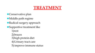
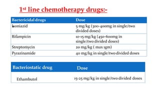
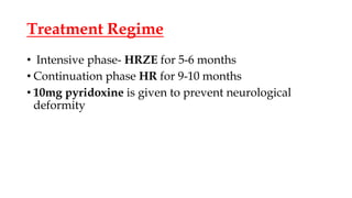
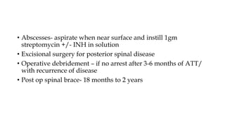
POTT’S SPINE
PRESENTED BY –
DR. VINAYAK MANI DWIVEDI
JR1
DEPARTMENT OF ORTHOPAEDICS
MODERATOR-
DR. ISHAN K. REDDY
SENIOR RESIDENT
EXAMINATION
Aim of examination is:
• to pick up findings suggestive of TB
• to localise the site of lesion(superficial,deep, rotatory palpation)
• find skip lesions (radiologically)
• to detect any associated complications
Physical general examination - to detect active or healed
primary lesion. The patient may have some other systemic
illness ( diabetes, HTN, jaundice etc)
• Gait - Patient walks with short steps in order to avoid jerking
the spine. In TB of cervical spine, the patient often supports
his head with both hands under the chin and twists his whole
body in order to look sideways. In Dorsal Spine TB, Military
attitude is seen.
• Attitude and deformity - Tb of cervical spine patient has a
stiff, straight neck. In dorsal spine TB, part of the spine
becomes prominent (gibbus or kyphus)
• Tenderness - Elicited by pressing upon the side of spinous
swelling on — the back, along the chest wall or spinous
process.
• Movement - Limited spinal movements.
• Para-vertebral swelling - A superficial cold abscess may
present
• Neurological examination - Thorough neurological
examination of the — limbs, upper or lower, depending on the
site of TB should be performed. In addition to motor, sensory
reflexes examination, an assessment should be made of urinary
or bowel functions.
1. whether or not there is any neurological compression
2. level of neurological compression
3. severity of neurological compression
•Pyogenic infections
•Tumorous condition
•Spinal
Osteochondrosis
•Spondylolisthesis
DIFFERENTIAL
DIAGNOSIS
Complications
• Paraplegia
• Cold Abscess
• Sinuses
• Secondary Infections
• Amyloid Disease
• Psoas Abscess
Psoas Abscess
• It is purulent collection within the psoas Muscle, which mostly
spreads through the hematogenous route.
•
Early onset paraplegia
(group A)
⚫Appears within 2 years of
onset – during the Active
phase
⚫Underlying pathology
-Inflammatory edema
-TB Granulation tissue
⚫Good prognosis
Late onset paraplegia
(Group B)
⚫Appears more than 2 years of
disease in vertebral column
⚫Underlying pathology –due to
mechanical pressure on cord
⚫Poor prognosis
Classification of TB Paraplegia
Classification of Tuberculous Paraplegia/Quadriplegia
(Based upon Motor Weakness)
Clinical features of Pott’s Paraplegia:-
⚫Spontaneous muscle twitching in lower limbs
⚫Clumsiness while walking
⚫Extensor plantar response
⚫Exaggerrated reflexes – Sustained clonus of patella and ankle
⚫Motor affected first – then Sensory
⚫Sense of position and vibration – last to disappear
TREATMENT
⚫Conservative plan
⚫Middle path regime
⚫Radical surgery approach
⚫Supportive treatment like
1)rest
2)braces
3)high protein diet
4)Urinary tract care
5) improve immune status
•
Bactericidal drugs Dose
Isoniazid 5 mg/kg (300-400mg in single/two
divided doses)
Rifampicin 10-15 mg/kg (450-600mg in
single/two divided doses)
Streptomycin 20 mg/kg ( max 1gm)
Pyrazinamide 40 mg/kg in single/two divided doses
Ethambutol 15-25 mg/kg in single/two divided doses
Bacteriostatic drug Dose
1st line chemotherapy drugs:-
Treatment Regime
• Intensive phase- HRZE for 5-6 months
• Continuation phase HR for 9-10 months
• 10mg pyridoxine is given to prevent neurological
deformity
Newer Anti-TB Drugs
• Amikacin, Kanamycin, Capriomycin
• Ciprofloxacin, Ofloxacin, Levofloxacin
• Rifabutin
• Clofazimine
• Cycloserine
• Bedaquiline (proton pump inhibitor)
• Protemanid, Delamanid (mycolic acid synthesis inhibitors)
Middle Path Regimen
• Rest in hard bed
• Chemotherapy
• X ray and ESR once in 3 months
• MRI/CT at 6 months interval for 2 yrs
• Gradual mobilization is encouraged in absence of neural deficit
with spinal braces and back extension exercises at 3 to 9 weeks
• Abscesses- aspirate when near surface and instill 1gm
streptomycin +/- INH in solution
• Excisional surgery for posterior spinal disease
• Operative debridement – if no arrest after 3-6 months of ATT/
with recurrence of disease
• Post op spinal brace- 18 months to 2 years
Indication for surgery in patients with
spinal TB & paraplegia
• Absolute Indications –
• Onset of paraplegia during conservative treatment
• Persistent or complete loss of motor power for 1 month despite
conservative treatment
• Paraplegia accompanied by uncontrolled spasticity
• Severe paraplegia of rapid onset
• Paraplegia of flexion
• Flaccid paraplegia
• Complete sensory or motor loss more than 6 months
• Relative Indications-
• Recurrent paraplegia
• Paraplegia with onset in old age
• Painful paraplegia
• Complications like UTI and Stones
Surgeries for Pott’s Paraplegia
• Antero-Lateral Decompression (M/c) – spine is opened up
from the lateral side and access is made to the front and side of
the cord. The cord is laid free from granulation tissue, caseous
material, bony spur or seqestrum.
• Posterior Decompression
• Costo-Transversectomy- removal of 2 inches of rib and
transverse process -> pus drained
Posterior Decompression
• Intra-operative picture
• Radical debridement and arthrodesis (Hongkong Operation)
• Laminectomy and posterior stabilisation – indicated in spinal
tumor syndrome and paraplegia resulting from posterior spinal
disease.
Spinal Braces
⚫Spinal braces are mostly used forambulation of cases of spinal
tuberculosis.
⚫Commonly used spinal braces for lesions from fourth dorsal to
second lumbarvertebra are jewett(milwaukee) brace, ASHE
(anteriorspinal hyperextension) brace , taylor brace.
MILWAUKEE BRACE OR JEWETT BRACE:-
ANTERIOR SPINAL HYPER EXTENSION
BRACE:- (ASHE BRACE)
•
SOMI BRACE( STERNAL OCCIPITAL
MANDIBULAR IMMOBILISER)
•
References -
• Tuberculosis of Skeletal System(6th edition) By – Dr. S.M. Tuli
• Newer Anti-TB Drugs and Regimen 2015 update –
http://www.ncbi.nlm.nih.gov> pmc
POTTS SPINE VINAYAK.pptx

POTTS SPINE VINAYAK.pptx

  • 1.
    POTT’S SPINE PRESENTED BY– DR. VINAYAK MANI DWIVEDI JR1 DEPARTMENT OF ORTHOPAEDICS MODERATOR- DR. ISHAN K. REDDY SENIOR RESIDENT
  • 2.
    EXAMINATION Aim of examinationis: • to pick up findings suggestive of TB • to localise the site of lesion(superficial,deep, rotatory palpation) • find skip lesions (radiologically) • to detect any associated complications Physical general examination - to detect active or healed primary lesion. The patient may have some other systemic illness ( diabetes, HTN, jaundice etc)
  • 3.
    • Gait -Patient walks with short steps in order to avoid jerking the spine. In TB of cervical spine, the patient often supports his head with both hands under the chin and twists his whole body in order to look sideways. In Dorsal Spine TB, Military attitude is seen. • Attitude and deformity - Tb of cervical spine patient has a stiff, straight neck. In dorsal spine TB, part of the spine becomes prominent (gibbus or kyphus)
  • 4.
    • Tenderness -Elicited by pressing upon the side of spinous swelling on — the back, along the chest wall or spinous process. • Movement - Limited spinal movements. • Para-vertebral swelling - A superficial cold abscess may present
  • 5.
    • Neurological examination- Thorough neurological examination of the — limbs, upper or lower, depending on the site of TB should be performed. In addition to motor, sensory reflexes examination, an assessment should be made of urinary or bowel functions. 1. whether or not there is any neurological compression 2. level of neurological compression 3. severity of neurological compression
  • 6.
  • 7.
    Complications • Paraplegia • ColdAbscess • Sinuses • Secondary Infections • Amyloid Disease • Psoas Abscess
  • 8.
    Psoas Abscess • Itis purulent collection within the psoas Muscle, which mostly spreads through the hematogenous route.
  • 9.
    • Early onset paraplegia (groupA) ⚫Appears within 2 years of onset – during the Active phase ⚫Underlying pathology -Inflammatory edema -TB Granulation tissue ⚫Good prognosis Late onset paraplegia (Group B) ⚫Appears more than 2 years of disease in vertebral column ⚫Underlying pathology –due to mechanical pressure on cord ⚫Poor prognosis Classification of TB Paraplegia
  • 10.
    Classification of TuberculousParaplegia/Quadriplegia (Based upon Motor Weakness)
  • 11.
    Clinical features ofPott’s Paraplegia:- ⚫Spontaneous muscle twitching in lower limbs ⚫Clumsiness while walking ⚫Extensor plantar response ⚫Exaggerrated reflexes – Sustained clonus of patella and ankle ⚫Motor affected first – then Sensory ⚫Sense of position and vibration – last to disappear
  • 12.
    TREATMENT ⚫Conservative plan ⚫Middle pathregime ⚫Radical surgery approach ⚫Supportive treatment like 1)rest 2)braces 3)high protein diet 4)Urinary tract care 5) improve immune status
  • 13.
    • Bactericidal drugs Dose Isoniazid5 mg/kg (300-400mg in single/two divided doses) Rifampicin 10-15 mg/kg (450-600mg in single/two divided doses) Streptomycin 20 mg/kg ( max 1gm) Pyrazinamide 40 mg/kg in single/two divided doses Ethambutol 15-25 mg/kg in single/two divided doses Bacteriostatic drug Dose 1st line chemotherapy drugs:-
  • 14.
    Treatment Regime • Intensivephase- HRZE for 5-6 months • Continuation phase HR for 9-10 months • 10mg pyridoxine is given to prevent neurological deformity
  • 15.
    Newer Anti-TB Drugs •Amikacin, Kanamycin, Capriomycin • Ciprofloxacin, Ofloxacin, Levofloxacin • Rifabutin • Clofazimine • Cycloserine • Bedaquiline (proton pump inhibitor) • Protemanid, Delamanid (mycolic acid synthesis inhibitors)
  • 16.
    Middle Path Regimen •Rest in hard bed • Chemotherapy • X ray and ESR once in 3 months • MRI/CT at 6 months interval for 2 yrs • Gradual mobilization is encouraged in absence of neural deficit with spinal braces and back extension exercises at 3 to 9 weeks
  • 17.
    • Abscesses- aspiratewhen near surface and instill 1gm streptomycin +/- INH in solution • Excisional surgery for posterior spinal disease • Operative debridement – if no arrest after 3-6 months of ATT/ with recurrence of disease • Post op spinal brace- 18 months to 2 years
  • 18.
    Indication for surgeryin patients with spinal TB & paraplegia • Absolute Indications – • Onset of paraplegia during conservative treatment • Persistent or complete loss of motor power for 1 month despite conservative treatment • Paraplegia accompanied by uncontrolled spasticity • Severe paraplegia of rapid onset • Paraplegia of flexion • Flaccid paraplegia • Complete sensory or motor loss more than 6 months
  • 19.
    • Relative Indications- •Recurrent paraplegia • Paraplegia with onset in old age • Painful paraplegia • Complications like UTI and Stones
  • 20.
    Surgeries for Pott’sParaplegia • Antero-Lateral Decompression (M/c) – spine is opened up from the lateral side and access is made to the front and side of the cord. The cord is laid free from granulation tissue, caseous material, bony spur or seqestrum. • Posterior Decompression • Costo-Transversectomy- removal of 2 inches of rib and transverse process -> pus drained
  • 21.
  • 22.
    • Radical debridementand arthrodesis (Hongkong Operation) • Laminectomy and posterior stabilisation – indicated in spinal tumor syndrome and paraplegia resulting from posterior spinal disease.
  • 23.
    Spinal Braces ⚫Spinal bracesare mostly used forambulation of cases of spinal tuberculosis. ⚫Commonly used spinal braces for lesions from fourth dorsal to second lumbarvertebra are jewett(milwaukee) brace, ASHE (anteriorspinal hyperextension) brace , taylor brace.
  • 24.
    MILWAUKEE BRACE ORJEWETT BRACE:-
  • 25.
    ANTERIOR SPINAL HYPEREXTENSION BRACE:- (ASHE BRACE) •
  • 26.
    SOMI BRACE( STERNALOCCIPITAL MANDIBULAR IMMOBILISER) •
  • 27.
    References - • Tuberculosisof Skeletal System(6th edition) By – Dr. S.M. Tuli • Newer Anti-TB Drugs and Regimen 2015 update – http://www.ncbi.nlm.nih.gov> pmc

Editor's Notes

  • #7 Typhoid spine Brucella Spondylitis Mycotic Spondylitis Syphilitic Primary malignant tumor Multiple Myeloma Lymphomas Secondary Histocytosis-X
  • #10 Type A – Abscess Caseous tissue Ischaemic lesion of cord (Rare) Type B - TB Debris TB Sequestra from body and disc Localized Internal gibbus Canal stenosis / Severe kyphotic deformity
  • #25 Recommended for tuberculous lesions in dorsal if the number vertebra involved is more than 2 or there is panvertebral disease
  • #26 MORE ACCEptable in young women and girls, as it gets hidden under clothes. It runs from symphysis pubis to manubrium sterni
  • #27 BECAUSE it leaves the Back , particularly free except the occipital pad.