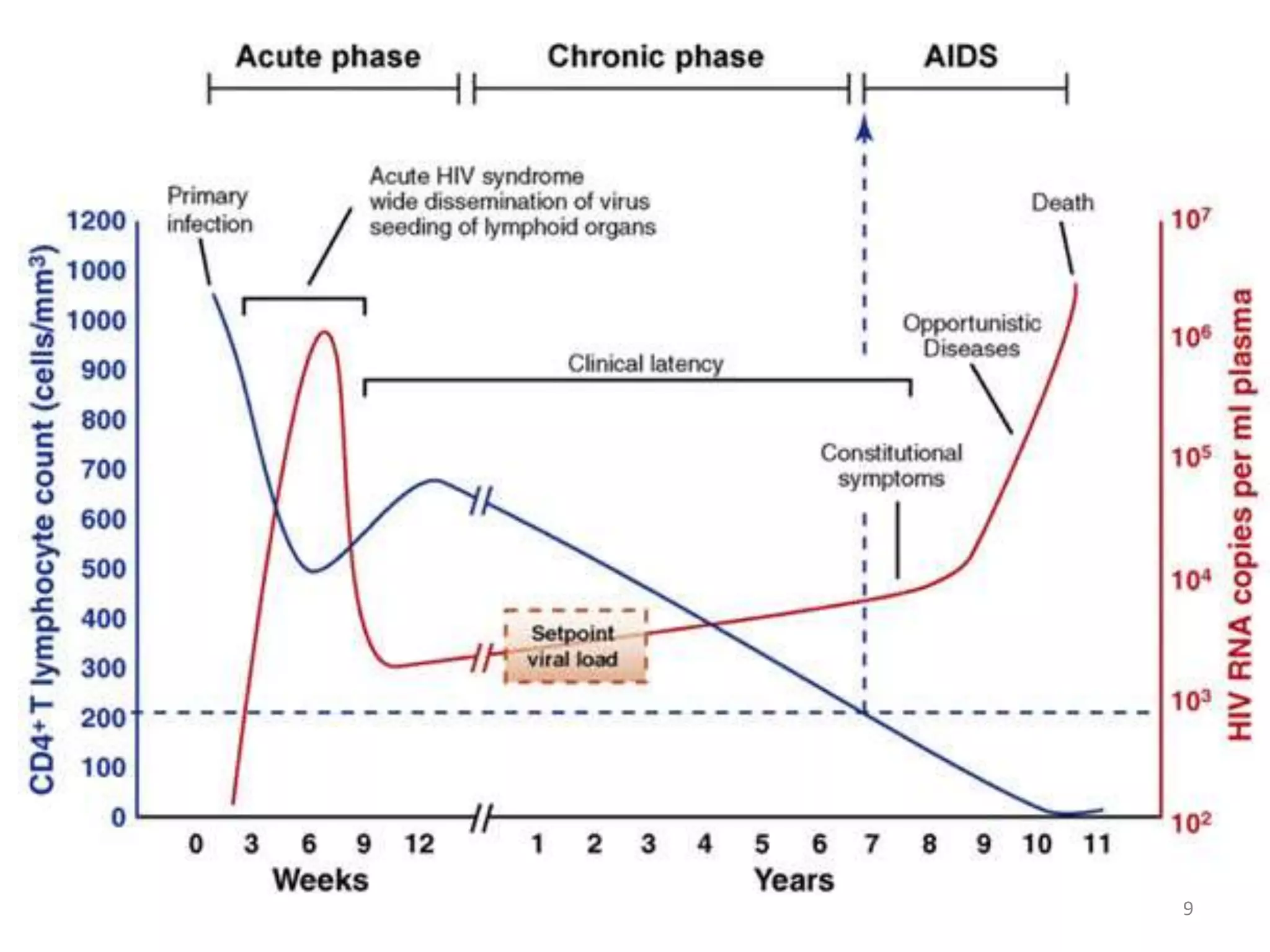
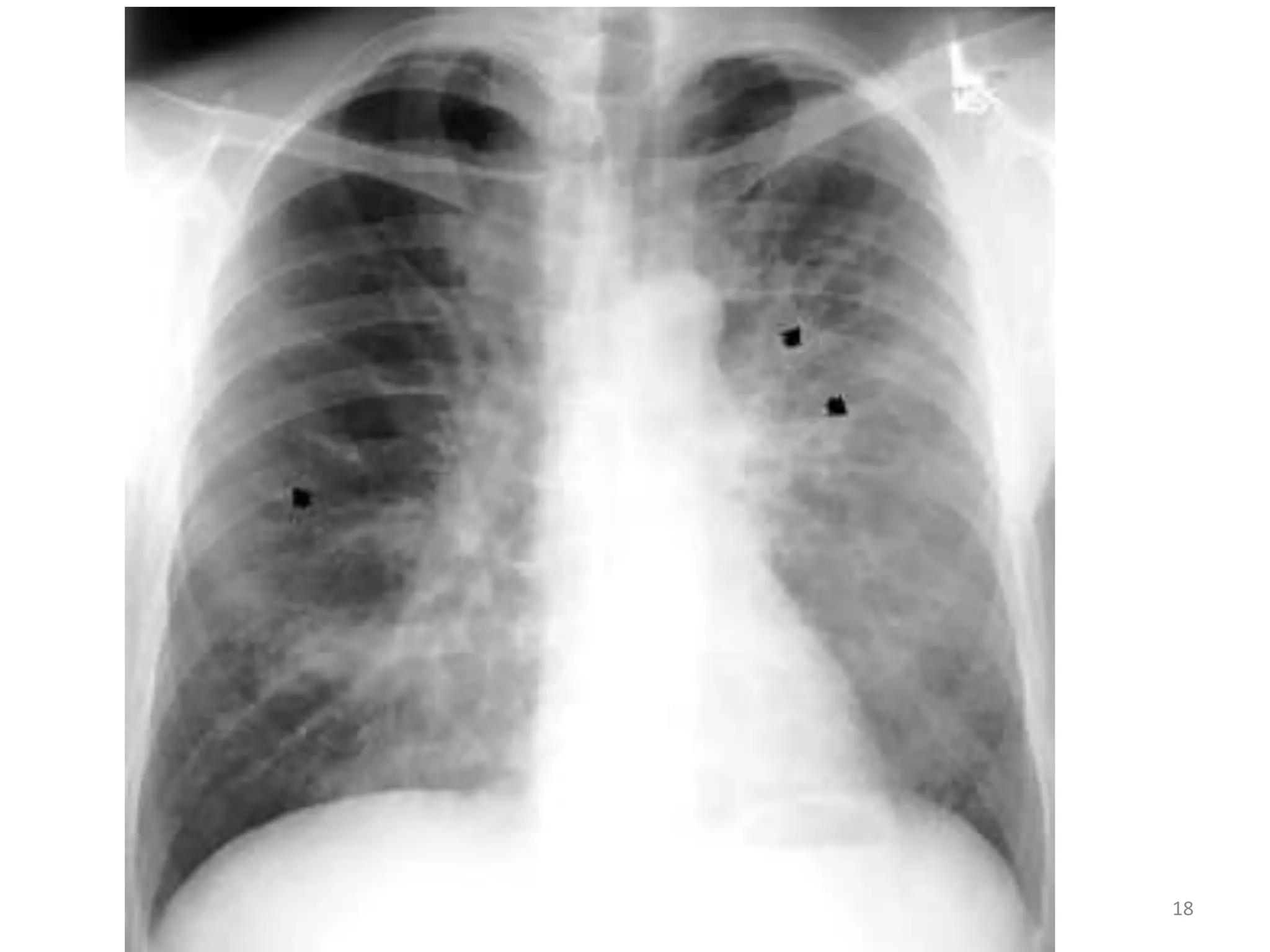
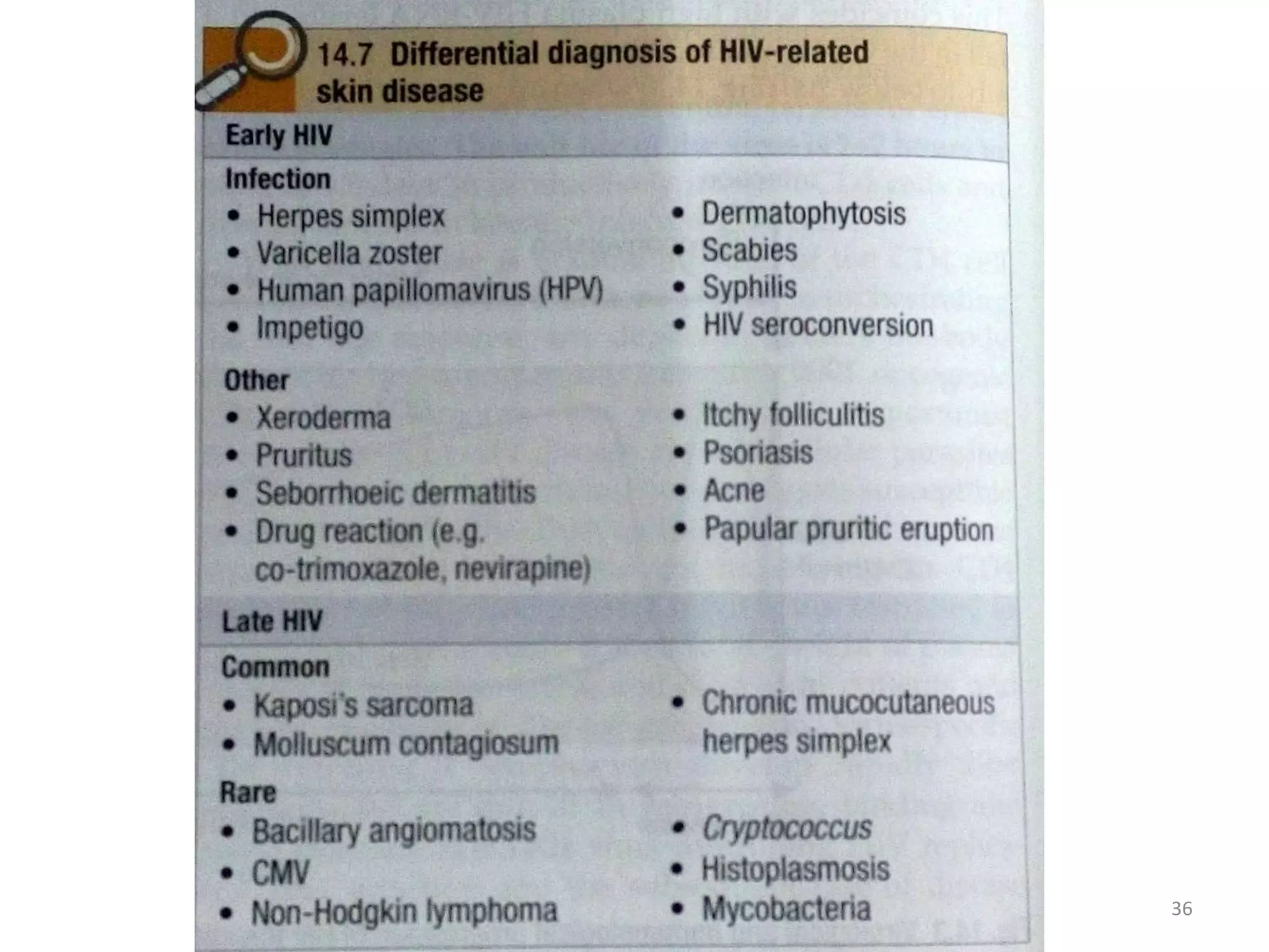
This document discusses the presenting problems in HIV infection. It describes the stages of infection from acute to asymptomatic to symptomatic disease. Common respiratory infections associated with HIV include pneumonia, Pneumocystis jiroveci pneumonia, tuberculosis, and atypical mycobacterial infections. Other organ systems affected by opportunistic infections in HIV patients with low CD4 counts include the cardiovascular and gastrointestinal systems. Mucocutaneous manifestations are also common.
This document discusses oral candidiasis, caused by the yeast Candida albicans. It presents various classifications and investigations for candidiasis, including smears, swabs, and cultures. Management involves identifying predisposing factors and using topical or systemic antifungal drugs like amphotericin B, nystatin, clotrimazole, ketoconazole, fluconazole, itraconazole or miconazole. For HIV patients, highly active antiretroviral therapy (HAART) is recommended along with antifungal drugs to treat oral candidiasis.
Ludwig's angina is a severe bacterial infection of the submandibular, sublingual, and submental spaces that can lead to airway obstruction if left untreated. It is usually caused by dental infections. Clinically, it presents with a firm, brawny swelling of the neck and floor of the mouth, accompanied by fever, malaise, and difficulty opening the mouth. Aggressive treatment involves securing the airway, administering IV antibiotics, and performing surgical incision and drainage. Without treatment, Ludwig's angina can prove fatal within 1-2 days due to asphyxiation or sepsis.
This document provides an overview of halitosis (bad breath), including its classification, etiology, diagnosis, and management. It discusses the role of volatile sulfur compounds and certain bacteria in causing halitosis. Diagnostic tools include organoleptic measurement, gas chromatography, and volatile sulfide monitoring to detect these compounds. Treatment involves identifying and addressing the underlying causes, such as periodontal disease, dry mouth, dental caries, or systemic conditions. Preventive measures focus on proper oral hygiene and avoiding foods that can cause temporary halitosis.
The document discusses various salivary gland disorders including infections, inflammatory conditions, cysts, tumors and other pathologies. It provides details on:
- Acute and chronic bacterial sialadenitis, most commonly caused by retrograde infection from the mouth. Acute infections more often affect the parotid gland.
- Viral infections like mumps can cause acute non-suppurative sialadenitis. Mumps is spread through droplets and involves the parotid glands.
- Sjögren's syndrome is an autoimmune condition characterized by lymphocytic destruction of exocrine glands causing dry mouth and eyes. Diagnosis involves labial biopsy.
- Common benign sal
This document provides information on periodontal management of HIV patients. It discusses the stages of HIV infection and common oral manifestations such as oral candidiasis, oral hairy leukoplakia, Kaposi's sarcoma, and non-Hodgkin's lymphoma. It also covers HIV-related periodontal diseases including linear gingival erythema, necrotizing ulcerative gingivitis, and necrotizing ulcerative periodontitis. Treatment protocols are provided for oral lesions and periodontal diseases seen in HIV patients.
This document summarizes common lesions of the oral cavity, including ulcers caused by infections (viral like herpes, bacterial like Vincent's infection, fungal like candidiasis), immune disorders (aphthous ulcers, Behcet's syndrome), trauma, skin disorders (lichen planus, pemphigus vulgaris), and submucous fibrosis caused by chewing areca nut. It describes the etiology, clinical features, and management of each condition. Major types of oral ulcers and lesions are infections, immune disorders, trauma, neoplasms, and skin disorders that may manifest in the oral cavity.
Candidiasis and its management in dentistryAshok Kumar
Candidiasis is a fungal infection caused by Candida species, most commonly Candida albicans. It can cause superficial infections of the skin, nails and mucous membranes, as well as systemic infections in severely immunocompromised individuals. Predisposing factors include antibiotic use, corticosteroid use, diabetes, and immunodeficiency. Diagnosis is made through microscopic examination, culture, or biopsy of infected tissues. Treatment involves topical or systemic antifungal medications. While orthodontic appliances do not typically cause Candida infections, carriers may be at higher risk of infection with appliances in place.
This document discusses diseases of the salivary glands, including sialadenitis (inflammation of the salivary glands), which can be caused by bacterial or viral infections. It also discusses Sjogren's syndrome, an autoimmune disease that causes dry mouth and dry eyes due to lymphocytic infiltration and destruction of the lacrimal and salivary glands. Obstructive diseases like salivary calculi (stones) are also covered. The document provides details on symptoms, diagnosis, and treatment of various salivary gland diseases.
Human Immunodeficiency Virus (HIV) can cause oral manifestations in 30-80% of infected individuals. Common oral issues include candidiasis, a fungal infection causing lesions; periodontitis with tissue destruction; and viral infections like herpes simplex causing vesicles and ulcers. Other conditions linked to HIV/AIDS are Kaposi's sarcoma, a cancer originating from blood vessels, and non-Hodgkin's lymphoma. Proper dental management of HIV-infected individuals includes monitoring for early signs of oral opportunistic infections and treating them promptly to support overall health.
This document discusses aphthous ulcers, also known as canker sores, which are characterized by recurring, painful ulcers in the mouth. It describes the three main types: minor aphthous ulcers, which are less than 10mm and heal within 2 weeks; major aphthous ulcers, which are larger than 10mm and can take over 6 weeks to heal, often leaving scars; and herpetiform ulcers, which present as multiple small ulcers that may join together into larger ones and persist for 1-3 years. The document provides details on symptoms, locations in the mouth, appearances, durations, and histopathology of the different types of aphthous ulcers.
Acute necrotizing ulcerative gingivitis (ANUG), also known as Vincent's gingivitis or Vincent's infection, is a polymicrobial infection of the gums that results in ulceration, necrosis, and pain. It is caused by an overgrowth of oral bacteria like spirochetes and fusiform bacteria when the immune system is weakened by factors like stress, malnutrition, or HIV infection. ANUG begins as small ulcers on the gums that can spread and deepen over time if left untreated, potentially resulting in bone and tissue loss. Symptoms include bleeding, foul taste, and pain when eating. Treatment focuses on improving oral hygiene and nutrition to restore immune function and resolve the
Not only the lesions in the body helps us to know about syphilis but also a minute nodule or lesion helps us to discover the syphilis. He who knows syphilis knows the medicine well. Earlier you found the disease the treatment and the prognosis will be good. Discover syphilis through your body's gateway.
Oral candidiasis is caused mainly by Candida albicans and presents in several forms depending on location and predisposing factors. Predisposing factors include antibiotics, immunosuppressants, and diseases that suppress the immune system. Diagnosis involves examining clinical signs, smears, cultures, and histology. Treatment focuses on reducing predisposing factors, improving oral hygiene, and using topical or systemic antifungal medications depending on the type and severity of infection.
This document discusses different types of pigmented cellular nevi:
- Intradermal (intramucosal) nevi occur within the dermis or mucosa and appear as raised or flat dark lesions.
- Junctional nevi are located at the dermal-epidermal junction and appear as flat brown-black macules, most often on the hard palate or gingiva.
- Compound nevi exhibit characteristics of both intradermal and junctional nevi, with nevus cells in the dermis and basal epidermis.
- Blue nevi appear as dark blue papules or macules on the skin or hard palate, composed of pigment
the most common viral infections that affects the maxillofacial area
sources( burket's oral medicine 11th ed,oral and maxillofacial pathology neville 2e )
This document discusses infections of the submandibular space, which is divided into two compartments by the mylohyoid muscle. Dental infections are the most common cause, with roots above or below the mylohyoid muscle leading to sublingual or submaxillary infections, respectively. Symptoms include odynophagia, trismus, and swelling of the submental and submandibular regions. Treatment involves systemic antibiotics, incision and drainage of any abscesses either intraorally or externally, and tracheostomy if the airway is endangered. Complications can arise from spread of infection or airway obstruction.
Erythema multiforme, Steven-Johnson syndrome and Toxic Epidermal NecrolysisBinaya Subedi
Erythema Multiforme is a common Vesiculobullous deramtological condition with mucosal manifestations trigged by Herpes virus infection and certain sulpha containing drugs.
This document provides an overview of several common bacterial infections that can affect the oral cavity. It discusses tuberculosis, which is caused by Mycobacterium tuberculosis and can spread through airborne droplets to the lungs. If the oral cavity is involved, it typically presents as indurated chronic ulcers. Syphilis is caused by Treponema pallidum and has primary, secondary, and tertiary stages marked by chancres, rashes, and potential neurological/cardiovascular effects. Actinomycosis is caused by Actinomyces israelii and usually appears in the jaw after trauma or infection, causing hard swellings that may drain through the gums. Gonorrhea, caused by Ne
- Herpes simplex virus types 1 and 2 are common oral viral infections, usually transmitted via saliva or direct contact. Both viruses establish latency in ganglia. HSV-1 typically causes oral lesions while HSV-2 typically causes genital lesions.
- Varicella zoster virus causes chickenpox in children and shingles in adults via reactivation. It establishes latency in ganglia.
- Epstein-Barr virus causes infectious mononucleosis, commonly known as "mono" or "kissing disease". It is transmitted through saliva and causes fever, lymphadenopathy and pharyngitis.
1. Oral manifestations are among the earliest signs of HIV infection. Conditions like oral candidiasis, oral hairy leukoplakia, and Kaposi's sarcoma are strongly associated with HIV.
2. Other less common conditions include necrotizing gingivitis/periodontitis, infections by Mycobacterium tuberculosis or M. avium-intracellulare, and salivary gland diseases.
3. The progression of oral lesions correlates with declining CD4 counts and worsening immune suppression in patients with HIV/AIDS.
This document discusses pemphigus vulgaris, an autoimmune disease characterized by the formation of blisters within the epidermis caused by autoantibodies against desmoglein proteins. It most commonly affects individuals in their 40s and 50s and is more prevalent in Ashkenazi Jews and those of Mediterranean descent. Clinical features include painful oral and skin ulcers. Diagnosis is confirmed through direct immunofluorescence detecting autoantibodies at the dermoepidermal junction. Treatment ranges from topical corticosteroids for mild cases to systemic immunosuppressants like prednisone and azathioprine for moderate to severe disease.
Candidiasis, also known as oral thrush, is a common fungal infection caused by various Candida species, most commonly Candida albicans. There are several types of oral candidiasis including chronic atrophic candidiasis seen in denture wearers, acute pseudomembranous candidiasis presenting as white plaques in the mouth, and acute atrophic candidiasis occurring after the plaques are lost. Treatment involves topical antifungal medications like clotrimazole or amphotericin B rinses, as well as systemic antifungals like ketoconazole, fluconazole, or nystatin for 2-3 weeks. Removing any predisposing
Oral submucous fibrosis (OSMF or OSF) is a chronic, complex, premalignant (1% transformation risk) condition of the oral cavity, characterized by juxta-epithelial inflammatory reaction and progressive fibrosis of the submucosal tissues (the lamina propria and deeper connective tissues). As the disease progresses, the jaws become rigid to the point that the person is unable to open the mouth.
The condition is remotely linked to oral cancers and is associated with areca nut or betel quid chewing, a habit similar to tobacco chewing, is practiced predominantly in Southeast Asia and India, dating back thousands of years.
Ludwig's angina is a serious neck infection that begins in the submandibular space and spreads to surrounding areas, potentially compromising the airway. It is usually caused by an odontogenic infection. Clinical features include swelling of the floor of mouth and neck, difficulty opening the mouth, dysphagia, and potential airway obstruction. Diagnosis is based on involvement of multiple deep neck spaces and spread via fascial planes rather than lymph nodes. Treatment requires securing the airway, typically via tracheostomy, and administering IV antibiotics.
This document provides an overview of erythema multiforme (EM), a self-limited inflammatory mucocutaneous disease that commonly affects the skin and oral mucosa. It discusses the etiology, pathogenesis, clinical features, classification variants, diagnosis and management of EM. Key points include: EM results from a hypersensitivity reaction, often to infections or drugs; it ranges from mild to severe based on extent of skin and mucosal involvement; diagnosis involves clinical exam, biopsy and ruling out other conditions; treatment depends on severity but may include antivirals, corticosteroids or immunosuppressants.
Acute necrotising ulcerative gingivitis is a rare condition characterized by necrosis of the gingiva and interdental papillae. It frequently occurs during times of stress and poor oral hygiene. The condition is caused by fusiform bacillus and Borrelia vincentii bacteria. Clinically, the gingiva becomes painful and develops punched-out ulcers. It can spread to other oral tissues and rarely the skin, causing further complications.
This document summarizes a seminar presentation on halitosis (bad breath). It defines halitosis and provides its history, epidemiology, classification, etiology, association with periodontal disease, diagnosis methods, and treatment approaches. Key causes of halitosis include oral diseases like gingivitis and periodontitis which produce volatile sulfur compounds from bacterial breakdown of proteins. Diagnosis involves self-assessment tests and objective tests like organoleptic measurement, gas chromatography, and sulfide monitoring. Treatment focuses on mechanical and chemical approaches to reduce oral bacteria and volatile compounds through techniques like tongue cleaning, tooth brushing, and using mouthwashes.
This document provides an overview of abscesses of the periodontium, specifically focusing on periodontal abscesses. It defines a periodontal abscess and classifies them based on location, course, number, affected tissue, and cause. Periodontal abscesses are most prevalent in molar sites and those with pre-existing periodontal pockets. They can be caused by factors like untreated periodontitis, foreign bodies, or changes after periodontal procedures or antibiotics. The pathogenesis involves bacterial entry triggering an inflammatory response that leads to tissue destruction and pus formation.
19. presenting problems in infectious diseasesAhmad Hamadi
This document discusses the evaluation and management of fever. It notes that the differential diagnosis for fever is broad and initial screening investigations should include blood tests, imaging, and cultures of potential sites of infection depending on symptoms. For patients where the cause is not obvious, further targeted investigations are needed. The document also discusses considerations for evaluating fever in people who inject drugs, including risks related to injection practices and common infections in this population.
This document discusses various lung disorders related to HIV infection. It notes that HIV-1 is more prevalent globally while HIV-2 is found principally in West Africa. Common transmission routes for HIV include sexual contact, intravenous drug use, and mother-to-child. Those at high risk include homosexuals/bisexuals, intravenous drug users, infants born to infected mothers, and recipients of blood/blood products. The document then discusses various classes of antiretroviral drugs and associated lung diseases like bacterial pneumonias, tuberculosis, and Pneumocystis jiroveci pneumonia, their symptoms, diagnoses and treatments.
Opportunistic infections are infections that occur more frequently and are more severe in people with weakened immune systems such as those with HIV/AIDS. These infections include fungal, bacterial, viral, and parasitic infections that typically do not seriously affect those with healthy immune systems. Common opportunistic infections in HIV/AIDS patients include Pneumocystis pneumonia, tuberculosis, candidiasis, toxoplasmosis, cryptococcus, and cytomegalovirus. Antiretroviral therapy has significantly reduced the rates of opportunistic infections by suppressing HIV and allowing immune recovery. HIV/AIDS remains a major global public health challenge.
HIV/AIDS was first identified in the 1980s but may have originated in Africa in the late 1950s. While drug treatments and education have slowed its spread in some areas, most people in Africa with HIV are unaware of their status. HIV is a retrovirus that can develop into AIDS if not properly managed, though it cannot be cured. It is most often transmitted through bodily fluids like blood, semen, and vaginal fluids or from mother to child. Prevention methods include abstinence, monogamy, condom use, sterile needles, and new blades. Sub-Saharan Africa has the most HIV cases, with 18% of new global infections. Efforts are needed to increase testing,
01.04 laboratory diagnosis and monitoring of hiv infectionDavid Ngogoyo
Laboratory tests play an important role in diagnosing and monitoring HIV infection. Tests used for diagnosis include ELISA, rapid tests, and confirmatory tests like Western Blot. CD4 counts and viral load are used to determine when to start ART, monitor disease progression and response to treatment. Other tests like hematology and biochemistry panels help monitor for side effects and coinfections. Proper use and interpretation of HIV laboratory tests is crucial for effective clinical management of patients.
Opportunistic Infection Among Hiv Infected ChildrenDang Thanh Tuan
The document provides guidelines for treating opportunistic infections among HIV-infected children from the CDC, NIH, and Infectious Diseases Society of America. It discusses recommendations for treating various bacterial, mycobacterial, fungal and viral infections including serious bacterial infections, syphilis, toxoplasmosis, cryptosporidiosis, tuberculosis, Pneumocystis jiroveci pneumonia, candidiasis, and cytomegalovirus. Treatment recommendations include antibiotics, antivirals, and antifungals with dosages provided for children.
Identification of AIDS? And what is HIV infection and mode of transmission?Hassan Shaker
This presentation includes the following:
1) What are viruses and its classification
2) Over view of HIV infection
3) Development of HIV infection into AIDS.
4) HIV infection's clinical features and its complications.
5) Life cycle of HIV infection.
6) Mode of transmission of HIV infection.
7) How to diagnose HIV infection.
8) How to manage HIV infection.
9) Explain different preventive measures to prevent sexually transmitted viral infection
2 natural history of hiv and who clinical staging naco lac mDrShruthi Pradeep
This document summarizes the natural history and clinical staging of HIV infection in 3 paragraphs. It describes the typical progression of untreated HIV infection from initial viral transmission and acute retroviral syndrome, to asymptomatic chronic infection lasting an average of 8 years, to symptomatic HIV infection and AIDS occurring on average 1.3 years later without treatment. It also outlines the WHO clinical staging system for classifying HIV patients based on their symptoms and disease progression into 4 stages, with stage 1 being asymptomatic and stage 4 involving advanced AIDS-defining illnesses. The document provides an overview of the modes of HIV transmission, pathogenesis, typical clinical course, and classification approach for monitoring HIV disease progression.
The document summarizes early signs and symptoms of AIDS, oral manifestations of HIV, and concludes with the importance of early testing. Early signs may include brief flu-like symptoms appearing 2-4 weeks after infection. As the virus destroys immune cells over years, mild infections and chronic symptoms develop, including swollen lymph nodes, diarrhea, weight loss and fever. Late-stage AIDS is marked by opportunistic infections causing symptoms like night sweats, cough, diarrhea and oral lesions. Oral manifestations include fungal, viral and bacterial lesions as well as neoplastic lesions like Kaposi's sarcoma. Early testing is key to stopping the spread of HIV and improving survival.
HIV infection
Mode of transmission, pathogenesis, clinical manifestations, laboratory diagnosis, treatment, prevention, prognosis, scope of AIDS vaccine.
Hiv infection progresses from asymptomatic infection to AIDS, the most severe stage. It is caused by the HIV virus which depletes CD4+ T cells, weakening the immune system. Left untreated, opportunistic infections develop. HIV is transmitted via blood, sex, or perinatally. Treatment involves antiretroviral therapy to suppress the virus indefinitely and treat any infections, with the goals of prolonging life, improving quality of life, and restoring immune function. Nursing care focuses on medication adherence, nutrition, symptom management, and psychological support.
This document provides a summary of key information about primary care approaches to treating HIV patients, including:
1) It discusses the history and epidemiology of HIV, modes of transmission, clinical presentations to different specialists, treatment with HAART, and baseline evaluations prior to treatment initiation.
2) Primary care providers should offer ART to patients with CD4 counts <200 or symptoms, consider treatment for counts 200-350, and can defer for asymptomatic patients with counts >350 and low viral loads.
3) When initiating ART, providers should evaluate readiness, ensure adherence, perform baseline testing, and select preferred first-line regimens consisting of 2 NRTIs combined with an NNRTI or PI.
The document summarizes the history, epidemiology, virology, immunology, transmission, clinical progression, and global impact of HIV/AIDS. It notes that HIV was first recognized in 1981 but has since been traced to 1959. It is caused by HIV-1 and HIV-2 viruses and is transmitted via bodily fluids. Left untreated, it progresses from primary infection to asymptomatic infection to AIDS as it depletes CD4 cells and allows opportunistic infections. Currently over 30 million people are living with HIV globally, with sub-Saharan Africa most severely impacted.
This document revises the WHO clinical staging system for HIV/AIDS in adults and adolescents aged 15 years or more. It outlines 4 clinical stages based on severity of conditions: asymptomatic, mild conditions (stage 1-2), advanced conditions where presumptive diagnosis can be made (stage 3), and severe conditions where confirmatory testing is required (stage 4). The revisions aim to standardize definitions, harmonize pediatric and adult staging, and support ART scale-up by guiding decisions on when to initiate treatment. Annexes provide more details on recognizing conditions. The staging system is intended to facilitate clinical management and monitoring of patients, as well as surveillance efforts.
This document discusses guidelines for consultations with patients who have tested positive for HIV. It outlines steps for the initial consultation 1-2 weeks after receiving a positive test result, including exploring the patient's understanding and feelings, addressing common questions, discussing support systems and lifestyle strategies, and making referrals. It also describes guidelines for continuing maintenance consultations, including examinations, tests, treatment, and prophylaxis based on immune status. The document provides information on contacting tracing, prevention, community education, and signs for when to refer a patient.
This document discusses tumors and cancers that are more common in HIV/AIDS patients. It states that those with HIV/AIDS have a higher risk of developing Kaposi sarcoma, non-Hodgkin lymphoma, and cervical cancer. It provides details on the symptoms, risk factors, and treatments for these types of cancers associated with HIV/AIDS. The survival rates for Kaposi sarcoma and outcomes for non-Hodgkin lymphoma have improved with newer HIV treatments.
HIV can increase the risk of developing lymphomas through sustained B-cell activation and high cytokine levels. AIDS-related lymphomas are categorized based on location into systemic, primary central nervous system, and body cavity-based types. The two major histological groups are immunoblastic lymphoma and small non-cleaved cell lymphoma (Burkitt lymphoma). Human herpes viruses have been linked to some forms of AIDS-related lymphomas.
CD4 counts give you and your doctor a good idea of how much damage HIV has done to your immune system. But you also need to know how fast that damage is happening. Viral load tests, which tell the doctor how much HIV is in your blood, are a very important clue to how quickly HIV is doing harm.
This document discusses pulmonary manifestations in HIV patients. It begins with an introduction to HIV transmission and risk groups. It then discusses how HIV affects the lungs, causing direct infection and immune dysfunction. Common pulmonary conditions in HIV patients are described, including opportunistic infections like Pneumocystis pneumonia and tuberculosis, which present differently based on CD4 count. Imaging findings for various lung diseases seen in HIV are provided, with examples of abnormalities seen on chest x-ray and CT for conditions like Pneumocystis pneumonia and bacterial/mycobacterial infections. Risk factors, diagnosis and treatment approaches are also summarized.
HIV CHEST AND OPPOTUNISTIC INFECTION IN AIDS.pptxJosephmwanika
HIV infection can directly infect lung cells and weaken the immune system's ability to fight pulmonary infections. Common lung manifestations of HIV/AIDS include opportunistic infections like Pneumocystis pneumonia, tuberculosis, and cytomegalovirus pneumonia. Chest imaging plays an important role in the diagnosis and management of these infections. On CT, Pneumocystis pneumonia typically appears as bilateral ground-glass opacity and septal thickening, while tuberculosis may show upper lobe cavitary lesions when CD4 counts are high and disseminated disease at low CD4 counts. Viral infections like CMV commonly cause ground-glass nodules in severely immunocompromised individuals.
Pneumocystis jirovecii is a fungus that causes pneumonia in immunocompromised patients. It is diagnosed through microscopic visualization of the organism in samples obtained through induced sputum or bronchoscopy with bronchoalveolar lavage. Real-time PCR assays have increased sensitivity over conventional staining but may produce false positives. Risk factors include HIV/AIDS with CD4 count <200 cells/uL, use of immunosuppressive drugs, hematologic malignancies, and organ transplantation. Presentation involves fever, cough, and dyspnea. Treatment involves trimethoprim-sulfamethoxazole.
This document discusses HIV related lung disorders, including:
- HIV-1 and HIV-2 viruses and their transmission routes such as sexual contact, blood transmission, and mother-to-child transmission.
- The significance of HIV and lung diseases, with pulmonary complaints often leading to HIV diagnosis.
- Various lung conditions that are more common in HIV patients like bacterial pneumonias, Pneumocystis jiroveci pneumonia, and tuberculosis.
- The interaction between tuberculosis and HIV, with TB being a leading opportunistic infection. Clinical presentations and treatments of TB in HIV patients are covered.
- Risk factors, clinical manifestations, diagnosis, and treatments of Pneumocystis jiroveci
This document discusses acute HIV infection and CDC criteria for diagnosis. It defines acute HIV infection as occurring within approximately six months of infection. Symptoms may include fever, lymphadenopathy, sore throat, rash, and others nonspecific symptoms. Diagnosis is made by detecting HIV RNA during the window period before antibodies develop or with a positive combination antigen/antibody test and negative antibody-only test. Early diagnosis is important for individual treatment and reducing transmission.
This document discusses HIV and its effects on the ENT system. It begins by explaining what HIV is and how it attacks the immune system. It then discusses the epidemiology of HIV and current global statistics. Various opportunistic infections that can affect the ENT system are described, including fungal infections of the ear, sinusitis, neoplasms like Kaposi's sarcoma, and lymphomas of the nose and oral cavity. Manifestations in different areas like the ear, nose, oral cavity and airways are summarized. Risk groups, disease progression, and treatment approaches are also briefly covered.
HIV AND LUNGSpulmonary infections in hiv paientsraajpatel7425
- 36.9 million people worldwide live with HIV. Pulmonary complications are common, occurring in up to 70% of patients. Lung infections are 25 times more common in those with HIV.
- HIV causes immune dysfunction including depletion of CD4+ T cells and abnormalities in innate immunity, increasing risk of various lung infections and cancers.
- Common pulmonary manifestations include Pneumocystis pneumonia, bacterial pneumonia, tuberculosis, nocardiosis, and Pneumocystis jirovecii. Evaluation involves physical exam, imaging like chest X-ray and CT, as well as microbiological testing of sputum and blood.
This document provides an overview of pneumonia, including its definition, classification, pathophysiology, clinical manifestations, diagnosis, treatment and antibiotic resistance. Pneumonia is an infection of the lungs that can be caused by bacteria, viruses or other pathogens. It is commonly classified as community-acquired or healthcare-associated pneumonia. Clinical diagnosis involves assessing symptoms and chest imaging, while etiologic diagnosis may involve sputum/blood cultures, antigen tests and PCR. Treatment depends on pneumonia severity and risk factors. Antibiotic resistance among pathogens like Streptococcus pneumoniae and gram-negative bacilli is an ongoing concern.
Secondary Immunodeficiency
By Dr. Usama Ragab Youssif
Reference: Included in Slides
Include causes of secondary immunodeficiency including AIDS and other viral infections
HIV causes AIDS by weakening the immune system. It is transmitted through bodily fluids and can be diagnosed by detecting HIV antibodies or antigens. The disease progresses from primary infection to asymptomatic infection and mild symptoms, and finally AIDS when opportunistic infections occur. Common signs include oral thrush, shingles, and weight loss. Treatment focuses on antiviral drugs and prophylaxis for opportunistic infections.
1. Acute rheumatic fever is an autoimmune disease that develops after a streptococcal infection and can affect the heart, joints, brain and skin. It is mainly seen in children aged 5-14 years from low socioeconomic backgrounds.
2. Bronchiectasis is a chronic lung condition caused by persistent lung infections that destroys the airways and causes them to dilate permanently. It can result from cystic fibrosis, primary ciliary dyskinesia or severe lung infections and treatments involve airway clearance and long-term antibiotics.
3. Both conditions involve recurrent infections and inflammation of the lungs and/or heart but acute rheumatic fever is self-limiting while bronchiectasis is
This document discusses the clinical manifestations of acute and early HIV infection. It begins by outlining the CDC stages of HIV disease progression. It then describes the symptoms of acute retroviral syndrome, which occurs in 50-70% of individuals within 3-6 weeks of initial HIV infection, including fever, lymphadenopathy, sore throat, rash, and constitutional symptoms. It also notes that opportunistic infections rarely occur during acute HIV infection but may include oral candidiasis or CMV. The document provides details on various oral manifestations of HIV infection and their diagnosis and treatment.
HIV associated Oppertunistic Infection. kajsdh kajs kjdsbfk ksjdbfkjs ksdf ksdbf ksjkjs kjshsa oefwhmashas aslasdh asnlasdh asblahdas snas lasn asd da sad s d asd qwhd as asd sd das ads sdaedfwa ag gsdg sd d ds dsf sfd sdf df s sd sd ds dg fd sa sd ge g ew ew dsv sdfkllSFKLKSF LSAFNLSDFNSC
1. Opportunistic infections associated with HIV can affect the gastrointestinal, respiratory, neurological, and mucocutaneous systems. Common gastrointestinal infections include Cryptosporidium, Microsporidia, and Cytomegalovirus, presenting with diarrhea, abdominal pain, and weight loss.
2. Frequent respiratory infections are Pneumocystis jirovecii pneumonia and bacterial pneumonias. Pneumocystis presents with cough and difficulty breathing, while bacterial pneumonias cause more acute symptoms.
3. Common neurological opportunistic infections are Toxoplasmosis, Cryptococcosis, and HIV-associated dementia. Toxoplasmosis and Cryptococcos
hiv AND ASSOASITE INFECTION KLASL SDHK OIUDQ OSAJS OSAIDOA S OSAIDJOAS OAISJQWU OASIdw8u sa oiSUDA S soaijoa S OAJSOq sd0qud ASCJwedfoWKD sdfiuwefok sojASOIJSDSD FOsfh sffuso iaSO SUIDisa siWUD[Qd sc sisiWUF CSS isuf[we fs ssduf sfs f9wufW SA wfuas CsiufWEF SDDFs9iFO F FIhfo FIf FIU 0s8fu0WEUFOKSDJHFOwifoSAJFUWEYf9ojssJDFH[9WEF 00UFOIsdf oiasfoiWUF ISAOFJ0q8wu ioasjf0 fsa fs fsd sdfa sd fds s df sdf dsf sdf d ds g df dgs sdg dg gd gr e ew ds adf f gd dg sd a d ds ds da dfa h htr yuklkkjkopopipuyu iuy ij
This document provides an overview of various pulmonary infections, including types of pneumonia (lobar, bronchopneumonia, interstitial) and infectious agents that can cause pneumonia (viral like influenza, bacterial, fungal, parasitic, rickettsial). It describes the typical clinical and radiographic presentation of different types of pneumonia and pulmonary infections. Radiography and CT are important diagnostic tools for evaluating pneumonia through findings like opacities, nodules, and ground glass abnormalities.
This document discusses cardiovascular diseases in HIV patients. It notes that cardiovascular disease is more common in HIV patients due to multiple potential factors, including traditional risk factors, HIV itself, antiretroviral therapy, and chronic inflammation. It also discusses specific cardiac complications in more detail, such as cardiomyopathy, pericardial effusion, endocarditis, pulmonary hypertension, vasculitis, and the possible association between viral infections and coronary artery disease.
TB remains an important disease condition globally, particularly with the high prevalence of HIV in many parts of the world. While there is interest in providing the adequate and often readily-available treatment, it might do more harm to the patient. In this presentation, I explored the concept of IRIS in the management of tuberculosis.
Similar to Presenting problems in HIV infection (20)
This document summarizes a journal club discussion on a clinical trial comparing active monitoring, surgery, and radiotherapy for treating clinically localized prostate cancer. The trial included over 2,600 men randomized to one of the three treatment groups or choosing their own treatment. Results found no difference in prostate cancer deaths between groups after 10 years. Exploratory analyses combining randomized and non-randomized cohorts found a lower risk of cancer death with radical treatment versus active monitoring. However, radical treatments were associated with higher rates of urinary incontinence, erectile dysfunction, and bowel issues compared to active monitoring. Limitations included potential for bias in the analyses and unknown long-term outcomes beyond 10 years.
COVID-19 has put tremendous pressure on the existing healthcare system. While , all resources are being diverted for COVID-19 treatment, Urology should put in effort for the same, meanwhile treating patients for their emergent urological problems. This presentation discusses the Dos and Donts of urological practice in the hero of COVID-19
1. Hematuria can signify serious underlying disease and should not be ignored.
2. A thorough history, physical exam, urinalysis, urine culture and renal function tests are recommended for initial evaluation of hematuria.
3. For microscopic hematuria, cystoscopy and CT urogram are usually recommended, while urine cytology is not routinely needed. Follow up depends on findings and persistence of hematuria. Gross hematuria warrants urgent urologic evaluation including cystoscopy and CT urogram.
This document discusses the approach to localized prostate cancer. It begins by outlining the various treatment options available after diagnosis. It then defines localized prostate cancer and discusses how widespread PSA screening has led to earlier detection and improved survival rates. Risk stratification methods like the D'Amico and EAU systems are introduced. Counseling patients involves shared decision making considering cancer severity, life expectancy, expected functional outcomes, and risks. Treatment methods like active surveillance, surgery, radiation, and androgen deprivation are covered at a high level. Follow up for active surveillance involves periodic PSA, DRE, and biopsy to monitor for cancer progression.
This document provides an introduction to nutrition and proteins. It defines nutrition as the science of food and its relationship to health. Nutrients include proteins, carbohydrates, fats, vitamins and minerals. Proteins are composed of amino acids and are important for growth, tissue repair, enzyme production and other bodily functions. Common sources of protein include foods from animals like meat and dairy, and plants like pulses, cereals and nuts. The document also outlines protein and nutrient requirements for different age groups and discusses protein metabolism and deficiency diseases.
Childbirth, labour, delivery, birth, partus, or parturition is the culmination of a pregnancy period with the expulsion of one or more newborn infants from a woman's uterus. The process of normal childbirth is categorized in three stages of labour: the shortening and dilation of the cervix, descent and birth of the infant, and birth of the placenta.
The document summarizes the treatment of portal hypertension and varices. It discusses treatments for variceal bleeding, ascites, and splenomegaly. For varices, it recommends primary prophylaxis with non-selective beta-blockers for medium/large varices without prior bleeding. For acute variceal hemorrhage, it recommends pharmacologic therapy, endoscopic band ligation or sclerotherapy, balloon tamponade, and TIPS or shunt surgery as rescue therapy. It also discusses preventing recurrent bleeding with beta-blockers, repeated endoscopic therapy, and TIPS or shunt surgery.
NAVIGATING THE HORIZONS OF TIME LAPSE EMBRYO MONITORING.pdfRahul Sen
Time-lapse embryo monitoring is an advanced imaging technique used in IVF to continuously observe embryo development. It captures high-resolution images at regular intervals, allowing embryologists to select the most viable embryos for transfer based on detailed growth patterns. This technology enhances embryo selection, potentially increasing pregnancy success rates.
Kosmoderma Academy, a leading institution in the field of dermatology and aesthetics, offers comprehensive courses in cosmetology and trichology. Our specialized courses on PRP (Hair), DR+Growth Factor, GFC, and Qr678 are designed to equip practitioners with advanced skills and knowledge to excel in hair restoration and growth treatments.
- Video recording of this lecture in English language: https://youtu.be/Pt1nA32sdHQ
- Video recording of this lecture in Arabic language: https://youtu.be/uFdc9F0rlP0
- Link to download the book free: https://nephrotube.blogspot.com/p/nephrotube-nephrology-books.html
- Link to NephroTube website: www.NephroTube.com
- Link to NephroTube social media accounts: https://nephrotube.blogspot.com/p/join-nephrotube-on-social-media.html
Breast cancer: Post menopausal endocrine therapyDr. Sumit KUMAR
Breast cancer in postmenopausal women with hormone receptor-positive (HR+) status is a common and complex condition that necessitates a multifaceted approach to management. HR+ breast cancer means that the cancer cells grow in response to hormones such as estrogen and progesterone. This subtype is prevalent among postmenopausal women and typically exhibits a more indolent course compared to other forms of breast cancer, which allows for a variety of treatment options.
Diagnosis and Staging
The diagnosis of HR+ breast cancer begins with clinical evaluation, imaging, and biopsy. Imaging modalities such as mammography, ultrasound, and MRI help in assessing the extent of the disease. Histopathological examination and immunohistochemical staining of the biopsy sample confirm the diagnosis and hormone receptor status by identifying the presence of estrogen receptors (ER) and progesterone receptors (PR) on the tumor cells.
Staging involves determining the size of the tumor (T), the involvement of regional lymph nodes (N), and the presence of distant metastasis (M). The American Joint Committee on Cancer (AJCC) staging system is commonly used. Accurate staging is critical as it guides treatment decisions.
Treatment Options
Endocrine Therapy
Endocrine therapy is the cornerstone of treatment for HR+ breast cancer in postmenopausal women. The primary goal is to reduce the levels of estrogen or block its effects on cancer cells. Commonly used agents include:
Selective Estrogen Receptor Modulators (SERMs): Tamoxifen is a SERM that binds to estrogen receptors, blocking estrogen from stimulating breast cancer cells. It is effective but may have side effects such as increased risk of endometrial cancer and thromboembolic events.
Aromatase Inhibitors (AIs): These drugs, including anastrozole, letrozole, and exemestane, lower estrogen levels by inhibiting the aromatase enzyme, which converts androgens to estrogen in peripheral tissues. AIs are generally preferred in postmenopausal women due to their efficacy and safety profile compared to tamoxifen.
Selective Estrogen Receptor Downregulators (SERDs): Fulvestrant is a SERD that degrades estrogen receptors and is used in cases where resistance to other endocrine therapies develops.
Combination Therapies
Combining endocrine therapy with other treatments enhances efficacy. Examples include:
Endocrine Therapy with CDK4/6 Inhibitors: Palbociclib, ribociclib, and abemaciclib are CDK4/6 inhibitors that, when combined with endocrine therapy, significantly improve progression-free survival in advanced HR+ breast cancer.
Endocrine Therapy with mTOR Inhibitors: Everolimus, an mTOR inhibitor, can be added to endocrine therapy for patients who have developed resistance to aromatase inhibitors.
Chemotherapy
Chemotherapy is generally reserved for patients with high-risk features, such as large tumor size, high-grade histology, or extensive lymph node involvement. Regimens often include anthracyclines and taxanes.
Travel vaccination in Manchester offers comprehensive immunization services for individuals planning international trips. Expert healthcare providers administer vaccines tailored to your destination, ensuring you stay protected against various diseases. Conveniently located clinics and flexible appointment options make it easy to get the necessary shots before your journey. Stay healthy and travel with confidence by getting vaccinated in Manchester. Visit us: www.nxhealthcare.co.uk
How to Control Your Asthma Tips by gokuldas hospital.Gokuldas Hospital
Respiratory issues like asthma are the most sensitive issue that is affecting millions worldwide. It hampers the daily activities leaving the body tired and breathless.
The key to a good grip on asthma is proper knowledge and management strategies. Understanding the patient-specific symptoms and carving out an effective treatment likewise is the best way to keep asthma under control.
Co-Chairs, Val J. Lowe, MD, and Cyrus A. Raji, MD, PhD, prepared useful Practice Aids pertaining to Alzheimer’s disease for this CME/AAPA activity titled “Alzheimer’s Disease Case Conference: Gearing Up for the Expanding Role of Neuroradiology in Diagnosis and Treatment.” For the full presentation, downloadable Practice Aids, and complete CME/AAPA information, and to apply for credit, please visit us at https://bit.ly/3PvVY25. CME/AAPA credit will be available until June 28, 2025.
Summer is a time for fun in the sun, but the heat and humidity can also wreak havoc on your skin. From itchy rashes to unwanted pigmentation, several skin conditions become more prevalent during these warmer months.
Test bank for karp s cell and molecular biology 9th edition by gerald karp.pdfrightmanforbloodline
Test bank for karp s cell and molecular biology 9th edition by gerald karp.pdf
Osteoporosis - Definition , Evaluation and Management .pdfJim Jacob Roy
Osteoporosis is an increasing cause of morbidity among the elderly.
In this document , a brief outline of osteoporosis is given , including the risk factors of osteoporosis fractures , the indications for testing bone mineral density and the management of osteoporosis
2. • The clinical consequences of HIV infection
encompass a spectrum ranging from an acute
syndrome associated with primary infection
through a prolonged asymptomatic state to an
advanced disease.
2
3. THE ACUTE HIV SYNDROME
• 50-70% of individuals with HIV infection
experience an acute clinical syndrome 3-6
weeks after primary infection.
• The syndrome is typical of an acute viral
syndrome .
• Symptoms persist for one to several weeks
and gradually subside as an immune response
to HIV develops.
3
6. • Lymphadenopathy occurs in -70% of
individuals with primary HIV infection.
• Most patients recover spontaneously from
this syndrome .
• Primary infection with or without the acute
syndrome is followed by a prolonged period of
clinical latency.
6
7. THE ASYMPTOMATIC STAGE- CLINICAL
LATENCY
• The median time of the asymptomatic stage for
untreated patients is about 10 years.
• HIV disease with active virus replication is
ongoing and progressive during this
asymptomatic period.
• The rate of disease progression is directly
correlated with HIV RNA levels.
• Some patients referred to as long-term non-progressors
show little decline in CD4+ T cell
counts over extended periods of time.
7
8. • During the asymptomatic period of HIV
infection, the average rate of CD4+ T cell
decline is ~50/μL per year.
• When the CD4+ T cell count falls to <200/μL,
the resulting state of immunodeficiency is
severe enough to place the patient at high risk
for opportunistic infection and neoplasms .
8
10. SYMPTOMATIC DISEASE
• Diagnosis of AIDS is made in anyone with HIV
infection and a CD4+ T cell count <200/ μL .
• Symptoms of HIV disease can appear at any
time during the course of HIV infection.
• severe and life-threatening complications of
HIV infection occur in patients with CD4+ T
cell counts <200/μL .
10
11. DISEASES OF THE RESPIRATORY
SYSTEM
• Acute bronchitis and sinusitis are prevalent
during all stages of HIV infection.
• Sinusitis presents as fever, nasal congestion,
and headache.
• The maxillary sinuses are most commonly
involved; however, ethmoid, sphenoid, and
frontal sinuses are also frequently involved.
11
12. • High incidence of sinusitis results from an
increased frequency of infection with
encapsulated organisms such as H. influenzae
and Streptococcus pneumoniae.
• patients with low CD4+ T cell counts may have
mucormycosis infections of the sinuses.
12
13. PNEUMONIA
• The most common manifestation of Pulmonary
disease is pneumonia.
• S. pneumoniae and H. influenzae are responsible
for most cases of bacterial pneumonia in patients
with AIDS.
• Consequence of altered B cell function and/or
defects in neutrophil function secondary to HIV
disease.
• Pneumonias due to S. aureus and P. aeruginosa
also occur with an increased frequency in
patients with HIV infection.
13
14. • Patients with untreated HIV infection have a six
fold increase in the incidence of pneumococcal
pneumonia and a 100-fold increase in the
incidence of pneumococcal bacteremia.
• inflammatory response to pneumococcal
infection is proportional to the CD4+ T cell count.
• Due to this high risk of pneumococcal disease,
immunization with pneumococcal polysaccharide
is generally recommended.
14
15. PNEUMOCYSTIS JIROVECI INFECTION
• PNEUMOCYSTIS Pneumonia (PCP) was once
the hallmark of AIDS.
• single most common cause of pneumonia in
patients with HIV and is likely the etiologic
agent in 25% of cases of pneumonia in
patients with HIV infection.
15
16. • PCP presents with non productive cough or
with scanty white sputum production.
• Patients complain of characteristic
retrosternal chest pain , described as sharp or
burning type, and worsens on inspiration.
• The disease usually has an indolent course
with weeks of vague symptoms.
16
17. • Patients receiving aerosolized pentamidine for
prophylaxis against PCP, show a variety of extra
pulmonary infections.
• Otic involvement may present as a polypoid
mass involving the external auditory canal.
• Others include ophthalmic lesions of the
choroid, necrotizing vasculitis , bone marrow
hypoplasia, and intestinal obstruction.
• Other organs involved include lymph nodes,
spleen, liver, kidney, pancreas, pericardium,
heart, thyroid, and adrenals.
17
19. TUBERCULOSIS
• Worldwide 1/3rd of the AIDS related deaths
are associated with TB.
• Patients with HIV infection are more likely to
have active TB by a factor of 100.
• Active TB often develops relatively early in the
course of HIV infection and may be an early
clinical sign of HIV disease.
19
20. • The clinical manifestations of TB in HIV-infected
patients are quite varied and
generally show different patterns as a function
of the CD4+ T count.
• In patients with relatively high CD4+ T cell
counts, the typical pattern of pulmonary
reactivation occurs.
• Patients present with fever, cough, dyspnea on
exertion, weight loss, night sweats, and a chest
x-ray revealing cavitary apical disease of the
upper lobes.
20
21. • In patients with lower CD4+ T cell counts,
disseminated disease is more common.
• In these patients the chest x-ray may reveal
diffuse or lower lobe bilateral reticulonodular
infiltrates consistent with miliary spread,
pleural effusions, and hilar or mediastinal
adenopathy.
• Infection may be present in bone, brain,
meninges, GI tract, lymph nodes and viscera.
21
22. ATYPICAL MYCOBACTERIAL INFECTION
• Atypical mycobacterial infections are also seen
with an increased frequency in patients with
HIV infection.
• MAC infection is a late complication of HIV
infection, occurring predominantly in patients
with CD4+ T cell counts of <50/μL.
• The most common atypical mycobacterial
infection is with M. avium or M. intracellulare
species—the Mycobacterium avium complex
(MAC). 22
23. • Prior infection with M. tuberculosis decreases
the risk of MAC infection.
• MAC infections arise from organisms that are
ubiquitous in the environment, including both
soil and water.
• There is also evidence for person-to-person
transmission of MAC infection.
• The presumed portals of entry are the
respiratory and GI tract.
23
24. • common presentation is disseminated disease
with fever, weight loss, and night
sweats,abdominal pain, diarrhea, and
lymphadenopathy.
• Bilateral, lower lobe infiltrate suggestive of
miliary spread.
• Alveolar or nodular infiltrates and hilar and/or
mediastinal adenopathy can also occur.
• Anemia and elevated liver alkaline phosphatase
are common.
24
26. OTHER RESPIRATORY INFECTIONS
• Rhodococcus equi is a gram positive,
pleomorphic, acid fast non- spore forming
bacillus that can cause pulmonary and
disseminated infection in HIV infected
patients.
• Fever and cough with expectoration are the
common presenting complaints.
• X-ray shows cavitary lesions and
consolidation.
26
27. • Coccidioides immitis is a mould that is endemic
in the southwest United States.
• It can cause a reactivation pulmonary
syndrome in patients with HIV infection.
• Most patients with this condition will have
CD4+ T cell counts <250/4.
• Patients present with fever, weight loss, cough,
and extensive, diffuse reticulonodular
infiltrates on chest x-ray.
• Nodules, cavities, pleural effusions, and hilar
adenopathy are also seen.
27
28. • Invasive aspergillosis is not an AIDS-defining
illness and is generally not seen in patients
with AIDS in the absence of neutropenia or
administration of glucocorticoids.
• Presents as pseudomembranous
tracheobronchitis.
• Primary pulmonary infection of the lung may
be seen with histoplasmosis.
28
30. IDOPATHIC INTERSTITIAL PNEUMONIA
• Two forms of idiopathic interstitial pneumonia:
a)lymphoid interstitial pneumonitis (LIP)
b)nonspecific interstitial pneumonitis (NIP).
• LIP is a common finding in children.
• This disorder is characterized by a benign
infiltrate of the lung and is due to the
polyclonal activation of lymphocytes.
• Transbronchial biopsy is diagnostic .
30
31. DISEASES OF THE CARDIOVASCULAR
SYSTEM
• Heart disease is a common postmortem
finding in HIV infected person.
• The most common heart disease is coronary
heart disease.
• Cardiovascular disease may result from the
classical risk factors, a direct consequence of
HIV infection or as a result of ART.
31
32. • Patients with HIV infection have higher levels
of triglycerides and lower levels of LDLs .
• Pathogenesis is likely related to the immune
activation and increased propensity for
coagulation seen as a consequence of HIV
replication.
• Exposure to HIV protease inhibitors and
certain reverse transcriptase inhibitors has
been associated with increase in total
cholesterol.
32
33. • Dilated cardiomyopathy associated with
congestive heart failure (CHF)in a HIV infected
patient is referred to as HIV-associated
cardiomyopathy.
• Generally occurs as a late complication of HIV
infection and, histologically, displays elements
of myocarditis.
• HIV can be directly demonstrated in cardiac
tissue in this setting.
• Patients present with typical findings of CHF
including edema and shortness of breath.
33
34. • Patients may also develop cardiomyopathy as
side effects of IFN-α or nucleoside analogue
therapy.
• KS, cryptococcosis, Chagas' disease, and
toxoplasmosis can involve the myocardium,
leading to cardiomyopathy.
• Pericardial effusions may be seen in the
setting of advanced HIV infection.
Predisposing factors include TB, CHF,
mycobacterial infection, cryptococcal
infection, pulmonary infection, lymphoma,
and KS.
34
37. • Dermatophyte infection involving skin hairs
and nails is common .
• 80% of the patients present with seborrhoeic
dermatitis.
• It presents as dry scaly erythematous plaques
on the face.
• M. furfur is the important causative organism.
37
39. • Major viral infections affecting the skin are
herpes zoster (VZV), human papillomavirus
(HPV) and molluscum contagiosum.
• Herpes simplex (type 1 or 2): Affect the lips,
mouth and skin or anogenital area .
In later-stage HIV, the lesions are usually
chronic, extensive, harder to treat and
recurrent.
Persistent and severe anogenital ulceration
is usually herpetic and a marker for underlying
HIV.
39
41. Varicella zoster:
• Presents with a dermatomal vesicular rash on
an erythematous base.
• It can occur at any stage but is more frequent
with failing immunity.
• The rash may be severe, multidermatomal,
persistent or recurrent, or may become
disseminated.
• Diagnosis of herpetic lesion can be confirmed
by culture, smear preparations ,characteristic
inclusion bodies .
41
42. • HPV infection is usually anogenital.
• Warts on hands and feet are also common.
• Molluscum contagiosum is found in about 10%
of the HIV infected patients. They present with
papules with central umbilications involving
the face , neck and scalp region.
• Scabies may cause intensely prutitic encrusted
papules ( NORWEGIAN Scabies)with secondary
infection affecting almost the whole of the
body.
42
45. CANDIDIASIS:
• Almost exclusively mucosal, affecting nearly all
patients with CD4 counts < 200/μL . Nearly
always caused by C. albicans.
• Pseudo membranous candidiasis presents as
white patches on the buccal mucosa that can
be scraped off to reveal a red raw surface .
• Tongue, palate and pharynx are involved.
• Hypertrophic candidiasis (leucoplakia-like
lesions which do not scrape off but respond to
antifungal treatment) and angular cheilitis may
also be present.
45
47. • Esophageal infection may coexist.
• Up to 80% of patients with pain on swallowing
have Candida esophagitis with pseudo
membranous plaques visible on barium
swallow and endoscopy .
• The pain is usually associated with dysphagia
and, when untreated, leads to weight loss.
47
49. ORAL HAIRY LEUCOPLAKIA:
• Appears as white plaques running vertically on
the sides of the tongue.
• EBV is implicated as the causative factor.
• Usually asymptomatic and doesn’t require any
treatment.
49
51. GASTROINTESTINAL DISEASES
• Pain on swallowing, weight loss and chronic
diarrhoea are common in the later stage of
HIV infection.
• A range of opportunistic infections and
tumours are also responsible for these
symptoms.
51
52. CYTOMEGALOVIRUS:
• Is only seen if the CD4+ count is less than
100/μL.
• Mainly affects the esophagus but may involve
the whole of the GIT.
• Presents as gradual onset of localized pain on
swallowing, retrosternal pain, dysphagia, fever
, weight loss, watery diarrhoea accompanied
with blood and colicky abdominal pain.
• Diagnosed by endoscopy, blood investigations
and tissue biopsy.
52
53. CRYPTOSPORIDIUM AND MICROSPORIDIUM:
• These are contagious zoonotic protozoal enteric
pathogens.
• They account for 20% of the cases of diarrhoea in
HIV infected individuals.
• Present as acute or sub acute onset of large
volume watery stools, vomiting and weight loss.
• Diagnosed by stool sample examination.
• Other protozoal infections include isospora,
cyclospora, cryptosporidium, Giardia and
Entamoeba hystolytica.
53
54. LIVER DISEASE
HEPATITIS B:
• Majority of HIV infection individuals show evidence
of HBV exposure.
• HBV carriage rate depends on the mode of
acquisition, place of birth and ethnic group ,
immunization history.
• Although HBV co-infected patients have more
aggressive disease, the immunosuppression seen in
more advanced HIV affords some protection to the
liver.
• Treatment with antivirals should be considered for
all patients who have active viral replication or
evidence of inflammation, fibrosis or scarring on
biopsy. 54
55. HEPATITIS C
• Most patients with HCV acquire their infection
from injection drug use .
• Only 15-20% of patients ever clear their initial
infection.
• HIV treatment is usually initiated first to
optimize the CD4 count to 350 cells/mm3.
• Because of interactions with ribavirin, some
nucleotide reverse transcriptase inhibitors (ZDV,
didanosine and possibly abacavir) should be
avoided if HAART is being co-administered.
55
56. NERVOUS SYSTEM AND EYE DISEASES
• Diseases of the central and peripheral
nervous system are common in HIV.
• This may be as a direct result of HIV infection
or as an indirect result of CD4+ cell depletion.
56
57. TOXOPLASMA GONDII:
• Results in mild subclinical illness in
immunocompromised with formation of latent
tissue cysts which persist for life.
• Acquired from ingestion of food contaminated by
cat feces or undercooked meat.
• Manifests when CD4+ cell count is below 100/μL.
• Presents with headache, fever, drowsiness, fits,
and focal neurological signs, retinitis may coexist.
• MRI shows multiple ring enhanced lesions in
cortical grey white matter.
57
59. PROGRESSIVE MULTOFOCAL LEUCOENCEPHALOPATHY
• Demyelinating disease caused by papavavirus.
• Occurs at very low cd4+ counts
• Presents with hemiperesis, visual/speech defects,
altered mood,ataxia and seizures.
• Diagnosis by MRI, viral particle detection in the CSF.
59
60. PRIMARY CNS LYMPHOMA:
• These are high grade ,diffuse, B- cell lymphomas
which occur in late stage HIV .
• History is 2-8 weeks of headaches focal features
and sometimes confusion; seizures occur in 15%
but fever is absent.
• Imaging demonstrates a large, single,
homogeneously enhancing periventricular lesion
with mild to moderate surrounding oedema and
mass effect.
• Biopsy is definitive, but carries a small risk of
morbidity.
60
62. HIV-ASSOCIATED ENCEPHALOPATHY
• HIV is a neurotropic virus and infects the CNS
early during infection.
• Aseptic meningitis or encephalitis may occur
at seroconversion, and minor cognitive defects
such as mental slowness and poor memory
may develop the disease progresses.
62
63. • Dementia occurs in late disease and is
characterised by global deterioration of
cognitive function, severe psychomotor
retardation, paraparesis, ataxia, and urinary
and faecal incontinence.
• Investigations show diffuse cerebral atrophy
with widened sulci and enlarged ventricles on
imaging, and a raised protein in the CSF.
63
65. CRYPTOCOCCOSIS :
• Caused by cryptococcus neoformans.
• At risk when CD4+ count is < 200/μL.
• Found in soil and spread through birds.
• Infection through inhalation with rapid
spread to the meninges.
65
66. • Presents with headache, fever, drowsiness,
confusion, photophobia, blurred vision and
seizures. meningism and papilledema are
usually absent.
• MRI shows meningeal enhancement with
evidence of raised ICP with occasion masses in
the Basal ganglia.
• Other tests are CSF analysis, blood
investigations and urine and stool culture.
66
67. SPINAL CORD, NERVE ROOT AND PERIPHERAL
NERVE DISEASE:
• Gullaian barre, transverse myelitis, facial palsy,
brachial neuritis, polyradiculitis and peripheral
neuropathy occur commonly in HIV infection.
• Vocuolar myelopathy is a slowly progressive
myelitis resulting in paraparesis with no sensory
level.
• Ataxia and incontinence occur in advanced cases.
• Hyperaesthesia, pain in the soles of the feet and
paraesthesia, with diminished pin-prick, light
touch and vibration sensation, and loss of ankle
reflexes (75%) are typical. 67
68. • Polyradiculitis occurs in late-stage HIV (CD4
count < 50 cells/μL) and is nearly always a
result of CMV.
• It causes rapidly progressive flaccid
paraparesis, saddle anesthesia, absent reflexes
and sphincter dysfunction.
68
69. RETINITIS:
• Usually caused by cytomegalovirus.
• At risk when CD4+ count < 50/μL.
• Causes necrosis and hemorrhage in the retina.
• Presents as sub acute history with flashing of
lights, floaters, field defects and reduced
visual acuity
• On fundoscopy well demarcated hemorrhagic
exudates along the vessels and the periphery
are seen.
69
71. PSYCHIATRIC DISEASE
• Anxiety and mood disturbance may be caused by
pre-test issues such as worries about being
infected and disclosure, receiving a positive result.
• Mild cognitive dysfunction is a common
occurrence in later-stage disease and usually
improves with HAART.
• Disorders of mental state may also result from
drugs directly (e.g. depression with efavirenz) or
indirectly .
71
72. DISEASES OF KIDNEY AND
GENITOURINARY SYSTEM
• Due to direct consequence of HIV infection,
due to oppurtunistic infection , neoplasms or
due to drug toxicity.
• HIV associated nephropathy presents with
proteinuria.
• Edema and hypertension are rare.
• Ultrasound examination shows enlarged and
hyperechoic kidneys.
• Definitive diagnosis is by renal biopsy.
72
73. • Focal segmental glomerulosclerosis is seen in
80% , and mesangial proliferation in 10-15 % of
the cases.
• Patients with HIV associated nephropathy
should be treated for HIV infection regardless of
the CD4+ cell count.
• Drug induced toxicity is due to pentamidine,
amphotericin B ,adefovir,tenofovir and
foscarnet.
• Cotrimoxazole may compete with tubular
secretion of creatinine and cause its increase in
the blood.
73
74. • Genitourinary tract infections are seen with a high
frequency in patients with HIV infection,
• They present with dysuria, hematuria and pyuria.
They may also present with skin lesions.
• Vulvovaginal candidiasis is a common problem in
women with HIV infection.
• Symptoms include pruritis,discomfort, dyspareunia
and dysuria.
• Vulval infection presents as morbilliform rash that
might extend upto the thighs.
• Vaginal infection presents with white discharge and
plaques may be seen along an erythematous
vaginal wall.
74
75. HAEMATOLOGICAL CONDITIONS
• All the three cell lines are affected by HIV.
• Anaemia is caused by bone marrow infiltration
with oppurtunistic infections, neoplasms, bone
marrow supression with drugs, as a direct affect
of HIV, blood loss from Kaposi sarcoma or
malabsorption as a result of a GI infection.
• Leucopenia results from bone marrow infiltration
or due to drug toxicity.lymphopenia is a good
marker of HIV.
• Thrombocytopenia occurs very early and may be
the first indiactor of HIV in some cases.
75
77. PATHOGENESIS
• Many are virally-induced cancers, but not all.
• Immune activation, inflammation and
decreased immune surveillance.
• HIV may activate cellular genes or proto-oncogenes
or inhibit tumor suppressor genes.
• HIV induces genetic instability.
• Increase susceptibility to effects of carcinogens
• Endothelial abnormalities may allow for cancer
development.
77
78. KAPOSI SARCOMA
• Appearance: Oral lesions appear as reddish
purple, raised or flat
• Size ranges from small to extensive.
• Behavior is unpredictable.
• Cutaneous lesions present as purple non pruritic
papules eapicially on the nose,legs and genitals
and crease line distribution over the
trunk.satellite lesion, brusing,local
lymphadenopathy and edema are typical.
78
79. • Oral and GI tract lesion present as purple
raised lesions at palate, gums, oesophagus,
stomach and large bowel.
Hepatospleenomegaly may be present.
• Pulmonary lesions present as breathlessness,
cough,hemoptysis, chest pain and fever.
79
82. • Definitive diagnosis: biopsy and histological
examination.
• No curative therapy-antiretroviral therapy,
radiation treatment, chemotherapy and
sclerosing agents have been, used to control
oral lesions .
82
83. AIDS-RELATED
NON-HODGKIN’S LYMPHOMA
• Small noncleaved-cell lymphoma
– Burkitt’s lymphoma and Burkitt-like lymphoma
• Immunoblastic lymphoma (primary CNS)
• Diffuse large-cell lymphoma (90% CD20+)
– Large noncleaved-cell lymphoma
– CD30+ anaplastic large B-cell lymphoma
• Plasmablastic lymphoma
• Extranodal involvement
– Central nervous system, liver, bone marrow,
gastrointestinal system.
83
Patients with HIV infection are particularly prone to infections with encapsulated organisms.
This is likely most effective if given while the CD4+ T cell count is >200/4, and, if given to patients with lower CD4+ T cell counts, should be repeated once the count has been above 200 for 6 months. Although clear guidelines do not exist, it also makes sense to repeat immunization every 5 years. The incidence of bacterial a pneumonia is cut in half when patients quit smoking.
. The standard treatment for PCP or disseminated pneumocystosis is trimetlaoprim/sulfamethoxazole (TMP/SMX). A high (20-85%) incidence of side effects, particularly skin rash and bone marrow suppression, is seen -with TMP/SIVIX in patients with HIV infection. Alternative treatments for mild to moderate PCP include dapsone/ trimethoprim, clindamycin/primaquine, and atovaquone. IV pentamidine is the treatment of choice for severe disease in the patient unable to tolerate TMP/SMX
X ray revealing bilateral, predominantly central, granular opacities and 3 thin-walled air-containing cysts (pneumatoceles) (arrows). This combination of findings is strongly suggestive of Pneumocystis jiroveci pneumonia, which was microscopically confirmed by examination of bronchoalveolar lavage fluid.
focal consolidation CXR (left), diffuse patchy infiltrates and cavities (right). The features resemble Mycobacterium tuberculosis ,nonspecific and the diagnosis is often delayed
Seborrhic dermatitis in a HIV infected patient presenting as itchy erythematous irregular papules and plaques over the shoulder and the chest.
Multiple fluid filled vescicles and papules with central umbilications
intensely prutitic encrusted papules in interdigital region containing millions of scabies mites. ( NORWEGIAN Scabies)
Ronald Mitsuyasu - Epi 227 - 3 May 2013
Pseudo membranous candidiasis presents as white patches that can be scraped off to reveal a red raw surface .
white plaques running vertically on the sides of the tongue.
T1 weighted MRI scan demonstrates peripheral enhancing lesion in the right frontal lobe with an eccentric nodular area of enhancement. ACCENTRIC TARGET SIGN
homogeneously enhancing periventricular lesion with mild to moderate surrounding oedema.