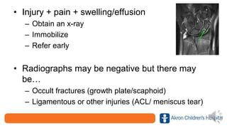
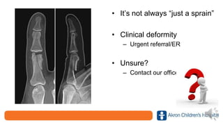
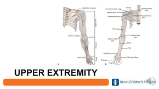
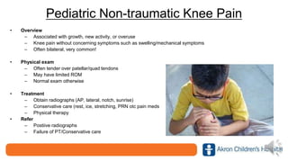
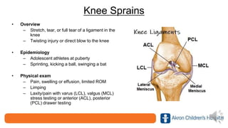
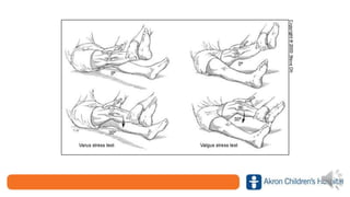
Recognition
and
Management
of Common
Pediatric
Sports
Injuries
John Polousky, MD
Objectives
• Recognize common
pediatric sports
injuries
• Discuss ambulatory
management when
appropriate
• Identify when to refer
to orthopedics
About Our Practice
• 24 Providers
– Various subspecialties including hand, sports, and spine
surgery
• Locations
– Akron, North Canton, Boston Heights, Medina, Mahoning
Valley, Mansfield, Norwalk
• Same-day appts
– Available Monday-Friday
• 330-543-3500
A FEW SPORTS INJURY
MANAGEMENT PEARLS
• Injury + pain + swelling/effusion
– Obtain an x-ray
– Immobilize
– Refer early
• Radiographs may be negative but there may
be…
– Occult fractures (growth plate/scaphoid)
– Ligamentous or other injuries (ACL/ meniscus tear)
• It’s not always “just a sprain”
• Clinical deformity
– Urgent referral/ER
• Unsure?
– Contact our office
UPPER EXTREMITY
Shoulder Dislocation
• Mechanism of Injury
– Direct blow or fall
• Epidemiology
– More common in adolescent
males over age 10
– Anterior dislocation most
common
• Physical exam
– Swelling, tenderness to
palpation, clinical deformity
• Treatment
– Early recognition, referral to
ED, reduction
• Pre and post-reduction
radiographs to confirm
reduction and
identify Hill-Sachs/Bony
Bankhart lesions
– Immobilization (sling and
swathe)
– PT after initial immobilization
– Activity restrictions (8-12
weeks depending)
• When to refer
– Within to 2 weeks post injury
– Ability to promptly obtain MR
arthrogram (labral pathology)
– Discussion of possible surgical
intervention
• 50-90% re-dislocation rate
in skeletally mature patients
AC Joint Injuries
• Mechanism of Injury
– Direct blow or fall
• Epidemiology
– More common in athletes and
males
• Physical exam
– Swelling, tenderness to palpation,
asymmetry
• Treatment
– Dependent upon displacement
• Simple sprain vs. dislocation
– Surgical intervention may be warranted
– Immobilization (sling and swathe)
– Activity restrictions (8-12 weeks
depending)
• When to refer
– Asymmetry
– Promptly (prior to 2 weeks post injury)
SC Joint Injuries • Mechanism of injury
– High energy trauma (contact
sports/MVA)
– Subluxation (atraumatic – arm
elevation)
• Physical exam
– Anterior Dislocation/Fracture
• Swelling, prominence, TTP
– Posterior Dislocation/Fracture
• Asymmetry, TTP
• Upper extremity paresthesia
• Diminished pulse compared to
contralateral side
• Dysphagia, dyspnea, tachypnea,
stridor
• Treatment
– Anterior
• Immobilization (sling and swathe)
• Activity restrictions (8-12 weeks depending)
• Some require surgical treatment (acute
dislocations)
– Posterior
• Conservative care versus urgent surgical reduction
• When to refer
– Emergent referral to ED
• Concern for posterior displacement and
vascular/pulmonary compromise
– Promptly (prior to 2 weeks post injury)
Clavicle fractures
• Mechanism of injury
– Fall or direct trauma
• Epidemiology
– More common in active males
– Most common are shaft fractures
• Physical exam
– Medial, distal, shaft
– Swelling, ecchymosis, asymmetry,
tenderness to palpation
– Skin tenting (impending open
fracture)
• Treatment
– Conservative care in the majority of cases
• Sling and swathe
• Activity restrictions (up to 12 weeks due to high risk of refracture)
– Operative
• Controversial
– Open, impending open fractures
– >2cm shortening
• When to refer
– Emergent referral to ED
• Concern for impending open fracture
• Neurovascular compromise
– Promptly (prior to 2 weeks post injury)
Little League Shoulder
• Mechanism of injury
– Overuse injury in skeletally immature child
• Epidemiology
– Proximal humeral epiphysiolysis
– Overhead athletes (pitching/tennis)
– Most common in males 9-15 years of age
• Physical exam/history
– Decreased pitch velocity/pain with throwing
– Mild swelling possible
– Tenderness over proximal humerus physis
– Pain/limited ROM (especially internal rotation)
• Treatment
– Obtain radiographs
• Contralateral if uncertain
– Sling for comfort, PRN
NSAIDs
– Rest with no pitching (up to
3 months)
– Physical therapy with
progressive throwing
program
• When to Refer
– Persistent
symptoms/positive
radiographs
– Discussion of long-term
complications of physeal
growth arrest and possible
deformity
Little League Elbow
• Mechanism of Injury
– Overuse/injury in skeletally immature child
• Epidemiology
– Medial elbow injury/stress
• Medial apophysitis, stress fracture, UCL injury, flexor pronator mass
strain
– Repetitive valgus elbow stress/microtrauma (pitching)
• Physical exam
– Mild swelling possible
– Tenderness over medial elbow
– Limited ROM
– Laxity with valgus stress testing
• Treatment
– Obtain radiographs
– Dependent upon diagnosis
• Sling for comfort, PRN NSAIDs
• Rest with no pitching (up to 3
months)
• Physical therapy with
progressive throwing program
• Surgical intervention
• When to Refer
– Persistent symptoms
– Radiographic abnormality
• Medial epicondyle widening,
avulsion fracture, capitellar
abnormality
– Concern for UCL injury
Wrist Sprain/Fractures
• Mechanism of Injury
– Fall, direct blow
• Physical exam
– Swelling, tenderness to palpation, limited ROM,
clinical deformity
• Treatment
– Gross deformity
• Refer to ED
– Splint
– Refer +/- radiographs
• When to Refer
– Positive radiographs/concern for occult fracture
– Within 1 week of injury
Finger Injuries/Sprains
• Mechanism of Injury
– Fall, direct blow,
“stoved finger”
• Physical exam
– Swelling,
tenderness to
palpation, limited
ROM, clinical
deformity
– Nail plate
deformity, bleeding
from the base of
the nail bed
Non-bony mallet
• Treatment
– Gross deformity +/- open
injury
• Refer to ED
– Splint
– Refer +/- radiographs
• When to Refer
– Positive
radiographs/concern for
occult fracture or non-
bony ligamentous injury
– Within 1 week of injury if
closed
– Urgently if concern for
open injury
Bony mallet
Seymour Fracture
• Mechanism of Injury
– Crush or direct trauma
• Physical exam
– Nail plate lying above
eponychium with bloody
drainage/scabbing
– Swelling, tenderness to
palpation, limited ROM
– Clinical deformity
• Flexion at the DIP joint with
inability to fully extend
finger
This is an OPEN fracture!
• Treatment
– Acute (within 24-48 hours)
• Refer to ED for I&D,
nailbed repair
• Possible surgical pinning
dependent upon deformity
• Oral antibiotics
– Chronic
• Refer to Orthopedics
• Complications
– Nail/finger deformity
– Chronic osteomyelitis
SPINE
Spondylolysis/Spondylolisthesis
• Overview
– Pars interarticularis fracture and
potential progression to slip
(spondylolisthesis)
• Epidemiology
– Adolescent athletes (gymnasts,
cheerleaders, football linemen, weight-
lifters)
• Physical exam/history
– Low back pain +/-radiculopathy
– Pain with back extension
– TTP lower lumbar spine/paraspinal
musculature
• Treatment
– Obtain radiographs
• PA/lateral only (no need for
obliques)
• May be negative with spondylolysis
only
– Rest, PT, as needed otc NSAIDS,
stretching (hamstrings)
– Injections
– Operative (fusion)
• When to Refer
– Persistent symptoms despite rest/PT
– Spondylolisthesis on radiographs
HIP
Avulsion Fractures
• Overview
– Indirect trauma with a sudden
forceful muscle contraction “snap or
pop”
• Epidemiology
– Adolescent athletes at puberty
– Sprinting, kicking a ball, swinging a
bat
• Physical exam
– Swelling, tenderness over affected
tendon attachment
– Weakness of affected muscle
– Difficulty with weight bearing/limping
• Treatment
– Obtain radiographs
– Protective weight bearing with
crutches
– Early ROM
– Physical therapy
– Surgical intervention for
significantly displaced
avulsion fractures-extremely
uncommon.
• When to Refer
– Positive radiographs
LOWER EXTREMITY
Pediatric Non-traumatic Knee Pain
• Overview
– Associated with growth, new activity, or overuse
– Knee pain without concerning symptoms such as swelling/mechanical symptoms
– Often bilateral, very common!
• Physical exam
– Often tender over patellar/quad tendons
– May have limited ROM
– Normal exam otherwise
• Treatment
– Obtain radiographs (AP, lateral, notch, sunrise)
– Conservative care (rest, ice, stretching, PRN otc pain meds
– Physical therapy
• Refer
– Postiive radiographs
– Failure of PT/Conservative care
Knee Sprains
• Overview
– Stretch, tear, or full tear of a ligament in the
knee
– Twisting injury or direct blow to the knee
• Epidemiology
– Adolescent athletes at puberty
– Sprinting, kicking a ball, swinging a bat
• Physical exam
– Pain, swelling or effusion, limited ROM
– Limping
– Laxity/pain with varus (LCL), valgus (MCL)
stress testing or anterior (ACL), posterior
(PCL) drawer testing
• Treatment
– Obtain radiographs
– Rest, ice, immobilization,
compression, as needed otc
pain medications
– Activity restrictions
– Physical therapy
• When to Refer
– Persistent symptoms despite
rest/PT
– Significant/persistent effusion
– Laxity on exam
ACL Tear
• Mechanism of Injury
– Non-contact hyperextension injury +/- rotation or
valgus force
– Pivoting with partially flexed knee
– “Popping” sensation may be noted
• Epidemiology
– ACL provides anterior and rotational stability
– Becoming more common (year-round/younger sport
participation)
• Physical Exam
– Effusion, tenderness to palpation, limited ROM
– Antalgia
– Positive Lachman/Anterior drawer/Pivot shift
• Treatment
– Radiographs
• Typically negative but may show
Segund fracture
– Immobilization, protected weight bearing
– Prompt referral!
• We will see as a same-day
appointment or within a couple days
• We will obtain prompt MRI
– Surgical management plus PT and
activity restrictions (9 months)
• Non-operative treatment leads to
functional instability, secondary
meniscal/cartilage damage, and
likely early osteoarthritis
Tibial Spine Fracture
• Overview
– Avulsion of the ACL off the tibial spine
– Non-contact hyperextension injury +/-
rotation or valgus force
– Pivoting with partially flexed knee
– “Popping” sensation may be noted
• Epidemiology
– Childhood equivalent of an ACL tear
– Most common between 8-14 years of
age
• Physical Exam
– Effusion, tenderness to palpation,
limited ROM (lack extension)
– Antalgia/inability to bear weight
• Treatment
– Radiographs
– Immobilization, protected
weight bearing
– Prompt referral!
• Within 1 week
– Management dependent upon
fracture displacement
• Casting versus surgical
management
• May have associated ACL
laxity
Tibial Tubercle Fracture
• Mechanism of Injury
– Non-contact injury when quadriceps
muscle is contracted during
jumping/knee extension
• Epidemiology
– Adolescent males near skeletal
maturity
• Physical Exam
– Effusion, tenderness to palpation over
tibial tubercle
– Inability or extensor lag with straight
leg raise
– Inability to bear weight
• Treatment
– Radiographs
• May be subtle
• Patella alta
– Immobilization, crutches
– Prompt referral!
• Within 1 week
– Management dependent
upon fracture displacement
• Casting versus surgical
management
• Activity restrictions
• Physical therapy
Meniscus Tears
• Mechanism of injury
– Most occur from twisting, non-contact injury
• Epidemiology
– Lateral tears most common
– May have associated ligamentous injury
• History
– Pain, swelling, reported mechanical
symptoms (locking, catching, “gives out”)
• Physical Exam
– Effusion
– Limited ROM
– Tenderness along the joint line
– Positive McMurray or Thessaly testing
McMurray Testing
• Treatment
– Obtain radiographs and refer early
• Typically normal
– Activity restriction
– Ice, rest, as needed otc pain
medication
– Treatment dependent upon type of tear
• Physical therapy/bracing
• Surgical intervention frequently
recommended
– Mensicectomy versus
meniscal repair
– Typically result in good
outcomes in pediatric
patients
• When to Refer
– Promptly (within 1-2 weeks)
– Ortho can initiate MRI
Ankle Sprains
• Mechanism of injury
– Most occur from a rotational, non-
contact injury
• Epidemiology
– Lateral sprains most common
– High ankle sprain versus low
– Grade 1-3 (stretch -> complete tear)
• Physical Exam
– Pain, inability to weight bear, antalgic
gait
– Ecchymosis, swelling
– Limited ROM
– Tenderness around affected ligament
– Positive anterior drawer testing
• Treatment
– Consider radiographs
– Immobilize, mobility
assistance if need be
(crutches)
– Rest, ice, compression, as
needed otc pain meds
– Bracing
– Physical therapy
• When to Refer
– Positive radiographs
– Persistent
symptoms/instability despite
conservative care/PT
– Recurrent sprains
Osteochondritis Dissecans (OCD) Lesions
• Overview
– Articular cartilage/subchondral bone lesion
• Epidemiology
– Most common in the knee but may also affect the elbow
and ankle
– More common in adolescence
• History
– Pain, swelling, reported mechanical symptoms
(locking/catching), limited ROM of affected joint
– Activity related pain
• Physical Exam
– Effusion
– Limited ROM
– Tenderness over affected bone
• Treatment
– Obtain radiographs
– Refer early
– Dependent upon stability of OCD
lesion based upon radiographs/MRI
• Stable
– Activity restriction, as
needed otc pain medication,
immobilization, restricted
weight bearing
• Unstable
– Surgical debridement,
microfracture, +/- fixation
• When to Refer
– Promptly (within 1-2 weeks)
– Ortho can initiate MRI
A FEW ORTHO
REFERRAL PEARLS
Scoliosis • If obtaining x-rays  PA of
the entire spine (lateral if first
imaging)
• Refer early and for
scoliometer readings > or = 5
degrees
• Low dose radiation x-rays
available in Akron (EOS)
Developmental Dysplasia of the Hip
• Do not obtain US prior to 6
weeks of age (corrected for
prematurity)
– Physiologic immaturity of the
hip
• Frank + Barlow/Ortolani
– Refer promptly/do not wait for
an US as we will promptly
initiate treatment (Pavlik
harness)
References
Deitch J, et al. Traumatic Anterior Shoulder Dislocation in Adolescents. Am J Sports Med. Sept-Oct, 2003;31(5):758-
763
Ghanem IB, Rizkallah M. Pediatric avulsion fractures of pelvis: current concepts. Curr Opin Pediatr. 2018 Feb;30(1):78-
83. doi: 10.1097/MOP.0000000000000575. PMID: 29176354.
Heyworth BE, Kramer DE, Martin DJ, Micheli LJ, Kocher MS, Bae DS. Trends in the Presentation, Management, and
Outcomes of Little League Shoulder. Am J Sports Med. 2016 Jun;44(6):1431-8. doi: 10.1177/0363546516632744.
Epub 2016 Mar 16. PMID: 26983458.
Klingele, K.E., Kocher, M.S. Little League Elbow. Sports Med 32, 1005–1015 (2002).
https://doi.org/10.2165/00007256200232150-00004
Shieh AK, Edmonds EW, Pennock AT. Revision Meniscal Surgery in Children and Adolescents: Risk Factors and
Mechanisms for Failure and Subsequent Management. Am J Sports Med. 2016 Apr;44(4):838-43.
Vanderhave KL, Moravek JE, Sekiya JK, Wojtys EM. Meniscus tears in the young athlete: results of arthroscopic repair.
J Pediatr Orthop. 2011 Jul-Aug;31(5):496-500.
Krusche-Mandl I, Kottstorfer J, Thalhammer G, Aldrian S, Erhart J, Platzer P. Seymour fractures: retrospective analysis
and therapeutic considerations. J Hand Surg Am. 2013;38(2):258-64.
Klingele, K.E., Kocher, M.S. Little League Elbow. Sports Med 32, 1005–1015 (2002).
https://doi.org/10.2165/00007256200232150-00004
LaFrance, Russell M.; Giordano, Brian; Goldblatt, John; Voloshin, Ilya; Maloney, Michael Less: Pediatric Tibial
Eminence Fractures: Evaluation and Management. Journal of the American Academy of Orthopaedic Surgeons.
18(7):395-405, July 2010.
Li X, et al. Management of Shoulder Instability in the Skeletally Immature Patient. J Am Acad Orthop Surg.
2013;21:529-537
Shieh AK, Edmonds EW, Pennock AT. Revision Meniscal Surgery in Children and Adolescents: Risk Factors and
Mechanisms for Failure and Subsequent Management. Am J Sports Med. 2016 Apr;44(4):838-43.
Vanderhave KL, Moravek JE, Sekiya JK, Wojtys EM. Meniscus tears in the young athlete: results of arthroscopic repair.
J Pediatr Orthop. 2011 Jul-Aug;31(5):496-500.

Polousky ortho

  • 1.
  • 2.
    Objectives • Recognize common pediatricsports injuries • Discuss ambulatory management when appropriate • Identify when to refer to orthopedics
  • 3.
    About Our Practice •24 Providers – Various subspecialties including hand, sports, and spine surgery • Locations – Akron, North Canton, Boston Heights, Medina, Mahoning Valley, Mansfield, Norwalk • Same-day appts – Available Monday-Friday • 330-543-3500
  • 4.
    A FEW SPORTSINJURY MANAGEMENT PEARLS
  • 5.
    • Injury +pain + swelling/effusion – Obtain an x-ray – Immobilize – Refer early • Radiographs may be negative but there may be… – Occult fractures (growth plate/scaphoid) – Ligamentous or other injuries (ACL/ meniscus tear)
  • 6.
    • It’s notalways “just a sprain” • Clinical deformity – Urgent referral/ER • Unsure? – Contact our office
  • 7.
  • 8.
    Shoulder Dislocation • Mechanismof Injury – Direct blow or fall • Epidemiology – More common in adolescent males over age 10 – Anterior dislocation most common • Physical exam – Swelling, tenderness to palpation, clinical deformity
  • 9.
    • Treatment – Earlyrecognition, referral to ED, reduction • Pre and post-reduction radiographs to confirm reduction and identify Hill-Sachs/Bony Bankhart lesions – Immobilization (sling and swathe) – PT after initial immobilization – Activity restrictions (8-12 weeks depending)
  • 10.
    • When torefer – Within to 2 weeks post injury – Ability to promptly obtain MR arthrogram (labral pathology) – Discussion of possible surgical intervention • 50-90% re-dislocation rate in skeletally mature patients
  • 11.
    AC Joint Injuries •Mechanism of Injury – Direct blow or fall • Epidemiology – More common in athletes and males • Physical exam – Swelling, tenderness to palpation, asymmetry
  • 12.
    • Treatment – Dependentupon displacement • Simple sprain vs. dislocation – Surgical intervention may be warranted – Immobilization (sling and swathe) – Activity restrictions (8-12 weeks depending) • When to refer – Asymmetry – Promptly (prior to 2 weeks post injury)
  • 13.
    SC Joint Injuries• Mechanism of injury – High energy trauma (contact sports/MVA) – Subluxation (atraumatic – arm elevation) • Physical exam – Anterior Dislocation/Fracture • Swelling, prominence, TTP – Posterior Dislocation/Fracture • Asymmetry, TTP • Upper extremity paresthesia • Diminished pulse compared to contralateral side • Dysphagia, dyspnea, tachypnea, stridor
  • 14.
    • Treatment – Anterior •Immobilization (sling and swathe) • Activity restrictions (8-12 weeks depending) • Some require surgical treatment (acute dislocations) – Posterior • Conservative care versus urgent surgical reduction • When to refer – Emergent referral to ED • Concern for posterior displacement and vascular/pulmonary compromise – Promptly (prior to 2 weeks post injury)
  • 15.
    Clavicle fractures • Mechanismof injury – Fall or direct trauma • Epidemiology – More common in active males – Most common are shaft fractures • Physical exam – Medial, distal, shaft – Swelling, ecchymosis, asymmetry, tenderness to palpation – Skin tenting (impending open fracture)
  • 16.
    • Treatment – Conservativecare in the majority of cases • Sling and swathe • Activity restrictions (up to 12 weeks due to high risk of refracture) – Operative • Controversial – Open, impending open fractures – >2cm shortening • When to refer – Emergent referral to ED • Concern for impending open fracture • Neurovascular compromise – Promptly (prior to 2 weeks post injury)
  • 17.
    Little League Shoulder •Mechanism of injury – Overuse injury in skeletally immature child • Epidemiology – Proximal humeral epiphysiolysis – Overhead athletes (pitching/tennis) – Most common in males 9-15 years of age • Physical exam/history – Decreased pitch velocity/pain with throwing – Mild swelling possible – Tenderness over proximal humerus physis – Pain/limited ROM (especially internal rotation)
  • 18.
    • Treatment – Obtainradiographs • Contralateral if uncertain – Sling for comfort, PRN NSAIDs – Rest with no pitching (up to 3 months) – Physical therapy with progressive throwing program • When to Refer – Persistent symptoms/positive radiographs – Discussion of long-term complications of physeal growth arrest and possible deformity
  • 19.
    Little League Elbow •Mechanism of Injury – Overuse/injury in skeletally immature child • Epidemiology – Medial elbow injury/stress • Medial apophysitis, stress fracture, UCL injury, flexor pronator mass strain – Repetitive valgus elbow stress/microtrauma (pitching) • Physical exam – Mild swelling possible – Tenderness over medial elbow – Limited ROM – Laxity with valgus stress testing
  • 20.
    • Treatment – Obtainradiographs – Dependent upon diagnosis • Sling for comfort, PRN NSAIDs • Rest with no pitching (up to 3 months) • Physical therapy with progressive throwing program • Surgical intervention • When to Refer – Persistent symptoms – Radiographic abnormality • Medial epicondyle widening, avulsion fracture, capitellar abnormality – Concern for UCL injury
  • 21.
    Wrist Sprain/Fractures • Mechanismof Injury – Fall, direct blow • Physical exam – Swelling, tenderness to palpation, limited ROM, clinical deformity • Treatment – Gross deformity • Refer to ED – Splint – Refer +/- radiographs • When to Refer – Positive radiographs/concern for occult fracture – Within 1 week of injury
  • 22.
    Finger Injuries/Sprains • Mechanismof Injury – Fall, direct blow, “stoved finger” • Physical exam – Swelling, tenderness to palpation, limited ROM, clinical deformity – Nail plate deformity, bleeding from the base of the nail bed Non-bony mallet
  • 23.
    • Treatment – Grossdeformity +/- open injury • Refer to ED – Splint – Refer +/- radiographs • When to Refer – Positive radiographs/concern for occult fracture or non- bony ligamentous injury – Within 1 week of injury if closed – Urgently if concern for open injury Bony mallet
  • 24.
    Seymour Fracture • Mechanismof Injury – Crush or direct trauma • Physical exam – Nail plate lying above eponychium with bloody drainage/scabbing – Swelling, tenderness to palpation, limited ROM – Clinical deformity • Flexion at the DIP joint with inability to fully extend finger This is an OPEN fracture!
  • 25.
    • Treatment – Acute(within 24-48 hours) • Refer to ED for I&D, nailbed repair • Possible surgical pinning dependent upon deformity • Oral antibiotics – Chronic • Refer to Orthopedics • Complications – Nail/finger deformity – Chronic osteomyelitis
  • 26.
  • 27.
    Spondylolysis/Spondylolisthesis • Overview – Parsinterarticularis fracture and potential progression to slip (spondylolisthesis) • Epidemiology – Adolescent athletes (gymnasts, cheerleaders, football linemen, weight- lifters) • Physical exam/history – Low back pain +/-radiculopathy – Pain with back extension – TTP lower lumbar spine/paraspinal musculature
  • 28.
    • Treatment – Obtainradiographs • PA/lateral only (no need for obliques) • May be negative with spondylolysis only – Rest, PT, as needed otc NSAIDS, stretching (hamstrings) – Injections – Operative (fusion) • When to Refer – Persistent symptoms despite rest/PT – Spondylolisthesis on radiographs
  • 29.
  • 30.
    Avulsion Fractures • Overview –Indirect trauma with a sudden forceful muscle contraction “snap or pop” • Epidemiology – Adolescent athletes at puberty – Sprinting, kicking a ball, swinging a bat • Physical exam – Swelling, tenderness over affected tendon attachment – Weakness of affected muscle – Difficulty with weight bearing/limping
  • 31.
    • Treatment – Obtainradiographs – Protective weight bearing with crutches – Early ROM – Physical therapy – Surgical intervention for significantly displaced avulsion fractures-extremely uncommon. • When to Refer – Positive radiographs
  • 32.
  • 33.
    Pediatric Non-traumatic KneePain • Overview – Associated with growth, new activity, or overuse – Knee pain without concerning symptoms such as swelling/mechanical symptoms – Often bilateral, very common! • Physical exam – Often tender over patellar/quad tendons – May have limited ROM – Normal exam otherwise • Treatment – Obtain radiographs (AP, lateral, notch, sunrise) – Conservative care (rest, ice, stretching, PRN otc pain meds – Physical therapy • Refer – Postiive radiographs – Failure of PT/Conservative care
  • 34.
    Knee Sprains • Overview –Stretch, tear, or full tear of a ligament in the knee – Twisting injury or direct blow to the knee • Epidemiology – Adolescent athletes at puberty – Sprinting, kicking a ball, swinging a bat • Physical exam – Pain, swelling or effusion, limited ROM – Limping – Laxity/pain with varus (LCL), valgus (MCL) stress testing or anterior (ACL), posterior (PCL) drawer testing
  • 37.
    • Treatment – Obtainradiographs – Rest, ice, immobilization, compression, as needed otc pain medications – Activity restrictions – Physical therapy • When to Refer – Persistent symptoms despite rest/PT – Significant/persistent effusion – Laxity on exam
  • 38.
    ACL Tear • Mechanismof Injury – Non-contact hyperextension injury +/- rotation or valgus force – Pivoting with partially flexed knee – “Popping” sensation may be noted • Epidemiology – ACL provides anterior and rotational stability – Becoming more common (year-round/younger sport participation) • Physical Exam – Effusion, tenderness to palpation, limited ROM – Antalgia – Positive Lachman/Anterior drawer/Pivot shift
  • 40.
    • Treatment – Radiographs •Typically negative but may show Segund fracture – Immobilization, protected weight bearing – Prompt referral! • We will see as a same-day appointment or within a couple days • We will obtain prompt MRI – Surgical management plus PT and activity restrictions (9 months) • Non-operative treatment leads to functional instability, secondary meniscal/cartilage damage, and likely early osteoarthritis
  • 41.
    Tibial Spine Fracture •Overview – Avulsion of the ACL off the tibial spine – Non-contact hyperextension injury +/- rotation or valgus force – Pivoting with partially flexed knee – “Popping” sensation may be noted • Epidemiology – Childhood equivalent of an ACL tear – Most common between 8-14 years of age • Physical Exam – Effusion, tenderness to palpation, limited ROM (lack extension) – Antalgia/inability to bear weight
  • 42.
    • Treatment – Radiographs –Immobilization, protected weight bearing – Prompt referral! • Within 1 week – Management dependent upon fracture displacement • Casting versus surgical management • May have associated ACL laxity
  • 43.
    Tibial Tubercle Fracture •Mechanism of Injury – Non-contact injury when quadriceps muscle is contracted during jumping/knee extension • Epidemiology – Adolescent males near skeletal maturity • Physical Exam – Effusion, tenderness to palpation over tibial tubercle – Inability or extensor lag with straight leg raise – Inability to bear weight
  • 45.
    • Treatment – Radiographs •May be subtle • Patella alta – Immobilization, crutches – Prompt referral! • Within 1 week – Management dependent upon fracture displacement • Casting versus surgical management • Activity restrictions • Physical therapy
  • 46.
    Meniscus Tears • Mechanismof injury – Most occur from twisting, non-contact injury • Epidemiology – Lateral tears most common – May have associated ligamentous injury • History – Pain, swelling, reported mechanical symptoms (locking, catching, “gives out”) • Physical Exam – Effusion – Limited ROM – Tenderness along the joint line – Positive McMurray or Thessaly testing McMurray Testing
  • 47.
    • Treatment – Obtainradiographs and refer early • Typically normal – Activity restriction – Ice, rest, as needed otc pain medication – Treatment dependent upon type of tear • Physical therapy/bracing • Surgical intervention frequently recommended – Mensicectomy versus meniscal repair – Typically result in good outcomes in pediatric patients • When to Refer – Promptly (within 1-2 weeks) – Ortho can initiate MRI
  • 48.
    Ankle Sprains • Mechanismof injury – Most occur from a rotational, non- contact injury • Epidemiology – Lateral sprains most common – High ankle sprain versus low – Grade 1-3 (stretch -> complete tear) • Physical Exam – Pain, inability to weight bear, antalgic gait – Ecchymosis, swelling – Limited ROM – Tenderness around affected ligament – Positive anterior drawer testing
  • 49.
    • Treatment – Considerradiographs – Immobilize, mobility assistance if need be (crutches) – Rest, ice, compression, as needed otc pain meds – Bracing – Physical therapy • When to Refer – Positive radiographs – Persistent symptoms/instability despite conservative care/PT – Recurrent sprains
  • 50.
    Osteochondritis Dissecans (OCD)Lesions • Overview – Articular cartilage/subchondral bone lesion • Epidemiology – Most common in the knee but may also affect the elbow and ankle – More common in adolescence • History – Pain, swelling, reported mechanical symptoms (locking/catching), limited ROM of affected joint – Activity related pain • Physical Exam – Effusion – Limited ROM – Tenderness over affected bone
  • 52.
    • Treatment – Obtainradiographs – Refer early – Dependent upon stability of OCD lesion based upon radiographs/MRI • Stable – Activity restriction, as needed otc pain medication, immobilization, restricted weight bearing • Unstable – Surgical debridement, microfracture, +/- fixation • When to Refer – Promptly (within 1-2 weeks) – Ortho can initiate MRI
  • 53.
  • 54.
    Scoliosis • Ifobtaining x-rays  PA of the entire spine (lateral if first imaging) • Refer early and for scoliometer readings > or = 5 degrees • Low dose radiation x-rays available in Akron (EOS)
  • 55.
    Developmental Dysplasia ofthe Hip • Do not obtain US prior to 6 weeks of age (corrected for prematurity) – Physiologic immaturity of the hip • Frank + Barlow/Ortolani – Refer promptly/do not wait for an US as we will promptly initiate treatment (Pavlik harness)
  • 56.
    References Deitch J, etal. Traumatic Anterior Shoulder Dislocation in Adolescents. Am J Sports Med. Sept-Oct, 2003;31(5):758- 763 Ghanem IB, Rizkallah M. Pediatric avulsion fractures of pelvis: current concepts. Curr Opin Pediatr. 2018 Feb;30(1):78- 83. doi: 10.1097/MOP.0000000000000575. PMID: 29176354. Heyworth BE, Kramer DE, Martin DJ, Micheli LJ, Kocher MS, Bae DS. Trends in the Presentation, Management, and Outcomes of Little League Shoulder. Am J Sports Med. 2016 Jun;44(6):1431-8. doi: 10.1177/0363546516632744. Epub 2016 Mar 16. PMID: 26983458. Klingele, K.E., Kocher, M.S. Little League Elbow. Sports Med 32, 1005–1015 (2002). https://doi.org/10.2165/00007256200232150-00004 Shieh AK, Edmonds EW, Pennock AT. Revision Meniscal Surgery in Children and Adolescents: Risk Factors and Mechanisms for Failure and Subsequent Management. Am J Sports Med. 2016 Apr;44(4):838-43. Vanderhave KL, Moravek JE, Sekiya JK, Wojtys EM. Meniscus tears in the young athlete: results of arthroscopic repair. J Pediatr Orthop. 2011 Jul-Aug;31(5):496-500.
  • 57.
    Krusche-Mandl I, KottstorferJ, Thalhammer G, Aldrian S, Erhart J, Platzer P. Seymour fractures: retrospective analysis and therapeutic considerations. J Hand Surg Am. 2013;38(2):258-64. Klingele, K.E., Kocher, M.S. Little League Elbow. Sports Med 32, 1005–1015 (2002). https://doi.org/10.2165/00007256200232150-00004 LaFrance, Russell M.; Giordano, Brian; Goldblatt, John; Voloshin, Ilya; Maloney, Michael Less: Pediatric Tibial Eminence Fractures: Evaluation and Management. Journal of the American Academy of Orthopaedic Surgeons. 18(7):395-405, July 2010. Li X, et al. Management of Shoulder Instability in the Skeletally Immature Patient. J Am Acad Orthop Surg. 2013;21:529-537 Shieh AK, Edmonds EW, Pennock AT. Revision Meniscal Surgery in Children and Adolescents: Risk Factors and Mechanisms for Failure and Subsequent Management. Am J Sports Med. 2016 Apr;44(4):838-43. Vanderhave KL, Moravek JE, Sekiya JK, Wojtys EM. Meniscus tears in the young athlete: results of arthroscopic repair. J Pediatr Orthop. 2011 Jul-Aug;31(5):496-500.