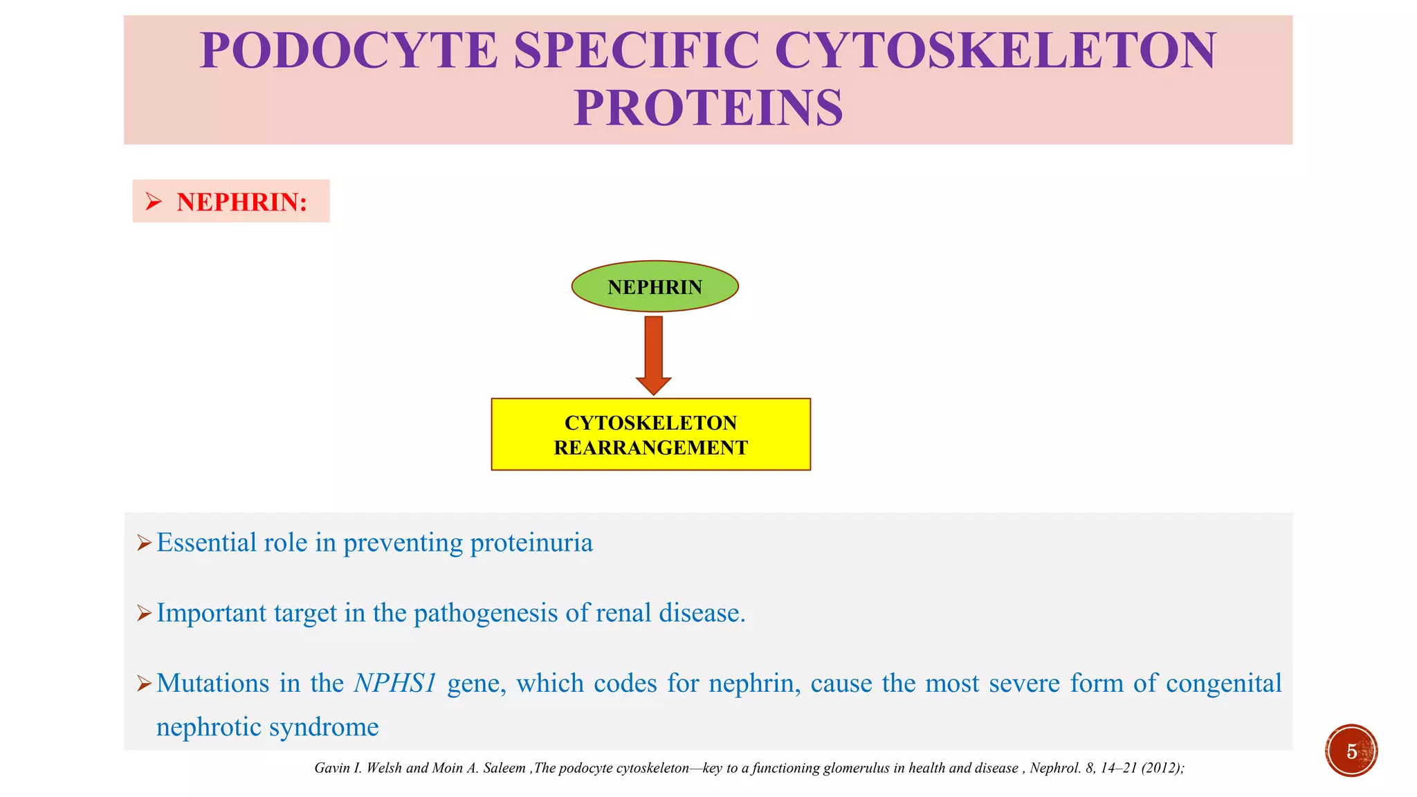
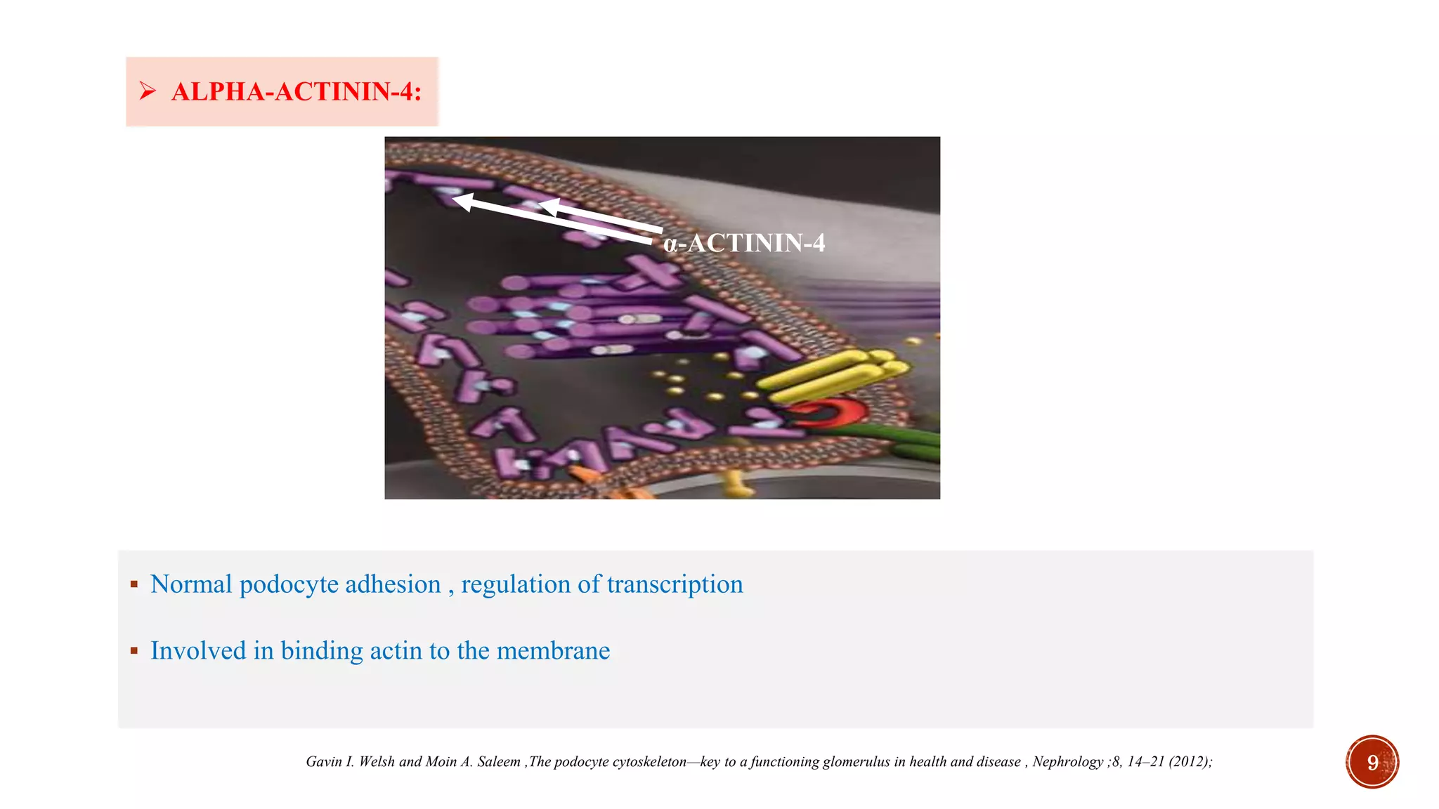
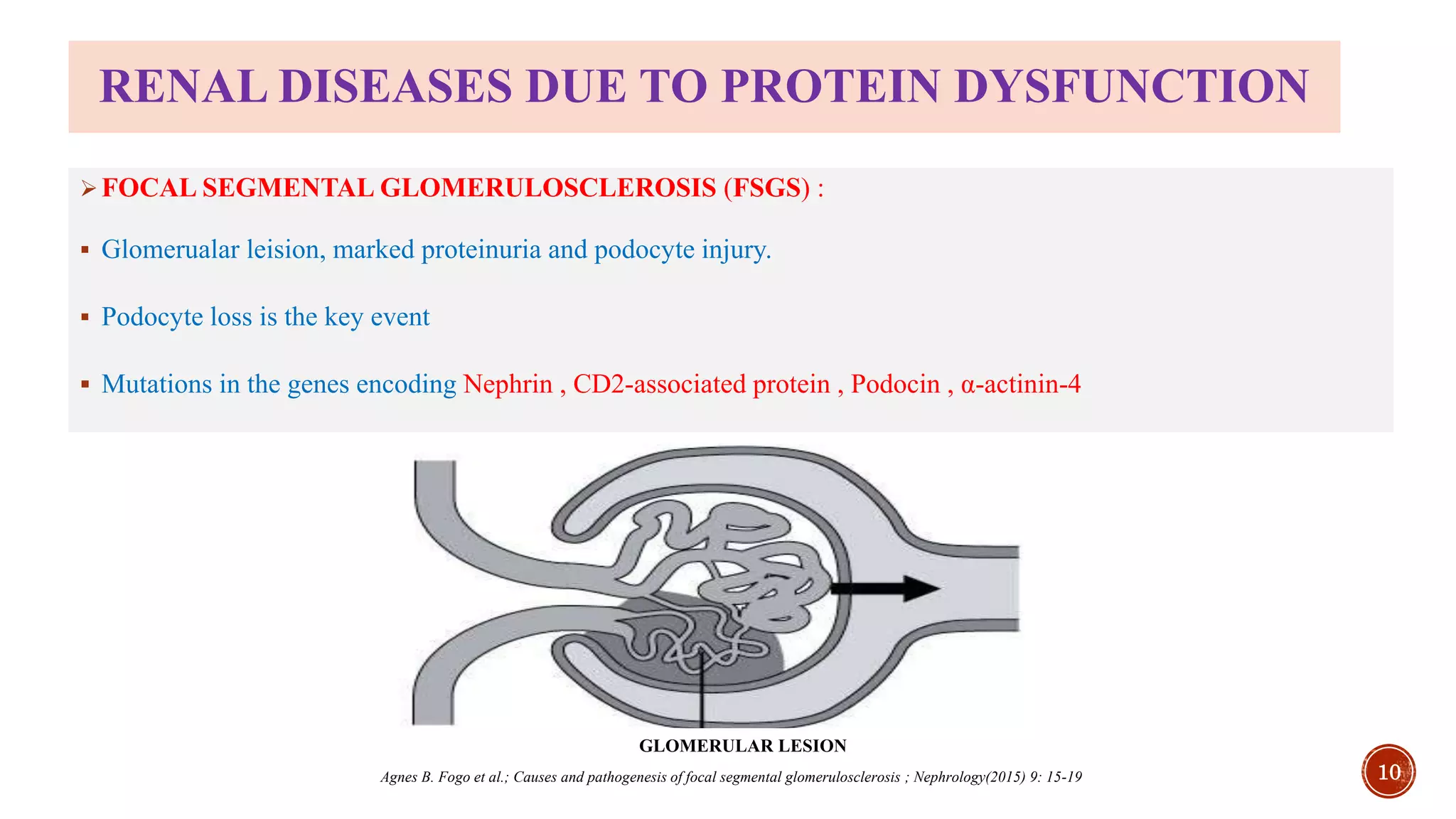
ROLE OF PODOCYTE CYTOSKELETON IN
PHYSIOLOGY AND RENAL DISEASES
Presented by:
UDDANDI SATYA KARTHIK
RT/2019/617
DEPT. OF REGULATORY TOXICOLOGY
Under guidance of:
Dr. ASHUTOSH KUMAR
HEAD OF DEPARTMENT
DEPT. OF PHARMACOLOGY & TOXICOLOGY
NIPER HYDERABAD
National Institute of Pharmaceutical Educational Education and Research
 WHAT ARE PODOCYTES?
 Cells that wrap around capillaries of the glomerulus
 Make up the epithelial lining of Bowman's capsule
 visceral epithelial cells
 FUNCTIONS:
 They are:
 GBM-turnover
 Support of the capillary tuft
 Regulation of glomerular filtration
 Immunological functions
INTRODUCTION
TEM view of podocytes in between the capillary profiles ( C )
1. Reiser J and Altintas MM. Podocytes , F1000 Research 2016, (F1000 Faculty Rev):114
2. Peter Mundel , Wilhelm Kriz,Structure and function of podocytes: an update, Anat Embryol (1995) 192:385-397
INTERDIGITATING
FOOT PROCESSES
PODOCYTE
CELLBODY
2
ULTRA STRUCTURE OF A PODOCYTE
FOOT PROCESS
GBM
ENDOTHELIUM
SLIT DIAPHRAGM
PODOCYTE CELLBODY
TRPC6 CD2AP PODOCALYXIN
ACTIN
α-ACTININ-4
SYNAPTOPODIN
NEPHRIN
SLIT DIAPHRAGM
Kirk N. Campbell et al;Protecting Podocytes: A Key Target for Therapy of Focal Segmental Glomerulosclerosis; American Journal of Nephrology, 2018;47(suppl 1):14–29
3
PODOCIN
IMPORTANCE OF PODOCYTE CYTOSKELETON
 Maintains filtration
 Podocyte shape and stability
 Foot processes increase filtration surface
 Attachment of podocyte to GBM
 Provide structural stability to the glomerular tuft
Dong Sun et al., Relationship between Urinary Podocytes and Kidney Diseases; Renal Failure, 34(3): 403–407, (2012)
4
PODOCYTE SPECIFIC CYTOSKELETON
PROTEINS
Essential role in preventing proteinuria
Important target in the pathogenesis of renal disease.
Mutations in the NPHS1 gene, which codes for nephrin, cause the most severe form of congenital
nephrotic syndrome
NEPHRIN
CYTOSKELETON
REARRANGEMENT
 NEPHRIN:
Gavin I. Welsh and Moin A. Saleem ,The podocyte cytoskeleton—key to a functioning glomerulus in health and disease , Nephrol. 8, 14–21 (2012);
5
Podocin localizes to lipid microdomains in the slit diaphragm where it co-localizes with nephrin.
Clusters and regulates the ion channel TRPC6
 Serve as a scaffold
 PODOCIN:
PODOCIN
Gavin I. Welsh and Moin A. Saleem ,The podocyte cytoskeleton—key to a functioning glomerulus in health and disease , Nephrology ,8, 14–21 (2012); 6
 SYNAPTOPODIN:
Synaptopodin was found to interact with a-actinin-4
Promotes formation of actin fibers
Helps in stabilization of the podocyte actin cytoskeleton and protection from proteinuria
SYNAPTOPODIN
7
Xuefei Tian et.al.;Targeting the podocyte cytoskeleton: from pathogenesis to therapy in proteinuric kidney disease; Nephrology Dialysis Transplant (2016) 31: 1577–1583
 PHALLADIN:
 In Stress fiber formation
 Co-localized with actin of podocytes
 N-terminal half of palladin helps in forming focal adhesions
 C-terminal half shows only a less cytosolic staining, but a very intense staining of the nucleus
PHALLADIN ACTIN PHALLADIN+ACTIN
Nicole Endlich et al.;Palladin is a dynamic actin-associated protein in podocytes, Kidney International (2009) 75, 214–226
8
 ΑLPHA-ACTININ-4:
 Normal podocyte adhesion , regulation of transcription
 Involved in binding actin to the membrane
α-ACTININ-4
9Gavin I. Welsh and Moin A. Saleem ,The podocyte cytoskeleton—key to a functioning glomerulus in health and disease , Nephrology ;8, 14–21 (2012);
RENAL DISEASES DUE TO PROTEIN DYSFUNCTION
FOCAL SEGMENTAL GLOMERULOSCLEROSIS (FSGS) :
 Glomerualar leision, marked proteinuria and podocyte injury.
 Podocyte loss is the key event
 Mutations in the genes encoding Nephrin , CD2-associated protein , Podocin , α-actinin-4
GLOMERULAR LESION
10Agnes B. Fogo et al.; Causes and pathogenesis of focal segmental glomerulosclerosis ; Nephrology(2015) 9: 15-19
PROGRESSION OF FSGS TO ESRD
CYTOSKELETON
INSTABILITY
MECHANICAL STRESS
HYPERFILTRATION
GLOMERULAR
HYPERTROPY
INFLAMMATORY
RESPONE
Monocyte
Macrophage
T cell
CYTOKINE RELEASE
TGF-β,VEGF,PDGF
ANGIOTENSIN
RELATIVE
PODOCYTE LOSS
COLLAGEN MATRIX
DEPOSITION AND
FIBROSIS
Endiothelial cell
proliferation
Tubular
inflammation
11
Kimberly Reidy et al.,Pathophysiology of focal segmental glomerulosclerosis, Pediatric Nephrology (2007) 22:350–354
 DIABETIC NEPHROPATHY
Chronic loss of kidney function occurring in those with diabetes mellitus characterized by:
 Persistent albuminuria (>300 mg/day)
 Progressive decline in GFR
 Elevated arterial blood pressure
12
HIGH GLUCOSE
HAEMODYNAMIC
ALTERATIONS
LOSS OF PODOCYTELOSS OF NEPHRIN
AND PODOCIN
IMPAIRED
PODOCYTE
INTEGRITY
METABOLIC
STRESS
PROTEINURIA
RENAL INJURY
Gavin I. Welsh and Moin A. Saleem ,The podocyte cytoskeleton—key to a functioning glomerulus in health and disease , Nephrology; 8, 14–21 (2012)
13
CONTD…
TGF-β
formation
APOPTOSIS
Local RAS
Podocyte stretch
Angiotensin
receptor
ANGIOTENSIN-II
Gavin I. Welsh and Moin A. Saleem ,The podocyte cytoskeleton—key to a functioning glomerulus in health and disease , Nephrology. 8, 14–21 (2012);
HYPERGLYCEMIA HAEMODYNAMIC
STRESS
 MINIMAL CHANGE DISEASE:
⮚ The three hallmarks of minimal change disease:
 Loss of podocyte foot processes
 Vacuolation
 Appearance of microvilli
Direct stimulation of
podocytes
Viral Bacterial
Allergen
T cell cytokines(IL-13)
HEALTY
PODOCYTE
Ineffective censoring of
podocyte CD80 due to
impaired podocyte
autoregulation
Reduced TGF-β response
CD80 expression
causes podocyte injury
Sustained podocyte
injury causes MCD
14Samuel Mon-Wei Yu et.al.,(2018) Proteinuric Kidney Diseases: A Podocyte’s Slit Diaphragm and Cytoskeleton Approach. Frontiers in Medicine 5:221.
SUMMARY
The podocyte cytoskeleton form the basis for the major renal functions such as glomerular
filtration and barrier to macromolecules
Several of the aforementioned proteins such as nephrin and podocin have been detected in
urine in disease conditions such as diabetic renal disease and focal glomerularsclerosis
The proteins can be used as major diagnostic markers in serval renal diseases
As several signalling pathways are interlinked with the cytoskeleton proteins, indirect targeting
of these signalling pathways preserve the podocyte structure and function
15
16

Podocyte mediated kidney diseases(diabetic nephropathy)

  • 1.
    ROLE OF PODOCYTECYTOSKELETON IN PHYSIOLOGY AND RENAL DISEASES Presented by: UDDANDI SATYA KARTHIK RT/2019/617 DEPT. OF REGULATORY TOXICOLOGY Under guidance of: Dr. ASHUTOSH KUMAR HEAD OF DEPARTMENT DEPT. OF PHARMACOLOGY & TOXICOLOGY NIPER HYDERABAD National Institute of Pharmaceutical Educational Education and Research
  • 2.
     WHAT AREPODOCYTES?  Cells that wrap around capillaries of the glomerulus  Make up the epithelial lining of Bowman's capsule  visceral epithelial cells  FUNCTIONS:  They are:  GBM-turnover  Support of the capillary tuft  Regulation of glomerular filtration  Immunological functions INTRODUCTION TEM view of podocytes in between the capillary profiles ( C ) 1. Reiser J and Altintas MM. Podocytes , F1000 Research 2016, (F1000 Faculty Rev):114 2. Peter Mundel , Wilhelm Kriz,Structure and function of podocytes: an update, Anat Embryol (1995) 192:385-397 INTERDIGITATING FOOT PROCESSES PODOCYTE CELLBODY 2
  • 3.
    ULTRA STRUCTURE OFA PODOCYTE FOOT PROCESS GBM ENDOTHELIUM SLIT DIAPHRAGM PODOCYTE CELLBODY TRPC6 CD2AP PODOCALYXIN ACTIN α-ACTININ-4 SYNAPTOPODIN NEPHRIN SLIT DIAPHRAGM Kirk N. Campbell et al;Protecting Podocytes: A Key Target for Therapy of Focal Segmental Glomerulosclerosis; American Journal of Nephrology, 2018;47(suppl 1):14–29 3 PODOCIN
  • 4.
    IMPORTANCE OF PODOCYTECYTOSKELETON  Maintains filtration  Podocyte shape and stability  Foot processes increase filtration surface  Attachment of podocyte to GBM  Provide structural stability to the glomerular tuft Dong Sun et al., Relationship between Urinary Podocytes and Kidney Diseases; Renal Failure, 34(3): 403–407, (2012) 4
  • 5.
    PODOCYTE SPECIFIC CYTOSKELETON PROTEINS Essentialrole in preventing proteinuria Important target in the pathogenesis of renal disease. Mutations in the NPHS1 gene, which codes for nephrin, cause the most severe form of congenital nephrotic syndrome NEPHRIN CYTOSKELETON REARRANGEMENT  NEPHRIN: Gavin I. Welsh and Moin A. Saleem ,The podocyte cytoskeleton—key to a functioning glomerulus in health and disease , Nephrol. 8, 14–21 (2012); 5
  • 6.
    Podocin localizes tolipid microdomains in the slit diaphragm where it co-localizes with nephrin. Clusters and regulates the ion channel TRPC6  Serve as a scaffold  PODOCIN: PODOCIN Gavin I. Welsh and Moin A. Saleem ,The podocyte cytoskeleton—key to a functioning glomerulus in health and disease , Nephrology ,8, 14–21 (2012); 6
  • 7.
     SYNAPTOPODIN: Synaptopodin wasfound to interact with a-actinin-4 Promotes formation of actin fibers Helps in stabilization of the podocyte actin cytoskeleton and protection from proteinuria SYNAPTOPODIN 7 Xuefei Tian et.al.;Targeting the podocyte cytoskeleton: from pathogenesis to therapy in proteinuric kidney disease; Nephrology Dialysis Transplant (2016) 31: 1577–1583
  • 8.
     PHALLADIN:  InStress fiber formation  Co-localized with actin of podocytes  N-terminal half of palladin helps in forming focal adhesions  C-terminal half shows only a less cytosolic staining, but a very intense staining of the nucleus PHALLADIN ACTIN PHALLADIN+ACTIN Nicole Endlich et al.;Palladin is a dynamic actin-associated protein in podocytes, Kidney International (2009) 75, 214–226 8
  • 9.
     ΑLPHA-ACTININ-4:  Normalpodocyte adhesion , regulation of transcription  Involved in binding actin to the membrane α-ACTININ-4 9Gavin I. Welsh and Moin A. Saleem ,The podocyte cytoskeleton—key to a functioning glomerulus in health and disease , Nephrology ;8, 14–21 (2012);
  • 10.
    RENAL DISEASES DUETO PROTEIN DYSFUNCTION FOCAL SEGMENTAL GLOMERULOSCLEROSIS (FSGS) :  Glomerualar leision, marked proteinuria and podocyte injury.  Podocyte loss is the key event  Mutations in the genes encoding Nephrin , CD2-associated protein , Podocin , α-actinin-4 GLOMERULAR LESION 10Agnes B. Fogo et al.; Causes and pathogenesis of focal segmental glomerulosclerosis ; Nephrology(2015) 9: 15-19
  • 11.
    PROGRESSION OF FSGSTO ESRD CYTOSKELETON INSTABILITY MECHANICAL STRESS HYPERFILTRATION GLOMERULAR HYPERTROPY INFLAMMATORY RESPONE Monocyte Macrophage T cell CYTOKINE RELEASE TGF-β,VEGF,PDGF ANGIOTENSIN RELATIVE PODOCYTE LOSS COLLAGEN MATRIX DEPOSITION AND FIBROSIS Endiothelial cell proliferation Tubular inflammation 11 Kimberly Reidy et al.,Pathophysiology of focal segmental glomerulosclerosis, Pediatric Nephrology (2007) 22:350–354
  • 12.
     DIABETIC NEPHROPATHY Chronicloss of kidney function occurring in those with diabetes mellitus characterized by:  Persistent albuminuria (>300 mg/day)  Progressive decline in GFR  Elevated arterial blood pressure 12 HIGH GLUCOSE HAEMODYNAMIC ALTERATIONS LOSS OF PODOCYTELOSS OF NEPHRIN AND PODOCIN IMPAIRED PODOCYTE INTEGRITY METABOLIC STRESS PROTEINURIA RENAL INJURY Gavin I. Welsh and Moin A. Saleem ,The podocyte cytoskeleton—key to a functioning glomerulus in health and disease , Nephrology; 8, 14–21 (2012)
  • 13.
    13 CONTD… TGF-β formation APOPTOSIS Local RAS Podocyte stretch Angiotensin receptor ANGIOTENSIN-II GavinI. Welsh and Moin A. Saleem ,The podocyte cytoskeleton—key to a functioning glomerulus in health and disease , Nephrology. 8, 14–21 (2012); HYPERGLYCEMIA HAEMODYNAMIC STRESS
  • 14.
     MINIMAL CHANGEDISEASE: ⮚ The three hallmarks of minimal change disease:  Loss of podocyte foot processes  Vacuolation  Appearance of microvilli Direct stimulation of podocytes Viral Bacterial Allergen T cell cytokines(IL-13) HEALTY PODOCYTE Ineffective censoring of podocyte CD80 due to impaired podocyte autoregulation Reduced TGF-β response CD80 expression causes podocyte injury Sustained podocyte injury causes MCD 14Samuel Mon-Wei Yu et.al.,(2018) Proteinuric Kidney Diseases: A Podocyte’s Slit Diaphragm and Cytoskeleton Approach. Frontiers in Medicine 5:221.
  • 15.
    SUMMARY The podocyte cytoskeletonform the basis for the major renal functions such as glomerular filtration and barrier to macromolecules Several of the aforementioned proteins such as nephrin and podocin have been detected in urine in disease conditions such as diabetic renal disease and focal glomerularsclerosis The proteins can be used as major diagnostic markers in serval renal diseases As several signalling pathways are interlinked with the cytoskeleton proteins, indirect targeting of these signalling pathways preserve the podocyte structure and function 15
  • 16.