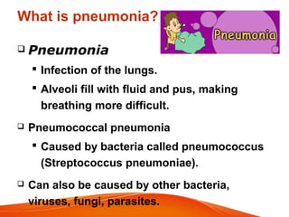
PNEUMONIAPNEUMONIA
Prepared by
ASWATHI C K
3rd YEAR BAMS
What is pneumonia?
 Pneumonia
 Infection of the lungs.
 Alveoli fill with fluid and pus, making
breathing more difficult.
 Pneumococcal pneumonia
 Caused by bacteria called pneumococcus
(Streptococcus pneumoniae).
 Can also be caused by other bacteria,
viruses, fungi, parasites.
EPIDEMOLOGICAL DETERMINANTS
• AGENT FACTOR
Bacteria : Most common
Streptococcus pneumoniae
Streptococcus pyogenes
Legionella pneumophila
Klebsiella pneumoniae
Virus : Respiratory syncycial virus
Influenza A,B
Parainfluenza 1-3
Adenoviruses
Other agents Clamydia type B
Coxiella burntti
Mycoplasma pneumoniae
AGENT......
• There are more than 100 strains of
infectious agents identified, only a few are
responsible for the majority of the cases
• Mixed infections with both viruses and
bacteria may occur in up to 45% of
infections in children
• agent may not be isolated in
approximately half of cases despite
careful testing
STREPTOCOCCUS PNEUMONIAE
• Gram-positive, alpha-hemolytic, facultative
anaerobic bacteria
• resides asymptomatically in the
nasopharynx of healthy carriers
Reservoir
• Exclusively a human pathogen; no animal or
environmental reservoir
Carrier
• Carried in the nasopharynx of 40-70% of
humans without symptoms.
Portal of entry
• Respiratory tract
Infecting body parts
• respiratory tract, sinuses, and nasal cavity
HOST FACTOR
• Case fatality rates are higher in young
infants & malnourished children
• Rate more in children <5yrs & adults older
than 75yrs
• In developing countries,rate is highest due
to malnutrition & low birth weight
• In developed countries RI are less fatal
RISK FACTORS
Developing countries : overcrowded dwellings, poor
nutrition
low birth weight, intense indoor smoke pollution
Developed countries : school going children to
- household
 People with some medical conditions are at higher
risk for pneumonia, including: heart disease, lung
disease, diabetes, etc.
 Smoking also increases risk of developing
pneumonia.
ENVIRONEMENT
• More common in winter & early spring
• Overcrowding
• Diminished host resistance
MODE OF TRANSMISSION
Direct transmission(person to person)
Normally by the airborne route(droplets)-
sneezing,coughing.....
INCUBATION PERIOD
1-4 Weeks
 Most cases of pneumonia are spread
person-to-person by coughing out of
tiny droplets.
 Some pathogens can live in nose and
throat without causing disease.
 But when inhaled into lungs, they can
cause pneumonia.
 While many people are exposed to
pneumococcus, usually only those with
underlying health issues develop
pneumonia.
PATHOGENESIS
• bacteria invade the spaces between cells
and between alveol
• macrophages and neutrophils attempt to
inactivate
• neutrophils cytokines activate immune
system fever, chills, and fatigue
• neutrophils, bacteria, and fluid from
surrounding blood vessels fill the alveoli,
resulting in the consolidation
TYPES OF PNEUMONIA
Pneumonia affects your lungs in two
Ways .
According to areas involved :
 Lobar pneumonia : affects a
section (lobe) of a lung.
 Bronchial pneumonia
(Bronchopneumonia) :
affects patches throughout both lungs.
LOBAR PNEUMONIA
BRONCHOPNEUMONIA
(Bronchitis and Pneumonia occur together)
CLINICAL MANIFESTATIONS
SIGNS AND SYMPTOMS
 High fever, Shaking Chills
 Shortness of breath (Dyspnoea)
 Increased breathing rate
 Chest pain when you breathe deeply or
cough
 Dusky or purplish skin colour (cyanosis)
from poorly oxygenated blood
 Fatigue and muscle aches
 Nausea, vomiting or diarrhoea
 Cough, particularly cough productive of
sputum
SIGNS AND SYMPTOMS
 Streptococcus pneumoniae: Rust-colored sputum
Pseudomonas, Haemophilus, and pneumococcal species:
May produce green sputum
Klebsiella species pneumonia: Red currant-jelly sputum
Anaerobic infections: Often produce foul-smelling or bad-
tasting sputum
Newborns and infants may not show any sign of the
infection. Or they may vomit, have a fever and cough,
appear restless or tired and without energy, or have
difficulty breathing and eating.
Older people who have pneumonia sometimes have
sudden changes in mental awareness.
COMPLICATIONS
 Bacteria in the bloodstream (bacteremia)
 Lung abscess.
 Build up of fluid in the space between the lung and chest wall
(pleural effusion).
 Difficulty breathing.
 Shock and respiratory failure
 Septic arthritis
 Endocarditis
DIAGNOSIS
• History taking and clinical assessment
• Physical examination
• X-ray findings of lobar consolidation
• Leukocytosis
• Bacteriological confirmation
Sputam-gram staining & culture
Blood culture
 PHYSICAL EXAMINATION
• Count the breath in one minute
• Look for chest indrawing
• Look & listen for stridor
• Look for wheeze
• See if the child is abnormally sleepy or
difficult to wake
• Feel for fever
• check for severe malnutrition
• Look for cyanosis
CONTROL
• Improving the primary medical care
services
• Developing better methods for early
detection,treatment & where possible
prevention
• Education of mother
PREVENTION
• Better nutrition
• Reduction of smoke pollution
• Better MCH care
• Immunization
Measles vaccine
Hib vaccine
Pneumococcal pneumonia vaccine
• Measles:
Dose : 0.5 ml age : 9th month
• Hib vaccine:
Dose : combined with DPT & polio myelitis
Age : 6,10,14th week
Booster dose:12-18 months
• Pneumococcal pneumonia vaccine
a.PPV23
b.PCV
PPV23(Pneumococcal Polysaccharide
Vaccine)
• protects against 23 types of pneumococcus
• Children under 2 yrs of age and immuno
-compromised individuals does not respond
• Recommended for selected groups - who
have undergone splenectomy/sickle cell
disease,
chronic disease of heart,lung,liver,.....DM,
alcoholism,generalized malignancies,organ
transplant,....
• Dose : 0.5ml(25microgrms of purified capsular
polysaccharide from each 23 serotypes)
• Site : intra-muscular ,deltoid or as
subcutaneous dose
Protective capsular type specific antibodies
develope by the 3rd week following vaccination
minor adverse effects such as transient
redness & pain at the site of injection occur in
30-50% cases
PCV(Pneumococcal conjugate vaccine)
• 2conjugate vaccines : PCV10 & PCV13
• 2 schedules 3 primary doses(3p+0)
2primarydoses+1booster
(2p+1)
• (3p+0) : from 6 weeks of age
interval : 4-8 weeks
given at 6,10,14 weeks or 2,4,6 months
• (2p+1) : 2 primary doses;as early as 6 weeks of
age in infant-interval-8weeks
• 4-8 weeks or more b/w primary doses for
infants≥7 months
• 1 booster dose should be given between
9-15 months of age
• mild reactions like erythema and
tenderness in 50% of cases
• HIV +ve and preterm babies : 3p doses in
12 month of age + booster dose in 2nd
yr of life
TREATMENT
• COTRIMOXAZOLE
• IM injection of benzyl pencillin,ampicillin or
chloramphenicol
• very severe : provision of oxygen therapy
IM injection of chloramphenicol
IM of Cloxacillin & Gentamycin
X-RAYS
Viral pneumonia x-ray
X-RAYS
Lobar pneumonia x-ray (RUL)
X-RAYS
bronchopneumonia x-ray
X-RAYS
Staph pneumonia x-ray
Pneumonia

Pneumonia

  • 1.
  • 2.
    What is pneumonia? Pneumonia  Infection of the lungs.  Alveoli fill with fluid and pus, making breathing more difficult.  Pneumococcal pneumonia  Caused by bacteria called pneumococcus (Streptococcus pneumoniae).  Can also be caused by other bacteria, viruses, fungi, parasites.
  • 3.
    EPIDEMOLOGICAL DETERMINANTS • AGENTFACTOR Bacteria : Most common Streptococcus pneumoniae Streptococcus pyogenes Legionella pneumophila Klebsiella pneumoniae Virus : Respiratory syncycial virus Influenza A,B Parainfluenza 1-3 Adenoviruses Other agents Clamydia type B Coxiella burntti Mycoplasma pneumoniae
  • 4.
    AGENT...... • There aremore than 100 strains of infectious agents identified, only a few are responsible for the majority of the cases • Mixed infections with both viruses and bacteria may occur in up to 45% of infections in children • agent may not be isolated in approximately half of cases despite careful testing
  • 5.
    STREPTOCOCCUS PNEUMONIAE • Gram-positive,alpha-hemolytic, facultative anaerobic bacteria • resides asymptomatically in the nasopharynx of healthy carriers
  • 6.
    Reservoir • Exclusively ahuman pathogen; no animal or environmental reservoir Carrier • Carried in the nasopharynx of 40-70% of humans without symptoms. Portal of entry • Respiratory tract Infecting body parts • respiratory tract, sinuses, and nasal cavity
  • 7.
    HOST FACTOR • Casefatality rates are higher in young infants & malnourished children • Rate more in children <5yrs & adults older than 75yrs • In developing countries,rate is highest due to malnutrition & low birth weight • In developed countries RI are less fatal
  • 8.
    RISK FACTORS Developing countries: overcrowded dwellings, poor nutrition low birth weight, intense indoor smoke pollution Developed countries : school going children to - household  People with some medical conditions are at higher risk for pneumonia, including: heart disease, lung disease, diabetes, etc.  Smoking also increases risk of developing pneumonia.
  • 9.
    ENVIRONEMENT • More commonin winter & early spring • Overcrowding • Diminished host resistance
  • 10.
    MODE OF TRANSMISSION Directtransmission(person to person) Normally by the airborne route(droplets)- sneezing,coughing..... INCUBATION PERIOD 1-4 Weeks
  • 11.
     Most casesof pneumonia are spread person-to-person by coughing out of tiny droplets.  Some pathogens can live in nose and throat without causing disease.  But when inhaled into lungs, they can cause pneumonia.  While many people are exposed to pneumococcus, usually only those with underlying health issues develop pneumonia.
  • 12.
    PATHOGENESIS • bacteria invadethe spaces between cells and between alveol • macrophages and neutrophils attempt to inactivate • neutrophils cytokines activate immune system fever, chills, and fatigue • neutrophils, bacteria, and fluid from surrounding blood vessels fill the alveoli, resulting in the consolidation
  • 13.
    TYPES OF PNEUMONIA Pneumoniaaffects your lungs in two Ways . According to areas involved :  Lobar pneumonia : affects a section (lobe) of a lung.  Bronchial pneumonia (Bronchopneumonia) : affects patches throughout both lungs.
  • 14.
  • 15.
  • 16.
  • 17.
    SIGNS AND SYMPTOMS High fever, Shaking Chills  Shortness of breath (Dyspnoea)  Increased breathing rate  Chest pain when you breathe deeply or cough  Dusky or purplish skin colour (cyanosis) from poorly oxygenated blood  Fatigue and muscle aches  Nausea, vomiting or diarrhoea  Cough, particularly cough productive of sputum
  • 18.
    SIGNS AND SYMPTOMS Streptococcus pneumoniae: Rust-colored sputum Pseudomonas, Haemophilus, and pneumococcal species: May produce green sputum Klebsiella species pneumonia: Red currant-jelly sputum Anaerobic infections: Often produce foul-smelling or bad- tasting sputum Newborns and infants may not show any sign of the infection. Or they may vomit, have a fever and cough, appear restless or tired and without energy, or have difficulty breathing and eating. Older people who have pneumonia sometimes have sudden changes in mental awareness.
  • 19.
    COMPLICATIONS  Bacteria inthe bloodstream (bacteremia)  Lung abscess.  Build up of fluid in the space between the lung and chest wall (pleural effusion).  Difficulty breathing.  Shock and respiratory failure  Septic arthritis  Endocarditis
  • 20.
    DIAGNOSIS • History takingand clinical assessment • Physical examination • X-ray findings of lobar consolidation • Leukocytosis • Bacteriological confirmation Sputam-gram staining & culture Blood culture
  • 21.
     PHYSICAL EXAMINATION •Count the breath in one minute • Look for chest indrawing • Look & listen for stridor • Look for wheeze • See if the child is abnormally sleepy or difficult to wake • Feel for fever • check for severe malnutrition • Look for cyanosis
  • 22.
    CONTROL • Improving theprimary medical care services • Developing better methods for early detection,treatment & where possible prevention • Education of mother
  • 23.
    PREVENTION • Better nutrition •Reduction of smoke pollution • Better MCH care • Immunization Measles vaccine Hib vaccine Pneumococcal pneumonia vaccine
  • 24.
    • Measles: Dose :0.5 ml age : 9th month • Hib vaccine: Dose : combined with DPT & polio myelitis Age : 6,10,14th week Booster dose:12-18 months • Pneumococcal pneumonia vaccine a.PPV23 b.PCV
  • 25.
    PPV23(Pneumococcal Polysaccharide Vaccine) • protectsagainst 23 types of pneumococcus • Children under 2 yrs of age and immuno -compromised individuals does not respond • Recommended for selected groups - who have undergone splenectomy/sickle cell disease, chronic disease of heart,lung,liver,.....DM, alcoholism,generalized malignancies,organ transplant,....
  • 26.
    • Dose :0.5ml(25microgrms of purified capsular polysaccharide from each 23 serotypes) • Site : intra-muscular ,deltoid or as subcutaneous dose Protective capsular type specific antibodies develope by the 3rd week following vaccination minor adverse effects such as transient redness & pain at the site of injection occur in 30-50% cases
  • 27.
    PCV(Pneumococcal conjugate vaccine) •2conjugate vaccines : PCV10 & PCV13 • 2 schedules 3 primary doses(3p+0) 2primarydoses+1booster (2p+1) • (3p+0) : from 6 weeks of age interval : 4-8 weeks given at 6,10,14 weeks or 2,4,6 months • (2p+1) : 2 primary doses;as early as 6 weeks of age in infant-interval-8weeks
  • 28.
    • 4-8 weeksor more b/w primary doses for infants≥7 months • 1 booster dose should be given between 9-15 months of age • mild reactions like erythema and tenderness in 50% of cases • HIV +ve and preterm babies : 3p doses in 12 month of age + booster dose in 2nd yr of life
  • 29.
    TREATMENT • COTRIMOXAZOLE • IMinjection of benzyl pencillin,ampicillin or chloramphenicol • very severe : provision of oxygen therapy IM injection of chloramphenicol IM of Cloxacillin & Gentamycin
  • 30.
  • 31.
  • 32.
  • 33.