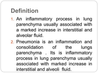
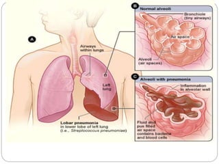
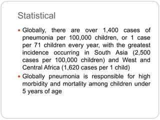
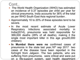
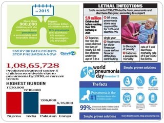
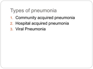
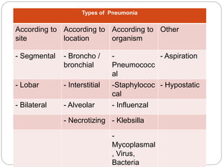
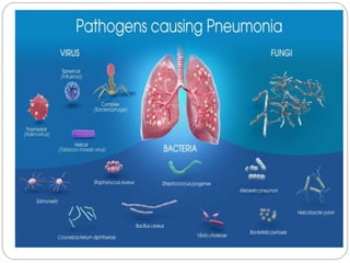
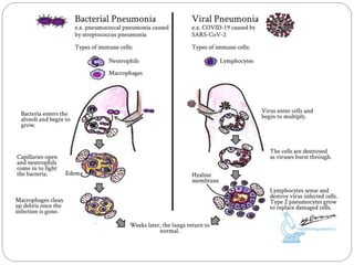
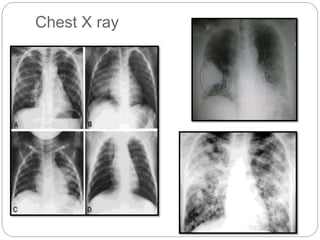
Mr. Rohan Phaphe
Asst .Prof.
Dept. Medical – Surgical Nursing
DYPCON, KOP
PNEUMONIA
Definition
1. An inflammatory process in lung
parenchyma usually associated with
a marked increase in interstitial and
alveolar fluid.
2. Pneumonia is an inflammation and
consolidation of the lungs
parenchyma . Its is inflammatory
process in lung parenchyma usually
associated with marked increase in
interstitial and alveoli fluid.
Statistical
 Globally, there are over 1,400 cases of
pneumonia per 100,000 children, or 1 case
per 71 children every year, with the greatest
incidence occurring in South Asia (2,500
cases per 100,000 children) and West and
Central Africa (1,620 cases per 1 child)
 Globally pneumonia is responsible for high
morbidity and mortality among children under
5 years of age
Cont..
 The World Health Organization (WHO) has estimated
an incidence of 0.37 episodes per child per year for
clinical pneumonia, India accounts for 36% of the total
as per WHO South East Asia regional burden .
 Approximately 10 to 20% of these episodes tend to be
severe.
 In the most recent estimate of Acute Lower
Respiratory Infections associated mortality in
India(2014), pneumonia was held responsible for
369,000 deaths (28% of all deaths), making it the
single most important killer in this age grou00,000
children)
 In Mumbai there were no confirmed cases of
pneumonia in the state last year,16th sep 2017 two
cases of the disease have been reported in the
fortnight from Jalgaon. The two patients, who are
suffering from pneumonia, were critical and were put
on a life-support system, said health department
Types of pneumonia
1. Community acquired pneumonia
2. Hospital acquired pneumonia
3. Viral Pneumonia
 Types of Pneumonia
Types of Pneumonia
According to
site
According to
location
According to
organism
Other
- Segmental - Broncho /
bronchial
-
Pneumococc
al
- Aspiration
- Lobar - Interstitial -Staphylococ
cal
- Hypostatic
- Bilateral - Alveolar - Influenzal
- Necrotizing - Klebsilla
-
Mycoplasmal
, Virus,
Bacteria
Etiology
 Bacteria- Streptococcus pneumonia ,
haemophilus influenza , staphylococcus
aureus
 Viruses- Coronavirus, Rhinovirus,
Influenzavirus
 Fungal agents– histoplasma capsulatum
 Mycoplasmas, & protozoa.
 Parasities – these parasites enters in body
through skin or the mouth , once enter in the
body,they travels through blood into the lungs.
Toxoplasma gondii, strongloids ,sterocoralis
 Aspiration of food, fluids, /vomit’s.
Risk Factors
 Advanced age.
 History of smoking.
 Upper respiratory infection.
 Tracheal intubation.
 Prolonged immobility.
 Immunosuppressive therapy.
 Nonfunctional immune system.
 Malnutrition.
 Dehydration.
 Chronic disease state.
 Cigarette smoking
 Recent viral respiratory infection (common
cold, laryngitis, influenza)
 Difficulty swallowing (due to stroke,
dementia, Parkinson's disease, or other
neurological conditions)
 Chronic lung disease (COPD, bronchiectasis,
cystic fibrosis)
 Cerebral palsy
 Other serious illnesses, such as heart
disease, liver cirrhosis, /diabetes mellitus
Pathophysiology:
Offending organism/agent.
Inflammatory pulmonary response.
Lose defense mechanisms of the lungs.
Allow organisms to penetrate the sterile LRT.
Develop inflammation.
Disruption of the mechanical defenses
(cough & ciliary motility)
Colonization of the lungs.
Inflamed & fluid-filled alveolar sacs.
Alveolar exudates tends to consolidate.
Difficult to expectorate.
Common Clinical Manifestation
Fever.
Chills.
 Sweats
 Pleuritic chest pain
 Cough.
 Sputum production.
 Hemoptysis.
 Dyspnea.
Headache & Fatigue
Clinical Manifestation as per Etiology
 Pneumococcal pneumonia:
 Caused by Streptococcus pneumonia.
 C/M:- high fever.
 Stabbing pleuritic chest pain.
 Malaise.
 Weakness.
 Elevated wbc count.
 Tachypnea.
 Dyspnea.
 Blood-streaked purulent sputum
 Staphylococcal pneumonia:
 Caused by Staphylococcus aureus.
C/M:- Fever with multiple chills.
 Pleuritic pain.
 Rales.
 Decreased breath sounds.
 Elevated wbc count.
 Dyspnea.
 Blood-streaked purulent
sputum
 Influenzal pneumonia:
 Caused by Haemophilus influenza.
 C/M:- High fever.
 Stabbing pleuritic chest pain.
 Malaise.
 Weakness.
 Elevated WBC count.
 Tachypnea.
 Dyspnea.
 Blood-tinged purulent sputum
 Gram-negative bacterial pneumonia:
 Caused by Klebsiella pneumonia.
 C/M:- Fever with multiple chills.
 Pleuritic pain.
 Rales.
 Cyanosis.
 Elevated wbc count.
 Dyspnea.
 Blood-streaked purulent sputum
 Anaerobic bacterial pneumonia:
 Caused by normal oral flora.
 C/M:- Low-grade fever.
 Hypertension.
 Crackles.
 Cyanosis.
 Elevated wbc count.
 Dyspnea.
 Foul smelling purulent sputum.
 Tachycardia.
 Legionnaires’ disease
 Caused by Legionella pneumophila.
 C/M:- Fever.
 Pleuritic pain.
 Nausea.
 Confusion.
 Elevated wbc count.
 Dyspnea.
 Blood-tinged sputum.
 Caused by Mycoplasma
microorganisms.
 C/M:- Slowly rising fever.
 Headache.
 Myalgia.
 Malaise.
 Normal WBC count.
 Scant mucoid sputum
 Viral pneumonia:
 Caused by Influenza A virus.
 C/M:- High fever.
 Headache.
 Myalgia.
 Dyspnea.
 Normal/slightly wbc count.
 Mucoid sputum.
 Normal breath sounds.
 Aspiration pneumonia:
 Caused by aspiration of gastric
contents.
 C/M:- Tachypnea.
 Apnea.
 Cyanosis.
 Hypotension.
 Leukocytosis.
 Adventitious lung sounds.
 Fever
Diagnostic Evaluation
 Chest auscultation.
 Sputum culture analysis & sensitivity/
serologic testing.
 Fiber optic bronchoscopy/ Transcutaneous
needle aspiration/ biopsy.
 Skin tests.
 Blood & urine cultures.
 Transcutaneous oxygen level analysis/ ABG
measurements.
 Chest X-ray examination
Chest X ray
ABG Analysis
Medical Management
Specific antibiotic therapy: Broad spectrum
antibiotics.
Respiratory support:
Administer oxygen.
Bronchodilator medications.
Postural drainage.
Chest physiotherapy.
Tracheal suctioning.
Nutritional support.
Fluid & electrolyte management
Nursing Management
 Asses for signs and symptoms of shock and
respiratory failure
 Administer intravenous fluids and medication and
respiratory support as ordered
 Initiate preventive measures for atelectasis
 Assess with the thoracentesis and monitor the
patient for pneumothorax after the procedure
 Monitor for Superinfection (rise of temperature ,
increased cough), and assist in therapy.
 Assess for confusion or cognitive changes and
underlying changes.
Health Education
 Instruct patient to continue taking antibiotics until
complete
 Advice patient to increase activites graduallyafter
fever subsides
 Advice patient that fatigue and weakness may
linger on]
 Encourage breathing exercise
 Instruct the patient to avoid fatigue , sudden
changes in temperature and excessive alcohol,
which lower resistance of pneumonia
 Recommend influenza vaccine and pneumovax
to all pateints at risk .
Nursing diagnosis
 Ineffective airway clearance R/t excessive
secretions & weak cough.
 Ineffective breathing pattern R/t tachypnea.
Activity intolerance R/t decreased oxygen
levels for metabolic demands.
 Deficient fluid volume R/t fever, diaphoresis,
& mouth breathing.
 Imbalanced nutrition: less than body
requirements R/t dyspnea.
 Pain R/t frequent coughing.
Pneumonia

Pneumonia

  • 1.
    Mr. Rohan Phaphe Asst.Prof. Dept. Medical – Surgical Nursing DYPCON, KOP PNEUMONIA
  • 3.
    Definition 1. An inflammatoryprocess in lung parenchyma usually associated with a marked increase in interstitial and alveolar fluid. 2. Pneumonia is an inflammation and consolidation of the lungs parenchyma . Its is inflammatory process in lung parenchyma usually associated with marked increase in interstitial and alveoli fluid.
  • 5.
    Statistical  Globally, thereare over 1,400 cases of pneumonia per 100,000 children, or 1 case per 71 children every year, with the greatest incidence occurring in South Asia (2,500 cases per 100,000 children) and West and Central Africa (1,620 cases per 1 child)  Globally pneumonia is responsible for high morbidity and mortality among children under 5 years of age
  • 6.
    Cont..  The WorldHealth Organization (WHO) has estimated an incidence of 0.37 episodes per child per year for clinical pneumonia, India accounts for 36% of the total as per WHO South East Asia regional burden .  Approximately 10 to 20% of these episodes tend to be severe.  In the most recent estimate of Acute Lower Respiratory Infections associated mortality in India(2014), pneumonia was held responsible for 369,000 deaths (28% of all deaths), making it the single most important killer in this age grou00,000 children)  In Mumbai there were no confirmed cases of pneumonia in the state last year,16th sep 2017 two cases of the disease have been reported in the fortnight from Jalgaon. The two patients, who are suffering from pneumonia, were critical and were put on a life-support system, said health department
  • 8.
    Types of pneumonia 1.Community acquired pneumonia 2. Hospital acquired pneumonia 3. Viral Pneumonia
  • 9.
     Types ofPneumonia Types of Pneumonia According to site According to location According to organism Other - Segmental - Broncho / bronchial - Pneumococc al - Aspiration - Lobar - Interstitial -Staphylococ cal - Hypostatic - Bilateral - Alveolar - Influenzal - Necrotizing - Klebsilla - Mycoplasmal , Virus, Bacteria
  • 11.
    Etiology  Bacteria- Streptococcuspneumonia , haemophilus influenza , staphylococcus aureus  Viruses- Coronavirus, Rhinovirus, Influenzavirus  Fungal agents– histoplasma capsulatum  Mycoplasmas, & protozoa.  Parasities – these parasites enters in body through skin or the mouth , once enter in the body,they travels through blood into the lungs. Toxoplasma gondii, strongloids ,sterocoralis  Aspiration of food, fluids, /vomit’s.
  • 12.
    Risk Factors  Advancedage.  History of smoking.  Upper respiratory infection.  Tracheal intubation.  Prolonged immobility.  Immunosuppressive therapy.  Nonfunctional immune system.  Malnutrition.  Dehydration.  Chronic disease state.
  • 13.
     Cigarette smoking Recent viral respiratory infection (common cold, laryngitis, influenza)  Difficulty swallowing (due to stroke, dementia, Parkinson's disease, or other neurological conditions)  Chronic lung disease (COPD, bronchiectasis, cystic fibrosis)  Cerebral palsy  Other serious illnesses, such as heart disease, liver cirrhosis, /diabetes mellitus
  • 14.
    Pathophysiology: Offending organism/agent. Inflammatory pulmonaryresponse. Lose defense mechanisms of the lungs. Allow organisms to penetrate the sterile LRT. Develop inflammation.
  • 15.
    Disruption of themechanical defenses (cough & ciliary motility) Colonization of the lungs. Inflamed & fluid-filled alveolar sacs. Alveolar exudates tends to consolidate. Difficult to expectorate.
  • 17.
    Common Clinical Manifestation Fever. Chills. Sweats  Pleuritic chest pain  Cough.  Sputum production.  Hemoptysis.  Dyspnea. Headache & Fatigue
  • 18.
    Clinical Manifestation asper Etiology  Pneumococcal pneumonia:  Caused by Streptococcus pneumonia.  C/M:- high fever.  Stabbing pleuritic chest pain.  Malaise.  Weakness.  Elevated wbc count.  Tachypnea.  Dyspnea.  Blood-streaked purulent sputum
  • 19.
     Staphylococcal pneumonia: Caused by Staphylococcus aureus. C/M:- Fever with multiple chills.  Pleuritic pain.  Rales.  Decreased breath sounds.  Elevated wbc count.  Dyspnea.  Blood-streaked purulent sputum
  • 20.
     Influenzal pneumonia: Caused by Haemophilus influenza.  C/M:- High fever.  Stabbing pleuritic chest pain.  Malaise.  Weakness.  Elevated WBC count.  Tachypnea.  Dyspnea.  Blood-tinged purulent sputum
  • 21.
     Gram-negative bacterialpneumonia:  Caused by Klebsiella pneumonia.  C/M:- Fever with multiple chills.  Pleuritic pain.  Rales.  Cyanosis.  Elevated wbc count.  Dyspnea.  Blood-streaked purulent sputum
  • 22.
     Anaerobic bacterialpneumonia:  Caused by normal oral flora.  C/M:- Low-grade fever.  Hypertension.  Crackles.  Cyanosis.  Elevated wbc count.  Dyspnea.  Foul smelling purulent sputum.  Tachycardia.
  • 23.
     Legionnaires’ disease Caused by Legionella pneumophila.  C/M:- Fever.  Pleuritic pain.  Nausea.  Confusion.  Elevated wbc count.  Dyspnea.  Blood-tinged sputum.
  • 24.
     Caused byMycoplasma microorganisms.  C/M:- Slowly rising fever.  Headache.  Myalgia.  Malaise.  Normal WBC count.  Scant mucoid sputum
  • 25.
     Viral pneumonia: Caused by Influenza A virus.  C/M:- High fever.  Headache.  Myalgia.  Dyspnea.  Normal/slightly wbc count.  Mucoid sputum.  Normal breath sounds.
  • 26.
     Aspiration pneumonia: Caused by aspiration of gastric contents.  C/M:- Tachypnea.  Apnea.  Cyanosis.  Hypotension.  Leukocytosis.  Adventitious lung sounds.  Fever
  • 27.
    Diagnostic Evaluation  Chestauscultation.  Sputum culture analysis & sensitivity/ serologic testing.  Fiber optic bronchoscopy/ Transcutaneous needle aspiration/ biopsy.  Skin tests.  Blood & urine cultures.  Transcutaneous oxygen level analysis/ ABG measurements.  Chest X-ray examination
  • 28.
  • 29.
  • 30.
    Medical Management Specific antibiotictherapy: Broad spectrum antibiotics. Respiratory support: Administer oxygen. Bronchodilator medications. Postural drainage. Chest physiotherapy. Tracheal suctioning. Nutritional support. Fluid & electrolyte management
  • 31.
    Nursing Management  Assesfor signs and symptoms of shock and respiratory failure  Administer intravenous fluids and medication and respiratory support as ordered  Initiate preventive measures for atelectasis  Assess with the thoracentesis and monitor the patient for pneumothorax after the procedure  Monitor for Superinfection (rise of temperature , increased cough), and assist in therapy.  Assess for confusion or cognitive changes and underlying changes.
  • 32.
    Health Education  Instructpatient to continue taking antibiotics until complete  Advice patient to increase activites graduallyafter fever subsides  Advice patient that fatigue and weakness may linger on]  Encourage breathing exercise  Instruct the patient to avoid fatigue , sudden changes in temperature and excessive alcohol, which lower resistance of pneumonia  Recommend influenza vaccine and pneumovax to all pateints at risk .
  • 33.
    Nursing diagnosis  Ineffectiveairway clearance R/t excessive secretions & weak cough.  Ineffective breathing pattern R/t tachypnea. Activity intolerance R/t decreased oxygen levels for metabolic demands.  Deficient fluid volume R/t fever, diaphoresis, & mouth breathing.  Imbalanced nutrition: less than body requirements R/t dyspnea.  Pain R/t frequent coughing.