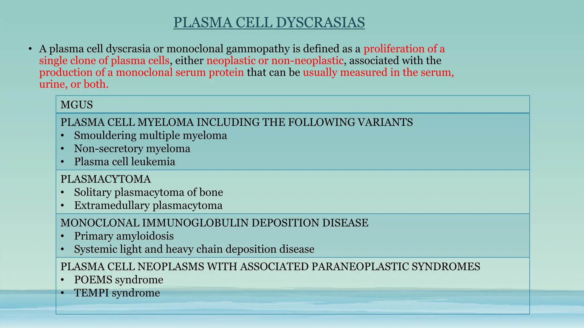
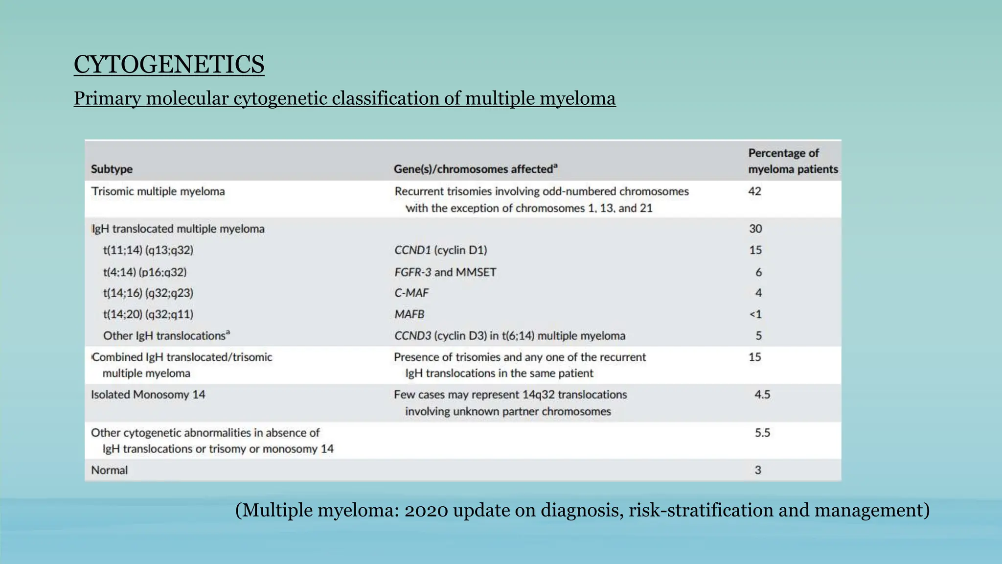
PLASMA CELL DYSCRASIAS
PRESENTED BY:
DR. GREESHMA GOPINATH
JR, DEPT. OF PATHOLOGY
AIIMS PATNA
MODERATED BY:
DR. SURABHI
ASST.PROFESSOR, DEPT.OF PATHOLOGY
AIIMS PATNA
PLASMA CELLS & ITS DEVELOPMENT
Plasma cells are terminally
differentiated B-lineage cells
that secrete Ig as a protective
antibody response
Normal stages of B cell development; adapted from Quick
reference handbook-Natasha Rekhtman
IMMUNOGLOBULIN(Ig) • Constitute 20-25 per cent of total serum proteins.
• Five classes: IgG, IgA, lgM, IgD and IgE.
• Four polypeptide chains
 Two identical heavy chains (H)
 Two identical light chains (L)
• Heavy chains are of five types:
• Light chains: kappa & lambda
• In humans, 60 percent of L chains are kappa
and 40 percent are lambda.
• Normal serum kappa/serum lambda
ratio=0.26 to 1.65
PLASMA CELL DYSCRASIAS
• A plasma cell dyscrasia or monoclonal gammopathy is defined as a proliferation of a
single clone of plasma cells, either neoplastic or non-neoplastic, associated with the
production of a monoclonal serum protein that can be usually measured in the serum,
urine, or both.
MGUS
PLASMA CELL MYELOMA INCLUDING THE FOLLOWING VARIANTS
• Smouldering multiple myeloma
• Non-secretory myeloma
• Plasma cell leukemia
PLASMACYTOMA
• Solitary plasmacytoma of bone
• Extramedullary plasmacytoma
MONOCLONAL IMMUNOGLOBULIN DEPOSITION DISEASE
• Primary amyloidosis
• Systemic light and heavy chain deposition disease
PLASMA CELL NEOPLASMS WITH ASSOCIATED PARANEOPLASTIC SYNDROMES
• POEMS syndrome
• TEMPI syndrome
MONOCLONAL GAMMOPATHY OF UNDETERMINED
SIGNIFICANCE (MGUS)
• Asymptomatic, pre-malignant clonal plasma cell proliferative disorder.
• Three types:
 IgM MGUS
 Non-IgM MGUS
 Light chain MGUS
Non IgM MGUS
• 80-85% of MGUS
• M>F, >70 years of age
• Diagnostic Criteria: All 3 must be met
• Serum M protein (non-lgM) concentration <30 g/L
• Clonal bone marrow plasma cells< 10%
• Absence of end-organ damage; e.g. hypercalcaemia, renal insufficiency, anaemia,
and bone lesions (CRAB) and amyloidosis attributable to the plasma cell
proliferative disorder
• M protein: IgG(60%)> IgA(15%)> Biclonal (3%)> IgD(1%) , IgE(1%)
• Flowcytometry: Shows two population of cells
• Shows aberrant Ag expression
• CD19-/CD56+ or CD19-/CD56-
• CD38 weak +
Monoclonal
population
• CD19+, CD56-
• CD38 bright +
• Polyclonal plasma cells are a consistent
finding in Non-IgM MGUS
Polyclonal
population
IgM MGUS
• Serum lgM monoclonal protein concentration < 30 g/L
• Bone marrow lymphoplasmacytic infiltration of <10%
• No evidence of anaemia, constitutional symptoms, hyperviscosity, lymphadenopathy,
hepatosplenomegaly, or other end-organ damage that can be attributed to the
underlying lymphoproliferative disorder.
• Flow cytometry: Non specific phenotype – CD19+, CD20+, CD10-, CD5-, CD103-, CD56-
• 15% cases of MGUS
• M>F
• >70 years of age
• Diagnostic criteria: All three must be met
Light Chain MGUS
• Diagnostic criteria:
 Abnormal free light chain ratio ( < 0.26 or> 1.65)
 Increased level of the involved free light chain
 No immunoglobulin heavy chain expression on immunofixation electrophoresis
 Urinary M protein < 500 mg/24 hours
 Clonal plasma cells < 10%
 Absence of end-organ damage (CRAB) and amyloidosis
TYPES RISK OF PROGRESSION
Non-IgM MGUS 1% per year risk of progression to plasma cell
myeloma, AL amyloidosis, or related disorder
IgM MGUS 1.5% per year risk of progression to
Lymphoplasmacytic lymphoma/
Waldenstrom’s macroglobulinemia or other B
cell neoplasms, primary amyloidosis. Rarely to
PCM.
Light chain MGUS Risk of progression to light chain myeloma and
AL amyloidosis. Rate of progression not
defined
MONOCLONAL GAMMOPATHY OF RENAL
SIGNIFICANCE (MGRS)
• Includes all kidney disorders caused by a monoclonal protein (M-protein) secreted by a small
plasma cell clone or other B-cell clones in patients who do not meet the diagnostic criteria for
multiple myeloma or other B-cell malignancies.
• MGRS includes:
 Light chain (AL), heavy chain (AH), and heavy and light chain (AHL) amyloidosis
 Monoclonal immunoglobulin deposition disease (MIDD)
 Proliferative glomerulonephritis with monoclonal immunoglobulin deposits
(PGNMID)
 Cryoglobulinaemia-associated glomerulonephritis (CGG)
 Monoclonal gammopathy-associated thrombotic microangiopathy (TMA)
 Monoclonal gammopathy-associated C3 glomerulopathy (C3GP)
 Light chain proximal tubulopathy (LCPT)
 Crystal-storing histiocytosis (CSH)
 Immunotactoid glomerulopathy (ITG)
Spectrum of monoclonal
gammopathy of renal
significance by localization
Source - Monoclonal gammopathy of renal significance (MGRS): histopathologic classification, diagnostic workup, and
therapeutic options - K. Amaador et. al
PLASMA CELL MYELOMA
• Bone marrow based multifocal neoplastic proliferation of plasma cells
characterized by a clinical pentad:
(1) anaemia
(2)a monoclonal protein in the serum or urine or both
(3)bone lesions and/ or bone pain
(4)hypercalcemia
(5)renal insufficiency
• 10-15% of haematopoietic neoplasms
• 90% cases occur >50 years(median age 70 years), M>F.
DIAGNOSTIC CRITERIA FOR PLASMA CELL MYELOMA
• Clonal bone marrow plasma cell percentage >=10% or biopsy-proven plasmacytoma and
>= 1 of the following myeloma-defining events
 End-organ damage attributable to the plasma cell proliferative disorder
• Hypercalcaemia: serum calcium > 1 mg/dL higher than the upper limit of normal or >
11 mg/dL
• Renal insufficiency: creatinine clearance<40ml/min or serum creatinine> 2 mg/dL
• Anaemia: a haemoglobin value of> 2g/dL below the lower limit of normal or a
haemoglobin value< 10g/dL
• Bone lesions: >= 1 osteolytic lesion on skeletal radiography, CT, or PET /CT
 >= 1 of the following biomarkers of malignancy
• Clonal bone marrow plasma cell percentage >= 60%
• An involved-to-uninvolved serum free light chain ratio >=100
• > 1 focal lesion on MRI
PATHOGENESIS
INITIATING EVENT:
IGH translocation
• Involves Cyclin D(CCND)
family – results in cell
progression
• MAF transcription factor
expression affected
Hyper diploidy of chromosome
3,5,7,9,11,15
MYC Locus rearrangements
Deletion 13q
Interaction of myeloma cells with bone
marrow microenvironment
Initiation of plasma cell proliferation in
microenvironment of bone marrow
MGUS Occurs
Myeloma cell growth and survival increased
The myeloma cells hijack the normal hematopoietic
niche to aid the growth & proliferation of tumor cells
Multiple Myeloma
MM cells + Mesenchymal stem
cells
Produce excess of MMP-
1,2,8,9,13
Main role of MMP-9
Secretes VEGF-A
Neo angiogenesis
Spread of MM
MM cells+ CD138 on their
surface(heparan sulfate
proteoglycan)
Binds to Type 1 collagen
Induces expression of MMP-1
Promotes tumor invasion, bone
resorption & angiogenesis
Resorption of ECM
Plasma cell leukemia
Myeloma cells secrete macrophage inflammatory protein-1alpha (MIP1alpha) and
MIP1ß
MIP1alpha binds to C-chemokine receptor 1 (CCR1) and CCR5 while MIP1ß binds
to CCR5 and CCR8 to induce osteoclast formation
Osteoclasts secrete IL6 - stimulate proliferation and growth of myeloma cells &
other osteoclasts
BMSC produce RANKL which binds with RANK receptor & stimulates osteoclastic
activation and differentiation
Bone lysis
• Osteoblasts produce
OPG(Osteoprotegerin)
• OPG competes with RANKL
for RANK
• Myeloma cells inhibit
osteoblast production &
activation
• Increase in ratio of
RANKL/OPG – Activation of
osteoclast
CLINICAL FEATURES
 Weakness, fatigue
 Pallor
 Bone pains
 Pathological fractures
 Recurrent infections
 Elevated ESR
 Hypercalcemia
 Bence Jones proteinuria
 Renal failure
 Amyloidosis
LABORATORY FINDINGS
• BLOOD
 Normocytic normochromic, macrocytes less
often
 Rouleaux formation
 Background of smear basophilic
 Leucoerythroblastic picture in some
 Coagulopathy
 Later stages- pancytopenia
• BIOCHEMICAL TESTS
 Liver function tests:
-Total protein: increased.
-Serum alkaline phosphatase: is normal or slightly increased
 Kidney function tests:
-Serum creatinine: is raised in the presence of renal insufficiency
-Serum calcium: hypercalcemia
• SERUM ELECTROPHORESIS
 Initial step in the identification of abnormal proteins
 A localized dense band with sharp margins in the γ region indicates M band
 In about 80% of patients with monoclonal gammopathies, M band will be
detected.
SERUM ELECTROPHORESIS
IN A NORMAL PERSON
SERUM ELECTROPHORESIS IN A PATIENT
OF MULTIPLE MYELOMA; M BAND PRSENT
• IMMUNOFIXATION ELECTROPHORESIS
 Gold standard for identification of nature of M protein
 In this technique, serum proteins are separated by electrophoresis
in a gel and monospecific antiserum (IgG, IgM, IgA, κ or λ light
chains) is applied directly over the surface of the gel.
 Immunoprecipitation band develops in the gel between
corresponding protein antigen and the monospecific antiserum.
• SERUM FREE LIGHT CHAIN(SFLC) ASSAY
 Sensitive marker than electrophoresis & immunofixation for detection of M protein
 Used as a substitute for 24 hr urine immunofixation
 Measured by immunonephelometry
 Clonal plasma cells produce>100mg/L of SFLC & affected SFLC/unaffected SFLC >100
 Free light chain assay measures amount of free kappa to lambda chain ratio (normal –
0.26 to 1.65)
 Useful in cases of
 Non-secretory myeloma
 Light chain myeloma
 MGUS
 Amyloidosis
 Smouldering MM
• QUANTITATIVE IMMUNOGLOBULIN ASSAY
 The quantitation of monoclonal and other immunoglobulins is necessary to assess the
disease severity and follow response to treatment
 M protein is considered measurable if it is >=1g/dL in the serum & or >=200mg/day in
urine.
 The height of the peak on serum protein electrophoresis is directly proportional to the
amount of M protein
 Exact quantification can be done by nephelometry
• BONE MARROW
 Hypercellular to normocellular
 Myeloid series, erythroid series & megakaryocytes morphology is normal
 Marrow infiltration by myeloma cells
 Monoclonal plasma cells varies between 10% to 90%
MATURE TYPE:
• Cytoplasm – abundant deeply
basophilic with a perinuclear clear
area/hof representing golgi zone
• Nucleus- Eccentrically placed nucleus
with coarse chromatin and no
nucleoli.
IMMATURE TYPE:
• Size larger
• Light blue abundant cytoplasm, hof can be
present
• Centrally/ eccentrically placed nucleus,
diffuse open chromatin, 1 or 2 prominent
nucleoli
• Pleomorphism may be seen
PLASMABLASTIC TYPE
• Large cells with central nuclei
having high N/C ratio
• Fine reticular chromatin &
prominent nucleoli
• No perinuclear hof
• Plasmablastic myeloma- >2%
marrow cells are plasmablasts
PLEOMORPHIC/ANAPLASTIC
TYPE:
• Marked pleomorphism
• Frequent multinucleated and
bizzare cells
• Nuclear lobulation
• Mitotic figures seen
OTHER VARIANTS:
Small cell variant:
Shows a lymphoplasmacytic
appearance, with a narrow rim
of basophilic cytoplasm and the
occasional perinuclear hof
Taken from: Plasma cell myeloma with lymphoplasmacytic morphology and cyclin D1 expression, an uncommon variant
Daniel A. Hale, MD, and John R. Krause, MD
Fig:
a) Scattered lymphoplasmacytic cells with occasional small
perinuclear hof and eccentric nuclei with rare definitive plasma
cell
b) Higher magnification shows small plasma cells with high N:C
ratio
c) Biopsy shows numerous lymphoplasmacytic cells
d) CD 138+
e) Cyclin D1+
PLASMA CELL INCLUSIONS
The cytoplasm of myeloma cells has abundant endoplasmic reticulum, which may contain condensed
or crystallized cytoplasmic Ig producing a variety of morphological findings
Mott cells (grape cells or morula forms)
• plasma cells filled with dense spherical
immunoglobulin inclusions
• these inclusions are colorless, pink, or blue
Russell bodies:
• Intracytoplasmic hyaline inclusions
• Appear as cherry-red refractive round
bodies
DUTCHER BODY:
• Intranuclear inclusions
FLAME CELLS:
• Peripheral rim of cytoplasm shows a
pinkish hue
• Glycogen rich fibrils
• Commonly seen in IgA myelomas
BONE MARROW BIOPSY:
Is of importance in
 Asymptomatic plasma cell myeloma
• Plasma cells <10% in aspirate
• BMB shows foci of plasma cells in marrow
 Myeloma cases with hypocellular marrow with inadequate aspirate
 Myeloma associated fibrosis (prognosis worse)
 Bone marrow necrosis in PCM
 Congo red staining in case of amyloidosis associated with PCM
 Proper assessment of hematopoietic reserve in non involved marrow
 Different infiltration pattern of myeloma cells can be understood
Interstitial infiltration Focal/nodular
Diffuse/packed Paratrabecular
IMMUNOPHENOTYPING BY FLOWCYTOMETRY
• Useful for diagnosis and monitoring of PCM
• MRD monitoring: FCM is a sensitive method, detects even one
myeloma cell among 10,000 total marrow cells.
NORMAL PLASMA CELLS NEOPLASTIC PLASMA CELLS
Polytypic cytoplasmic Igs with
kappa:lambda in range of 1:1 to
2:1
Monotypic cytoplasmic Ig and lacks
surface Ig
CD 38 and CD 138+ CD138 +(brighter) CD38+ (dimmer)
CD19 + CD19-
CD45+ CD45+ in early plasma cells and
CD45- in mature ones
CD56- & CD20- CD56 + & CD20+(~20% cases)
Additional findings
• CD27 & CD 81 +
• CD117, CD200,CD28 -
Aberrant expression in ~90% cases
• CD117, CD200, CD28+
• CD10+
• Myeloid and monocytic antigens
• CD27- & CD81-
Blue – Normal plasma cells
Brown - Neoplastic plasma cells
Wang H-W, and Lin P. Flow Cytometric Immunophenotypic Analysis in the Diagnosis and
Prognostication of Plasma Cell Neoplasms. Cytometry Part B 2019; 96B: 338–350
Adapted from:
IMMUNOHISTOCHEMISTRY
• On marrow biopsies, IHC is the method of choice for clonality and enumeration of plasma cells.
• Plasma cell number – CD38/CD138
• For characterizing clonal nature & distinguishing from reactive plasmacytosis – Anti-kappa &
Anti-lambda.
• CD138 IHC most a sensitive method to assess volume pre-treatment or posttreatment PCM
• Presence of microaggregates of plasma cells(>10 in non-perivascular location) in a post
remission BMB indicates early relapse
Cyclin D1 positive cases
carry good prognosis
CD138 positivity
(membrane positivity)
Clonality – Anti-kappa
(cytoplasmic positivity)
CYTOGENETICS
Primary molecular cytogenetic classification of multiple myeloma
(Multiple myeloma: 2020 update on diagnosis, risk-stratification and management)
Cytogenetic abnormalities on clinical course and prognosis in multiple myeloma
STANDARD RISK HIGH RISK
Trisomies
• Standard risk MM
• Median OS- 7 to 10 years
t(4;14) (p16;q32)
• Median OS 5 y
t(11;14) (q13;q32)
• Standard risk MM
• Median OS- 7 to 10 years
t(14;16) (q32;q23)
• Median OS 5 y
• Associated with high levels of FLC
t(6;14) (p21;q32)
• Standard-risk MM
• Median OS 7-10 years
t(14;20) (q32;q11)
• Median OS 5 y
Normal
• Good prognosis
• Probably reflecting low tumor burden
• Median OS >7-10 y
Del(17p) & Gain(1q21)
• Median OS 5 y
Trisomies plus any one of the IgH
translocations
• May ameliorate adverse prognosis conferred
by high risk IgH translocations, and del 17p
Revised international staging system for myeloma
(Multiple myeloma: 2020 update on diagnosis, risk-stratification and management)
VARIANTS OF PLASMA CELL MYELOMA
1. SMOLDERING PLASMA CELL MYELOMA
• Represents progression from MGUS with a greater burden of plasma cells in the bone
marrow(>10%) & a higher risk of transformation to MM.
• Diagnostic criteria(IMWG 2014): Both criteria must be met
 Serum M protein (lgG or lgA) >=30 g/L or urinary M protein >= 500 mg/24hrs
and/or clonal bone marrow plasma cell percentage of 10-60%.
 Absence of myeloma-defining events or amyloidosis
• Light chain SPCM: 10-60% bone marrow clonal plasma cells & urinary light chain M
protein excretion of >=0.5mg/24hrs.
• Risk factors for early progression to symptomatic PCM:
 The presence of both > 10% bone marrow plasma cells and > 30 g/L M protein
 Detection of bone lesions by MRI
 High percentage of bone marrow plasma cells with an aberrant
immunophenotype
 Abnormal serum free light chain ratio
 High-risk gene expression profile
 High plasma cell proliferation rate, and circulating plasma cells.
2. NON SECRETORY MYELOMA
• ~1% cases of PCM
• Absence of an M protein by serum & urine immunofixation electrophoresis
• 85% cases show cytoplasmic M protein by IHC indicating impaired secretion of Ig
• 15% cases are non-producer myelomas(no cytoplasmic Ig)
• Elevated serum free light chain &/or an abnormal free light chain ratio by
immunofixation electrophoresis – oligosecretory myeloma
3. PLASMA CELL LEUKEMIA
• PCM in which clonal plasma cells constitute > 20% of total leukocytes in the blood or the
absolute count is >2000/ul
• The bone marrow is usually extensively and diffusely infiltrated.
• Primary PCL- A de novo disease with no evidence of PCM & Secondary PCL – leukemic
transformation of PCM
• Extramedullary involvement like in liver, spleen, spinal fluid, body cavity effusions seen
• A higher proportion of cases of light chain- only, lgE, and lgD myelomas present as PCL
compared with lgG or lgA myelomas.
• PCL – CD20+ & CD56- (~80% cases)
• Patients with PCL have aggressive disease, poor response to therapy, and a relatively short
survival.
• Single localized tumors consisting of monoclonal plasma cells
• No clinical features of plasma cell myeloma (PCM)
• No physical or radiographical evidence of additional plasma cell tumor
• Two types:
 Solitary plasmacytoma of bone (1-2% of plasma cell neoplasms)
 Extraosseous(extramedullary) plasmacytoma (1% of plasma cell neoplasms)
PLASMACYTOMA
SOLITARY PLASMACYTOMA
• A localized tumor consisting of monoclonal plasma cells with no clinical features of PCM
• M/c site: Thoracic vertebrae>lumbar/cervical>ribs>skull>pelvis>femur>humerus> clavicle
• IMWG diagnostic criteria for solitary plasmacytoma:
Solitary plasmacytoma of rib
EXTRAOSSEOUS PLASMACYTOMA
• Localized plasma cell neoplasms that arise in tissues other than bone.
• M/C site: mucous membrane of upper airway passages.
• Other sites: gastrointestinal tract, lymph nodes, bladder, breasts, thyroid, testes, parotid
glands, skin, and CNS
• M protein present in 20% cases in low levels
• Morphology: Usually mature plasma cells seen.
• Immunophenotype:
 Plasma cell markers & cytoplasmic Ig light chains are positive
 CD56 positivity less common, weak
 Cyclin D1 negative
• Molecular alterations: Same as PCM except that t(11;14) translocation and MYC
rearrangement are absent.
• Good outcome, progression to PCM in 15% cases.
• D/D:
 Extraosseous infiltrates of plasma cell myeloma
 MALT lymphoma with plasma cell differentiation
 Lymphoplasmacytic lymphoma
 Plasmablastic lymphoma
EXTRAOSSEOUS PLASMACYTOMA OF TESTIS
Diffuse sheets of immature
plasma cells
Extraosseous infiltrates
of PCM
Plasmablastic lymphoma Primary
extraosseous
plasmacytoma
Clinical features
& predisposing
factors
• Usually in PCM
• Relapse after treatment
• HIV infection
• Iatrogenic
immunosuppression
• Elderly immunocompetent
patients
No known predisposing
factors
Location Any site Predominantly extranodal,
GIT, oral cavity, skin, LNs
80% in head & neck
region,mostly extranodal
Osteolytic
lesions
Common, disseminated Rare Rare local
infiltration(skull)
M protein >95% Rare 20%, low level
Bone marrow
involvement
Yes Rare No manifested
involvement
Morphology Plasmablastic/ plasmacytic Immunoblastic/plasmablastic,
occasionally plasmacytic
component
Usually plasmacytic
Extraosseous
infiltrates of PCM
Plasmablastic
lymphoma
Primary extraosseous
plasmacytoma
Immunophenotype • PC markers +
• Cyt Ig light chains +
• CD 56 +
• PC markers +
• Cyt Ig light chains +
• CD 56 + (10 to 30 %)
• B cell markers negative
• PC markers +
• Cyt Ig light chains +
• CD 56 weak +
• Cyclin D1 -ve
Molecular
alterations
• MYC rearrangement
frequent with PB
morphology
• PCM cytogenetics
• 50% MYC rearrangement
• PCM type translocations
absent
• MYC rearrangement
absent
• t(11;14)
EBV infection Absent 50 – 75% Rare
Outcome Poor Poor Good, progression to PCM
in 15%
WHO classification of tumors of hematopoietic & lymphoid tissue
MONOCLONAL IMMUNOGLOBULIN
DEPOSITION DISEASE
• Closely related disorders characterized by visceral and soft tissue deposition of
aberrant lg, resulting in compromised organ function
• Underlying disorder: a plasma cell neoplasm or rarely a lymphoplasmacytic neoplasm
• Two major categories:
 Primary amyloidosis
 Light chain and heavy chain deposition diseases.
PRIMARY AMYLOIDOSIS
• Disorder in which the monoclonal plasma cells secrete intact or fragments of abnormal Ig
light chains that deposit in various tissues and form a beta-pleated sheet structure
(amyloid light chain).
• M>F
• >40 years of age
• Diagnostic biopsy site: Abdominal subcutaneous fat pad or bone marrow
• M protein detected in serum: IgG> only light chain(Lambda>kappa)> IgA> IgM> IgD
• Gross: Amyloid has dense waxy appearance
• On H&E: Amyloid is a pink, amorphous, waxy-looking substance with a characteristic
cracking artefact.
• Macrophages and foreign-body giant cells may be found around deposits.
Bone marrow showing extensive involvement by
amyloidosis
Pulmonary blood vessel with amyloid deposition,
showing Congo red staining
• Congo red stains amyloid pink to red by standard light microscopy, and under polarized
light produces a characteristic apple-green birefringence
LIGHT CHAIN & HEAVY CHAIN DEPOSITION DISEASES
• Plasma cell or (rarely) lymphoplasmacytic neoplasms that secrete an abnormal light or (less
often) heavy chain, or both, which deposit in tissues, causing organ dysfunction.
• They do not form amyloid beta-pleated sheets, bind Congo red stain, or contain an amyloid
P component.
• 3 disorders:
 Light chain deposition disease (LCDD),
 Heavy chain deposition disease (HCDD)
 Light and heavy chain deposition disease (LHCDD)
• M>F
• 30 – 80 Years
• Deposition: m/c kidney, others- liver, heart. blood vessels, nerves
• Detectable M protein seen in 85% cases
• HCDD – m/c IgG3/ IgG1 type & LCDD – m/c kappa chain
• Microscopy:
 Deposition m/c in renal biopsies- seen as
amorphous eosinophilic material non-amyloid,
non-fibrillary. No congo red staining.
 LCDD can present as nodular sclerosing GN
• IF: hallmark of LCDD - prominent, smooth, ribbon-like linear
peritubular deposits of monotypic lg along the outer edge of the
tubular basement membrane
• EM: deposits are non-fibrillary, powdery, and electron dense,
with an absence of the beta-pleated sheet structure by X-ray
diffraction
PLASMA CELL NEOPLASMS WITH ASSOCIATED
PARANEOPLASTIC SYNDROME
POEMS SYNDROME TEMPI SYNDROME
P- Polyneuropathy
O- Organomegaly
E- Endocrinopathy
M- Monoclonal gammopathy
S- Skin changes
T- Telangiectasias
E- Elevated erythropoietin & erythrocytosis
M- Monoclonal gammopathy
P- Perinephric fluid collection
I- Intrapulmonary shunting
Neoplastic plasma cells are IgG or IgA type
& are lambda restricted
Mostly IgG kappa
VEGF levels are elevated markedly Normal VEGF levels
Characteristic BM finding is a
single/multiple osteosclerotic plasmacytoma
No specific morphologic findings
BM clonal plasma cells are <5% but can be
>50% also
Usually <10%
HEAVY CHAIN DISEASES
ALPHA (M/C) GAMMA MU
Definition • A/k/a Immunoproliferative
small intestinal disease,
Mediterranean lymphoma.
• a variant of extra nodal
marginal zone MALToma
Small B cell neoplasm with
plasmacytic differentiation
B cell neoplasm
resembling CLL
Age 2nd to 3rd decade 60 years Adults( median age-
60)
Cause Chronic intestinal infections-
C.jejuni
Localiza
tion
GIT + Mesenteric lymph nodes LN, GIT, BM, Waldeyer ring,
liver, spleen, blood
Spleen, liver, BM,
blood
Clinical
features
Malabsorption, diarrhoea, pain
abdomen, hypocalcemia, fever
Systemic symptoms+
Autoimmune manifestations+
generalized lymphadenopathy
Hepatosplenomegaly+
absence of
lymphadenopathy
ALPHA GAMMA MU
Microscopy Lamina propria infiltrated
by plasma cells & small
lymphocytes, villous
atrophy can be seen.
Marginal zone B cells +/-
LN: polymorphous
proliferation of admixed
lymphocytes,
plasmacytoid
lymphocytes, plasma cells,
immunoblasts, histiocytes
& eosinophils
BM: Vacuolated
plasma cells + small
round lymphocytes
Immunophenotyping • Marginal zone B cells-
CD20+ & CD10,CD5 –
• Plasma cells – CD20-
CD138+
• Monoclonal
cytoplasmic alpha
chains without light
chains
• CD79a +, CD5 & CD10-
• CD138 + on plasma
cells
• CD20+ on lymphocytes
• Kappa & lambda -
• monoclonal
cytoplasmic mu
heavy chain positive
• CD20,CD79a +
• CD10 & CD5 -
BMA shows plasma cells with prominent
cytoplasmic vacuoles along with small mature
lymphocytes – Mu HCD
A) LP shows dense inflammatory infiltrates
B) Infiltrates composed of atypical plasma cells
C) IgA positive on IHC
D) IgM negative
Alpha HCD
Polymorphous population of cells in LN – Gamma
HCD
Taken from:
The Heavy Chain Diseases: Clinical and Pathologic Features
Giada Bianchi, MD, Kenneth C. Anderson, MD, Nancy Lee Harris, MD, Aliyah R. Sohani, MD
WALDENSTROM MACROGLOBULINEMIA
• A malignancy of mature B cells characterized by a
monoclonal IgM in the serum and the presence of
lymphoplasmacytic lymphoma (LPL) in the bone
marrow.
• Classical clinical findings include anemia,
organomegaly, lymphadenopathy, and
hyperviscosity.
• WM cells are characterized by the surface
expression of CD19, CD20, CD22 & are light chain
restricted, and commonly express CD79a. CD10 &
CD23 negative
• CD138 expressed in plasma cells
Lymphoplasmacytic morphology of the clonal cells of
WM
RECENT CASES
CASE 1
• 56 year/F
• H/o backpain since last 4 years
• Advised for CBC & other biochemical tests
• Radiological investigations done-
revealed lytic lesions in skull and L4
vertebra
• Bone marrow aspiration studies and
serum electrophoresis and
immunofixation was advised
DIAGNOSIS: Plasma Cell Myeloma
CONGO RED
UNDER POLARIZED LIGHT
• 65/F, back pain for last 3 months
• Routine laboratory test done
• Radiological investigations were done
• Bone marrow aspiration, biopsy from lytic
lesions & other biochemical tests were
advised
CASE 2
CD138
Kappa
Lambda
NON-SECRETORY MYELOMA
CASE 3:
• 5/M presented with right sided hard palate
mass
• Routine investigations and biopsy from the
mass was advised.
• Biopsy report: Poorly differentiated
malignancy
• Immunohistochemistry done: CD138+,
CD45 -, CD20-, Ki67 high
CD138
• Bone marrow aspiration & biopsy
done – Trilineage hematopoiesis with
no atypical lymphoid infiltration.
PLASMABLASTIC LYMPHOMA
REFERENCES:
• WHO classification of tumors of hematopoietic and lymphoid tissues, revised 4th edition.
• Wintrobe’s Clinical Hematology, 13th Edition
• Bone marrow pathology- Barbara J Bain
• Quick Reference Handbook for Surgical Pathologists - Natasha Rekhtman
• Wang H-W, and Lin P. Flow Cytometric Immunophenotypic Analysis in the Diagnosis and
Prognostication of Plasma Cell Neoplasms. Cytometry Part B 2019; 96B: 338–350
• The Longevity of the Humoral Immune Response: Survival of Long-lived Plasma Cells - Pei Xiong Liew
• Multiple myeloma: 2020 update on diagnosis, risk-stratification and management
• Monoclonal gammopathy of renal significance (MGRS): histopathologic classification, diagnostic
workup, and therapeutic options- K. Amaador et. al
• The Heavy Chain Diseases: Clinical and Pathologic Features - Giada Bianchi, MD, Kenneth C. Anderson,
MD, Nancy Lee Harris, MD, Aliyah R. Sohani, MD
PLASMA CELL DYSCRASIAS Overview.....pptx

PLASMA CELL DYSCRASIAS Overview.....pptx

  • 1.
    PLASMA CELL DYSCRASIAS PRESENTEDBY: DR. GREESHMA GOPINATH JR, DEPT. OF PATHOLOGY AIIMS PATNA MODERATED BY: DR. SURABHI ASST.PROFESSOR, DEPT.OF PATHOLOGY AIIMS PATNA
  • 2.
    PLASMA CELLS &ITS DEVELOPMENT Plasma cells are terminally differentiated B-lineage cells that secrete Ig as a protective antibody response Normal stages of B cell development; adapted from Quick reference handbook-Natasha Rekhtman
  • 3.
    IMMUNOGLOBULIN(Ig) • Constitute20-25 per cent of total serum proteins. • Five classes: IgG, IgA, lgM, IgD and IgE. • Four polypeptide chains  Two identical heavy chains (H)  Two identical light chains (L) • Heavy chains are of five types: • Light chains: kappa & lambda • In humans, 60 percent of L chains are kappa and 40 percent are lambda. • Normal serum kappa/serum lambda ratio=0.26 to 1.65
  • 4.
    PLASMA CELL DYSCRASIAS •A plasma cell dyscrasia or monoclonal gammopathy is defined as a proliferation of a single clone of plasma cells, either neoplastic or non-neoplastic, associated with the production of a monoclonal serum protein that can be usually measured in the serum, urine, or both. MGUS PLASMA CELL MYELOMA INCLUDING THE FOLLOWING VARIANTS • Smouldering multiple myeloma • Non-secretory myeloma • Plasma cell leukemia PLASMACYTOMA • Solitary plasmacytoma of bone • Extramedullary plasmacytoma MONOCLONAL IMMUNOGLOBULIN DEPOSITION DISEASE • Primary amyloidosis • Systemic light and heavy chain deposition disease PLASMA CELL NEOPLASMS WITH ASSOCIATED PARANEOPLASTIC SYNDROMES • POEMS syndrome • TEMPI syndrome
  • 5.
    MONOCLONAL GAMMOPATHY OFUNDETERMINED SIGNIFICANCE (MGUS) • Asymptomatic, pre-malignant clonal plasma cell proliferative disorder. • Three types:  IgM MGUS  Non-IgM MGUS  Light chain MGUS
  • 6.
    Non IgM MGUS •80-85% of MGUS • M>F, >70 years of age • Diagnostic Criteria: All 3 must be met • Serum M protein (non-lgM) concentration <30 g/L • Clonal bone marrow plasma cells< 10% • Absence of end-organ damage; e.g. hypercalcaemia, renal insufficiency, anaemia, and bone lesions (CRAB) and amyloidosis attributable to the plasma cell proliferative disorder • M protein: IgG(60%)> IgA(15%)> Biclonal (3%)> IgD(1%) , IgE(1%)
  • 7.
    • Flowcytometry: Showstwo population of cells • Shows aberrant Ag expression • CD19-/CD56+ or CD19-/CD56- • CD38 weak + Monoclonal population • CD19+, CD56- • CD38 bright + • Polyclonal plasma cells are a consistent finding in Non-IgM MGUS Polyclonal population
  • 8.
    IgM MGUS • SerumlgM monoclonal protein concentration < 30 g/L • Bone marrow lymphoplasmacytic infiltration of <10% • No evidence of anaemia, constitutional symptoms, hyperviscosity, lymphadenopathy, hepatosplenomegaly, or other end-organ damage that can be attributed to the underlying lymphoproliferative disorder. • Flow cytometry: Non specific phenotype – CD19+, CD20+, CD10-, CD5-, CD103-, CD56- • 15% cases of MGUS • M>F • >70 years of age • Diagnostic criteria: All three must be met
  • 9.
    Light Chain MGUS •Diagnostic criteria:  Abnormal free light chain ratio ( < 0.26 or> 1.65)  Increased level of the involved free light chain  No immunoglobulin heavy chain expression on immunofixation electrophoresis  Urinary M protein < 500 mg/24 hours  Clonal plasma cells < 10%  Absence of end-organ damage (CRAB) and amyloidosis
  • 10.
    TYPES RISK OFPROGRESSION Non-IgM MGUS 1% per year risk of progression to plasma cell myeloma, AL amyloidosis, or related disorder IgM MGUS 1.5% per year risk of progression to Lymphoplasmacytic lymphoma/ Waldenstrom’s macroglobulinemia or other B cell neoplasms, primary amyloidosis. Rarely to PCM. Light chain MGUS Risk of progression to light chain myeloma and AL amyloidosis. Rate of progression not defined
  • 11.
    MONOCLONAL GAMMOPATHY OFRENAL SIGNIFICANCE (MGRS) • Includes all kidney disorders caused by a monoclonal protein (M-protein) secreted by a small plasma cell clone or other B-cell clones in patients who do not meet the diagnostic criteria for multiple myeloma or other B-cell malignancies. • MGRS includes:  Light chain (AL), heavy chain (AH), and heavy and light chain (AHL) amyloidosis  Monoclonal immunoglobulin deposition disease (MIDD)  Proliferative glomerulonephritis with monoclonal immunoglobulin deposits (PGNMID)  Cryoglobulinaemia-associated glomerulonephritis (CGG)  Monoclonal gammopathy-associated thrombotic microangiopathy (TMA)  Monoclonal gammopathy-associated C3 glomerulopathy (C3GP)  Light chain proximal tubulopathy (LCPT)  Crystal-storing histiocytosis (CSH)  Immunotactoid glomerulopathy (ITG)
  • 12.
    Spectrum of monoclonal gammopathyof renal significance by localization Source - Monoclonal gammopathy of renal significance (MGRS): histopathologic classification, diagnostic workup, and therapeutic options - K. Amaador et. al
  • 13.
    PLASMA CELL MYELOMA •Bone marrow based multifocal neoplastic proliferation of plasma cells characterized by a clinical pentad: (1) anaemia (2)a monoclonal protein in the serum or urine or both (3)bone lesions and/ or bone pain (4)hypercalcemia (5)renal insufficiency • 10-15% of haematopoietic neoplasms • 90% cases occur >50 years(median age 70 years), M>F.
  • 14.
    DIAGNOSTIC CRITERIA FORPLASMA CELL MYELOMA • Clonal bone marrow plasma cell percentage >=10% or biopsy-proven plasmacytoma and >= 1 of the following myeloma-defining events  End-organ damage attributable to the plasma cell proliferative disorder • Hypercalcaemia: serum calcium > 1 mg/dL higher than the upper limit of normal or > 11 mg/dL • Renal insufficiency: creatinine clearance<40ml/min or serum creatinine> 2 mg/dL • Anaemia: a haemoglobin value of> 2g/dL below the lower limit of normal or a haemoglobin value< 10g/dL • Bone lesions: >= 1 osteolytic lesion on skeletal radiography, CT, or PET /CT  >= 1 of the following biomarkers of malignancy • Clonal bone marrow plasma cell percentage >= 60% • An involved-to-uninvolved serum free light chain ratio >=100 • > 1 focal lesion on MRI
  • 15.
    PATHOGENESIS INITIATING EVENT: IGH translocation •Involves Cyclin D(CCND) family – results in cell progression • MAF transcription factor expression affected Hyper diploidy of chromosome 3,5,7,9,11,15 MYC Locus rearrangements Deletion 13q Interaction of myeloma cells with bone marrow microenvironment Initiation of plasma cell proliferation in microenvironment of bone marrow MGUS Occurs Myeloma cell growth and survival increased The myeloma cells hijack the normal hematopoietic niche to aid the growth & proliferation of tumor cells Multiple Myeloma
  • 16.
    MM cells +Mesenchymal stem cells Produce excess of MMP- 1,2,8,9,13 Main role of MMP-9 Secretes VEGF-A Neo angiogenesis Spread of MM MM cells+ CD138 on their surface(heparan sulfate proteoglycan) Binds to Type 1 collagen Induces expression of MMP-1 Promotes tumor invasion, bone resorption & angiogenesis Resorption of ECM Plasma cell leukemia
  • 17.
    Myeloma cells secretemacrophage inflammatory protein-1alpha (MIP1alpha) and MIP1ß MIP1alpha binds to C-chemokine receptor 1 (CCR1) and CCR5 while MIP1ß binds to CCR5 and CCR8 to induce osteoclast formation Osteoclasts secrete IL6 - stimulate proliferation and growth of myeloma cells & other osteoclasts BMSC produce RANKL which binds with RANK receptor & stimulates osteoclastic activation and differentiation Bone lysis • Osteoblasts produce OPG(Osteoprotegerin) • OPG competes with RANKL for RANK • Myeloma cells inhibit osteoblast production & activation • Increase in ratio of RANKL/OPG – Activation of osteoclast
  • 18.
    CLINICAL FEATURES  Weakness,fatigue  Pallor  Bone pains  Pathological fractures  Recurrent infections  Elevated ESR  Hypercalcemia  Bence Jones proteinuria  Renal failure  Amyloidosis
  • 19.
    LABORATORY FINDINGS • BLOOD Normocytic normochromic, macrocytes less often  Rouleaux formation  Background of smear basophilic  Leucoerythroblastic picture in some  Coagulopathy  Later stages- pancytopenia
  • 20.
    • BIOCHEMICAL TESTS Liver function tests: -Total protein: increased. -Serum alkaline phosphatase: is normal or slightly increased  Kidney function tests: -Serum creatinine: is raised in the presence of renal insufficiency -Serum calcium: hypercalcemia • SERUM ELECTROPHORESIS  Initial step in the identification of abnormal proteins  A localized dense band with sharp margins in the γ region indicates M band  In about 80% of patients with monoclonal gammopathies, M band will be detected.
  • 21.
    SERUM ELECTROPHORESIS IN ANORMAL PERSON SERUM ELECTROPHORESIS IN A PATIENT OF MULTIPLE MYELOMA; M BAND PRSENT
  • 22.
    • IMMUNOFIXATION ELECTROPHORESIS Gold standard for identification of nature of M protein  In this technique, serum proteins are separated by electrophoresis in a gel and monospecific antiserum (IgG, IgM, IgA, κ or λ light chains) is applied directly over the surface of the gel.  Immunoprecipitation band develops in the gel between corresponding protein antigen and the monospecific antiserum.
  • 23.
    • SERUM FREELIGHT CHAIN(SFLC) ASSAY  Sensitive marker than electrophoresis & immunofixation for detection of M protein  Used as a substitute for 24 hr urine immunofixation  Measured by immunonephelometry  Clonal plasma cells produce>100mg/L of SFLC & affected SFLC/unaffected SFLC >100  Free light chain assay measures amount of free kappa to lambda chain ratio (normal – 0.26 to 1.65)  Useful in cases of  Non-secretory myeloma  Light chain myeloma  MGUS  Amyloidosis  Smouldering MM
  • 24.
    • QUANTITATIVE IMMUNOGLOBULINASSAY  The quantitation of monoclonal and other immunoglobulins is necessary to assess the disease severity and follow response to treatment  M protein is considered measurable if it is >=1g/dL in the serum & or >=200mg/day in urine.  The height of the peak on serum protein electrophoresis is directly proportional to the amount of M protein  Exact quantification can be done by nephelometry
  • 25.
    • BONE MARROW Hypercellular to normocellular  Myeloid series, erythroid series & megakaryocytes morphology is normal  Marrow infiltration by myeloma cells  Monoclonal plasma cells varies between 10% to 90%
  • 26.
    MATURE TYPE: • Cytoplasm– abundant deeply basophilic with a perinuclear clear area/hof representing golgi zone • Nucleus- Eccentrically placed nucleus with coarse chromatin and no nucleoli. IMMATURE TYPE: • Size larger • Light blue abundant cytoplasm, hof can be present • Centrally/ eccentrically placed nucleus, diffuse open chromatin, 1 or 2 prominent nucleoli • Pleomorphism may be seen
  • 27.
    PLASMABLASTIC TYPE • Largecells with central nuclei having high N/C ratio • Fine reticular chromatin & prominent nucleoli • No perinuclear hof • Plasmablastic myeloma- >2% marrow cells are plasmablasts PLEOMORPHIC/ANAPLASTIC TYPE: • Marked pleomorphism • Frequent multinucleated and bizzare cells • Nuclear lobulation • Mitotic figures seen
  • 28.
    OTHER VARIANTS: Small cellvariant: Shows a lymphoplasmacytic appearance, with a narrow rim of basophilic cytoplasm and the occasional perinuclear hof Taken from: Plasma cell myeloma with lymphoplasmacytic morphology and cyclin D1 expression, an uncommon variant Daniel A. Hale, MD, and John R. Krause, MD Fig: a) Scattered lymphoplasmacytic cells with occasional small perinuclear hof and eccentric nuclei with rare definitive plasma cell b) Higher magnification shows small plasma cells with high N:C ratio c) Biopsy shows numerous lymphoplasmacytic cells d) CD 138+ e) Cyclin D1+
  • 29.
    PLASMA CELL INCLUSIONS Thecytoplasm of myeloma cells has abundant endoplasmic reticulum, which may contain condensed or crystallized cytoplasmic Ig producing a variety of morphological findings Mott cells (grape cells or morula forms) • plasma cells filled with dense spherical immunoglobulin inclusions • these inclusions are colorless, pink, or blue Russell bodies: • Intracytoplasmic hyaline inclusions • Appear as cherry-red refractive round bodies
  • 30.
    DUTCHER BODY: • Intranuclearinclusions FLAME CELLS: • Peripheral rim of cytoplasm shows a pinkish hue • Glycogen rich fibrils • Commonly seen in IgA myelomas
  • 31.
    BONE MARROW BIOPSY: Isof importance in  Asymptomatic plasma cell myeloma • Plasma cells <10% in aspirate • BMB shows foci of plasma cells in marrow  Myeloma cases with hypocellular marrow with inadequate aspirate  Myeloma associated fibrosis (prognosis worse)  Bone marrow necrosis in PCM  Congo red staining in case of amyloidosis associated with PCM  Proper assessment of hematopoietic reserve in non involved marrow  Different infiltration pattern of myeloma cells can be understood
  • 32.
  • 33.
    IMMUNOPHENOTYPING BY FLOWCYTOMETRY •Useful for diagnosis and monitoring of PCM • MRD monitoring: FCM is a sensitive method, detects even one myeloma cell among 10,000 total marrow cells. NORMAL PLASMA CELLS NEOPLASTIC PLASMA CELLS Polytypic cytoplasmic Igs with kappa:lambda in range of 1:1 to 2:1 Monotypic cytoplasmic Ig and lacks surface Ig CD 38 and CD 138+ CD138 +(brighter) CD38+ (dimmer) CD19 + CD19- CD45+ CD45+ in early plasma cells and CD45- in mature ones CD56- & CD20- CD56 + & CD20+(~20% cases) Additional findings • CD27 & CD 81 + • CD117, CD200,CD28 - Aberrant expression in ~90% cases • CD117, CD200, CD28+ • CD10+ • Myeloid and monocytic antigens • CD27- & CD81-
  • 34.
    Blue – Normalplasma cells Brown - Neoplastic plasma cells Wang H-W, and Lin P. Flow Cytometric Immunophenotypic Analysis in the Diagnosis and Prognostication of Plasma Cell Neoplasms. Cytometry Part B 2019; 96B: 338–350 Adapted from:
  • 35.
    IMMUNOHISTOCHEMISTRY • On marrowbiopsies, IHC is the method of choice for clonality and enumeration of plasma cells. • Plasma cell number – CD38/CD138 • For characterizing clonal nature & distinguishing from reactive plasmacytosis – Anti-kappa & Anti-lambda. • CD138 IHC most a sensitive method to assess volume pre-treatment or posttreatment PCM • Presence of microaggregates of plasma cells(>10 in non-perivascular location) in a post remission BMB indicates early relapse
  • 36.
    Cyclin D1 positivecases carry good prognosis CD138 positivity (membrane positivity) Clonality – Anti-kappa (cytoplasmic positivity)
  • 37.
    CYTOGENETICS Primary molecular cytogeneticclassification of multiple myeloma (Multiple myeloma: 2020 update on diagnosis, risk-stratification and management)
  • 38.
    Cytogenetic abnormalities onclinical course and prognosis in multiple myeloma STANDARD RISK HIGH RISK Trisomies • Standard risk MM • Median OS- 7 to 10 years t(4;14) (p16;q32) • Median OS 5 y t(11;14) (q13;q32) • Standard risk MM • Median OS- 7 to 10 years t(14;16) (q32;q23) • Median OS 5 y • Associated with high levels of FLC t(6;14) (p21;q32) • Standard-risk MM • Median OS 7-10 years t(14;20) (q32;q11) • Median OS 5 y Normal • Good prognosis • Probably reflecting low tumor burden • Median OS >7-10 y Del(17p) & Gain(1q21) • Median OS 5 y Trisomies plus any one of the IgH translocations • May ameliorate adverse prognosis conferred by high risk IgH translocations, and del 17p
  • 39.
    Revised international stagingsystem for myeloma (Multiple myeloma: 2020 update on diagnosis, risk-stratification and management)
  • 40.
    VARIANTS OF PLASMACELL MYELOMA 1. SMOLDERING PLASMA CELL MYELOMA • Represents progression from MGUS with a greater burden of plasma cells in the bone marrow(>10%) & a higher risk of transformation to MM. • Diagnostic criteria(IMWG 2014): Both criteria must be met  Serum M protein (lgG or lgA) >=30 g/L or urinary M protein >= 500 mg/24hrs and/or clonal bone marrow plasma cell percentage of 10-60%.  Absence of myeloma-defining events or amyloidosis • Light chain SPCM: 10-60% bone marrow clonal plasma cells & urinary light chain M protein excretion of >=0.5mg/24hrs.
  • 41.
    • Risk factorsfor early progression to symptomatic PCM:  The presence of both > 10% bone marrow plasma cells and > 30 g/L M protein  Detection of bone lesions by MRI  High percentage of bone marrow plasma cells with an aberrant immunophenotype  Abnormal serum free light chain ratio  High-risk gene expression profile  High plasma cell proliferation rate, and circulating plasma cells.
  • 42.
    2. NON SECRETORYMYELOMA • ~1% cases of PCM • Absence of an M protein by serum & urine immunofixation electrophoresis • 85% cases show cytoplasmic M protein by IHC indicating impaired secretion of Ig • 15% cases are non-producer myelomas(no cytoplasmic Ig) • Elevated serum free light chain &/or an abnormal free light chain ratio by immunofixation electrophoresis – oligosecretory myeloma
  • 43.
    3. PLASMA CELLLEUKEMIA • PCM in which clonal plasma cells constitute > 20% of total leukocytes in the blood or the absolute count is >2000/ul • The bone marrow is usually extensively and diffusely infiltrated. • Primary PCL- A de novo disease with no evidence of PCM & Secondary PCL – leukemic transformation of PCM • Extramedullary involvement like in liver, spleen, spinal fluid, body cavity effusions seen • A higher proportion of cases of light chain- only, lgE, and lgD myelomas present as PCL compared with lgG or lgA myelomas. • PCL – CD20+ & CD56- (~80% cases) • Patients with PCL have aggressive disease, poor response to therapy, and a relatively short survival.
  • 44.
    • Single localizedtumors consisting of monoclonal plasma cells • No clinical features of plasma cell myeloma (PCM) • No physical or radiographical evidence of additional plasma cell tumor • Two types:  Solitary plasmacytoma of bone (1-2% of plasma cell neoplasms)  Extraosseous(extramedullary) plasmacytoma (1% of plasma cell neoplasms) PLASMACYTOMA
  • 45.
    SOLITARY PLASMACYTOMA • Alocalized tumor consisting of monoclonal plasma cells with no clinical features of PCM • M/c site: Thoracic vertebrae>lumbar/cervical>ribs>skull>pelvis>femur>humerus> clavicle • IMWG diagnostic criteria for solitary plasmacytoma:
  • 46.
  • 47.
    EXTRAOSSEOUS PLASMACYTOMA • Localizedplasma cell neoplasms that arise in tissues other than bone. • M/C site: mucous membrane of upper airway passages. • Other sites: gastrointestinal tract, lymph nodes, bladder, breasts, thyroid, testes, parotid glands, skin, and CNS • M protein present in 20% cases in low levels • Morphology: Usually mature plasma cells seen. • Immunophenotype:  Plasma cell markers & cytoplasmic Ig light chains are positive  CD56 positivity less common, weak  Cyclin D1 negative
  • 48.
    • Molecular alterations:Same as PCM except that t(11;14) translocation and MYC rearrangement are absent. • Good outcome, progression to PCM in 15% cases. • D/D:  Extraosseous infiltrates of plasma cell myeloma  MALT lymphoma with plasma cell differentiation  Lymphoplasmacytic lymphoma  Plasmablastic lymphoma
  • 49.
    EXTRAOSSEOUS PLASMACYTOMA OFTESTIS Diffuse sheets of immature plasma cells
  • 50.
    Extraosseous infiltrates of PCM Plasmablasticlymphoma Primary extraosseous plasmacytoma Clinical features & predisposing factors • Usually in PCM • Relapse after treatment • HIV infection • Iatrogenic immunosuppression • Elderly immunocompetent patients No known predisposing factors Location Any site Predominantly extranodal, GIT, oral cavity, skin, LNs 80% in head & neck region,mostly extranodal Osteolytic lesions Common, disseminated Rare Rare local infiltration(skull) M protein >95% Rare 20%, low level Bone marrow involvement Yes Rare No manifested involvement Morphology Plasmablastic/ plasmacytic Immunoblastic/plasmablastic, occasionally plasmacytic component Usually plasmacytic
  • 51.
    Extraosseous infiltrates of PCM Plasmablastic lymphoma Primaryextraosseous plasmacytoma Immunophenotype • PC markers + • Cyt Ig light chains + • CD 56 + • PC markers + • Cyt Ig light chains + • CD 56 + (10 to 30 %) • B cell markers negative • PC markers + • Cyt Ig light chains + • CD 56 weak + • Cyclin D1 -ve Molecular alterations • MYC rearrangement frequent with PB morphology • PCM cytogenetics • 50% MYC rearrangement • PCM type translocations absent • MYC rearrangement absent • t(11;14) EBV infection Absent 50 – 75% Rare Outcome Poor Poor Good, progression to PCM in 15% WHO classification of tumors of hematopoietic & lymphoid tissue
  • 52.
    MONOCLONAL IMMUNOGLOBULIN DEPOSITION DISEASE •Closely related disorders characterized by visceral and soft tissue deposition of aberrant lg, resulting in compromised organ function • Underlying disorder: a plasma cell neoplasm or rarely a lymphoplasmacytic neoplasm • Two major categories:  Primary amyloidosis  Light chain and heavy chain deposition diseases.
  • 53.
    PRIMARY AMYLOIDOSIS • Disorderin which the monoclonal plasma cells secrete intact or fragments of abnormal Ig light chains that deposit in various tissues and form a beta-pleated sheet structure (amyloid light chain). • M>F • >40 years of age • Diagnostic biopsy site: Abdominal subcutaneous fat pad or bone marrow • M protein detected in serum: IgG> only light chain(Lambda>kappa)> IgA> IgM> IgD • Gross: Amyloid has dense waxy appearance • On H&E: Amyloid is a pink, amorphous, waxy-looking substance with a characteristic cracking artefact. • Macrophages and foreign-body giant cells may be found around deposits.
  • 54.
    Bone marrow showingextensive involvement by amyloidosis Pulmonary blood vessel with amyloid deposition, showing Congo red staining • Congo red stains amyloid pink to red by standard light microscopy, and under polarized light produces a characteristic apple-green birefringence
  • 55.
    LIGHT CHAIN &HEAVY CHAIN DEPOSITION DISEASES • Plasma cell or (rarely) lymphoplasmacytic neoplasms that secrete an abnormal light or (less often) heavy chain, or both, which deposit in tissues, causing organ dysfunction. • They do not form amyloid beta-pleated sheets, bind Congo red stain, or contain an amyloid P component. • 3 disorders:  Light chain deposition disease (LCDD),  Heavy chain deposition disease (HCDD)  Light and heavy chain deposition disease (LHCDD) • M>F • 30 – 80 Years • Deposition: m/c kidney, others- liver, heart. blood vessels, nerves • Detectable M protein seen in 85% cases
  • 56.
    • HCDD –m/c IgG3/ IgG1 type & LCDD – m/c kappa chain • Microscopy:  Deposition m/c in renal biopsies- seen as amorphous eosinophilic material non-amyloid, non-fibrillary. No congo red staining.  LCDD can present as nodular sclerosing GN • IF: hallmark of LCDD - prominent, smooth, ribbon-like linear peritubular deposits of monotypic lg along the outer edge of the tubular basement membrane • EM: deposits are non-fibrillary, powdery, and electron dense, with an absence of the beta-pleated sheet structure by X-ray diffraction
  • 57.
    PLASMA CELL NEOPLASMSWITH ASSOCIATED PARANEOPLASTIC SYNDROME POEMS SYNDROME TEMPI SYNDROME P- Polyneuropathy O- Organomegaly E- Endocrinopathy M- Monoclonal gammopathy S- Skin changes T- Telangiectasias E- Elevated erythropoietin & erythrocytosis M- Monoclonal gammopathy P- Perinephric fluid collection I- Intrapulmonary shunting Neoplastic plasma cells are IgG or IgA type & are lambda restricted Mostly IgG kappa VEGF levels are elevated markedly Normal VEGF levels Characteristic BM finding is a single/multiple osteosclerotic plasmacytoma No specific morphologic findings BM clonal plasma cells are <5% but can be >50% also Usually <10%
  • 58.
    HEAVY CHAIN DISEASES ALPHA(M/C) GAMMA MU Definition • A/k/a Immunoproliferative small intestinal disease, Mediterranean lymphoma. • a variant of extra nodal marginal zone MALToma Small B cell neoplasm with plasmacytic differentiation B cell neoplasm resembling CLL Age 2nd to 3rd decade 60 years Adults( median age- 60) Cause Chronic intestinal infections- C.jejuni Localiza tion GIT + Mesenteric lymph nodes LN, GIT, BM, Waldeyer ring, liver, spleen, blood Spleen, liver, BM, blood Clinical features Malabsorption, diarrhoea, pain abdomen, hypocalcemia, fever Systemic symptoms+ Autoimmune manifestations+ generalized lymphadenopathy Hepatosplenomegaly+ absence of lymphadenopathy
  • 59.
    ALPHA GAMMA MU MicroscopyLamina propria infiltrated by plasma cells & small lymphocytes, villous atrophy can be seen. Marginal zone B cells +/- LN: polymorphous proliferation of admixed lymphocytes, plasmacytoid lymphocytes, plasma cells, immunoblasts, histiocytes & eosinophils BM: Vacuolated plasma cells + small round lymphocytes Immunophenotyping • Marginal zone B cells- CD20+ & CD10,CD5 – • Plasma cells – CD20- CD138+ • Monoclonal cytoplasmic alpha chains without light chains • CD79a +, CD5 & CD10- • CD138 + on plasma cells • CD20+ on lymphocytes • Kappa & lambda - • monoclonal cytoplasmic mu heavy chain positive • CD20,CD79a + • CD10 & CD5 -
  • 60.
    BMA shows plasmacells with prominent cytoplasmic vacuoles along with small mature lymphocytes – Mu HCD A) LP shows dense inflammatory infiltrates B) Infiltrates composed of atypical plasma cells C) IgA positive on IHC D) IgM negative Alpha HCD Polymorphous population of cells in LN – Gamma HCD Taken from: The Heavy Chain Diseases: Clinical and Pathologic Features Giada Bianchi, MD, Kenneth C. Anderson, MD, Nancy Lee Harris, MD, Aliyah R. Sohani, MD
  • 61.
    WALDENSTROM MACROGLOBULINEMIA • Amalignancy of mature B cells characterized by a monoclonal IgM in the serum and the presence of lymphoplasmacytic lymphoma (LPL) in the bone marrow. • Classical clinical findings include anemia, organomegaly, lymphadenopathy, and hyperviscosity. • WM cells are characterized by the surface expression of CD19, CD20, CD22 & are light chain restricted, and commonly express CD79a. CD10 & CD23 negative • CD138 expressed in plasma cells Lymphoplasmacytic morphology of the clonal cells of WM
  • 62.
  • 63.
    CASE 1 • 56year/F • H/o backpain since last 4 years • Advised for CBC & other biochemical tests • Radiological investigations done- revealed lytic lesions in skull and L4 vertebra • Bone marrow aspiration studies and serum electrophoresis and immunofixation was advised DIAGNOSIS: Plasma Cell Myeloma
  • 64.
  • 65.
    • 65/F, backpain for last 3 months • Routine laboratory test done • Radiological investigations were done • Bone marrow aspiration, biopsy from lytic lesions & other biochemical tests were advised CASE 2
  • 66.
  • 67.
    CASE 3: • 5/Mpresented with right sided hard palate mass • Routine investigations and biopsy from the mass was advised. • Biopsy report: Poorly differentiated malignancy • Immunohistochemistry done: CD138+, CD45 -, CD20-, Ki67 high CD138 • Bone marrow aspiration & biopsy done – Trilineage hematopoiesis with no atypical lymphoid infiltration. PLASMABLASTIC LYMPHOMA
  • 68.
    REFERENCES: • WHO classificationof tumors of hematopoietic and lymphoid tissues, revised 4th edition. • Wintrobe’s Clinical Hematology, 13th Edition • Bone marrow pathology- Barbara J Bain • Quick Reference Handbook for Surgical Pathologists - Natasha Rekhtman • Wang H-W, and Lin P. Flow Cytometric Immunophenotypic Analysis in the Diagnosis and Prognostication of Plasma Cell Neoplasms. Cytometry Part B 2019; 96B: 338–350 • The Longevity of the Humoral Immune Response: Survival of Long-lived Plasma Cells - Pei Xiong Liew • Multiple myeloma: 2020 update on diagnosis, risk-stratification and management • Monoclonal gammopathy of renal significance (MGRS): histopathologic classification, diagnostic workup, and therapeutic options- K. Amaador et. al • The Heavy Chain Diseases: Clinical and Pathologic Features - Giada Bianchi, MD, Kenneth C. Anderson, MD, Nancy Lee Harris, MD, Aliyah R. Sohani, MD

Editor's Notes

  • #16 MAF- minor allele frequency. MAF transcription factor expression affected: increased expression of adhesion molecules
  • #25 IgG: 0.56 to1.8g/dL • IgM:0.45 to 2.50g/dL • IgA:0.1 to 0.4g/dL
  • #50 diffuse sheets of immature plasma cells. Entrapped seminiferous tubules lined by only Sertoli cells were seen at the periphery of the tumor.