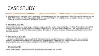
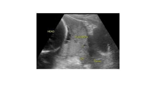
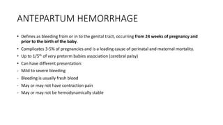
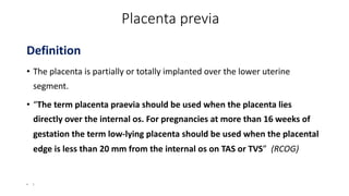
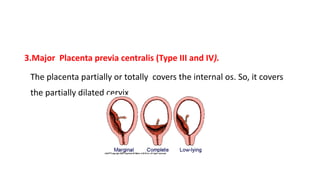
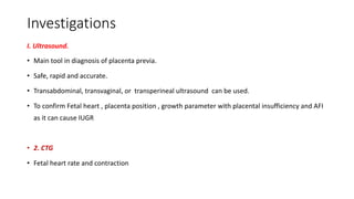
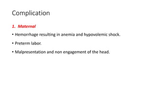
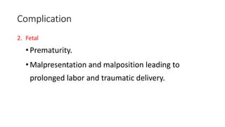
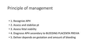
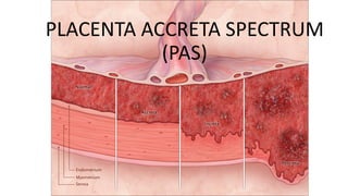
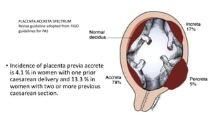
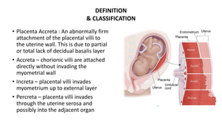
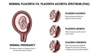
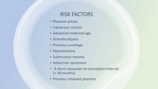
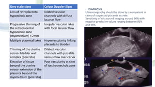
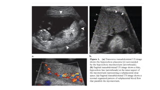
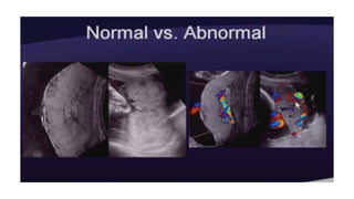
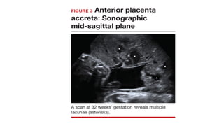
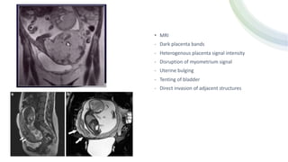
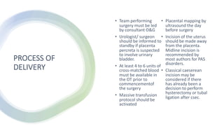
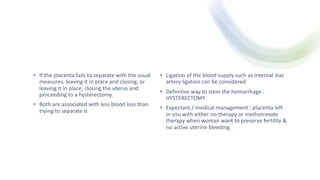
Placenta Previa
/ Placenta
Accreta
Spectrum
NAJMI SHAFIZ BIN ROZMAN
MENTOR DR SALBIAH
• “Placenta praevia and placenta accreta are associated with high maternal and neonatal
morbidity and mortality. The rates of placenta praevia and accreta have increased and will
continue to do so as a result of rising rates of caesarean deliveries, increased maternal age and
use of assisted reproductive technology (ART), placing greater demands on maternity-related
resources. The highest rates of complication for both mother and newborn are observed when
these conditions are only diagnosed at delivery”.
RCOG Royal College of Obstetricians and Gynaecologists
Placenta Praevia and Placenta Accreta: Diagnosis and Management 2018
CASE STUDY
• Fatimah Hussin is a 40 year old G3, P1+1 who is 32 weeks gestation. She awakened at 0200 thinking that she had wet the
bed. When she arose she discovered her bed was covered in bright red blood (painless pv bleed). Denies any trauma
recently , no urti/uti symptoms, good fetal movement
• ANTENATAL HISTORY :
Her records indicate she is carrying a singleton pregnancy in the vertex presentation.Sure of date . Antenatally patient had
history of incomplete miscarriage at 10 weeks (First pregnancy), D+C was done , uneventful.. Her second pregnancy pt
went for Elective C section for placenta previa type 3 in labor at 37 weeks gestation. This pregnancy has been uneventful
up until this time
• PAST MEDICAL HISTORY :
Her past medical history is uncomplicated, she has no allergies, and she takes no medications other than prenatal
vitamins. She admits to smoking less than one-half pack per day of cigarettes. Her prenatal labs are normal and her
pregnancy has been uncomplicated except for intermittent spotting in the last six weeks.
• ON EXAMINATION
Alert , pink not pale , warm peripheries , good pulse volume vItal sign is stable
What would you assess first?
• What would be one of the first questions you would ask?
• What would you not do?
.After recording your assessment data (from your facilitator) proceed with your care.
Per abdomen:
Soft, non tender
Uterus at 30-32 w
Singleton, cephalic
EFW: 2.8-3.0 kg
Per speculum:
VVNAD
Cx: healthy, noted cervical
erosion, minimal blood-stained, no active bleeding seen
Os: closed
Time contraction: 1:10:15sec
CTG: reactive with baseline FHR
150bpm, variability >5bpm and
acceleration. No deceleration.
TAS: follows parameters (30-32
weeks)
Placenta: low lying
AFI: 12cm
No retro-placental haemorrhage.
ANTEPARTUM HEMORRHAGE
• Defines as bleeding from or in to the genital tract, occurring from 24 weeks of pregnancy and
prior to the birth of the baby.
• Complicates 3-5% of pregnancies and is a leading cause of perinatal and maternal mortality.
• Up to 1/5th of very preterm babies association (cerebral palsy)
• Can have different presentation:
- Mild to severe bleeding
- Bleeding is usually fresh blood
- May or may not have contraction pain
- May or may not be hemodynamically stable
Placenta previa
Definition
• The placenta is partially or totally implanted over the lower uterine
segment.
• “The term placenta praevia should be used when the placenta lies
directly over the internal os. For pregnancies at more than 16 weeks of
gestation the term low-lying placenta should be used when the placental
edge is less than 20 mm from the internal os on TAS or TVS” (RCOG)
• I
• Determining placental location is one of the first aims of routine midpregnancy (18+6 to 21+6 weeks of gestation)
transabdominal obstetric ultrasound examination.
• Placenta praevia was originally defined using transabdominal scan (TAS) as a placenta developing within the lower
uterine segment and graded according to the relationship and/or the distance between the lower placental edge
and the internal os of the uterine cervix.
• If the placenta is thought to be low lying (less than 20 mm from the internal os) or praevia (covering the os) at the
routine fetal anomaly scan, a follow-up ultrasound examination including a TVS is recommended at 32 weeks of
gestation to diagnose persistent low-lying placenta and/or placenta praevia
• Clinicians should be aware that TVS for the diagnosis of placenta previa or a low-lying placenta is superior to
transabdominal and transperineal approaches, and is safe.
• In women with a persistent low-lying placenta or placenta previa at 32 weeks of gestation who remain asymptomatic, an
additional TVS is recommended at around 36 weeks of gestation to inform discussion about mode of delivery.
Type—I (Low-lying)
Type—II (Marginal)
Type—III (Incomplete or
partial central)
Type—IV (Central or
total)
Grade I or minor praevia is defined as a
lower edge inside the lower uterine
segment;
grade II or marginal praevia as a lower edge
reaching the internal os;
grade III or partial praevia when the
placenta partially covers the cervix;
grade IV or complete praevia when the
placenta completely covers the cervix.
Grades I and II are also often defined as
‘minor’ placenta praevia whereas
grades III and IV are referred to as ‘major’
placenta praevia.
Cause
• Placental abruption.
• Trauma.
• Tumors (cancer cervix, cervical polyp).
• Other rare causes include: Vaginal varicosities, severe vaginal infections, rupture uterus, cervical
erosion, placental edge bleeding.
Incidence
• 1/200 of pregnancies beyond 24 weeks of gestations.
• More in multiparas.
Etiology
A. Maternal
• Uterine scarring
• Prior Caesarean Section : 4 times more common.
• Prior D&C.
• Prior surgical evacuation for abortion.
• Increased age and parity. 9 fold increase in women > 40 years.
• Anemia.
• Previous history of placenta previa.
• Smoking.
• Assisted conception
B. Fetal
• Multiple pregnancy.
• Placenta membranecea.
• Large placenta (maternal diabetes)
Types
1. Low lying placenta, or placenta previa lateralis (Type I).
The placental edge encroaches upon the lower uterine segment, but not be
reaching the margin of the internal os.
2. Placenta previa marginalis
(Type II).
The lower edge of the placenta is reaching the margins of the internal os. It
may be implanted anteriorly (type II anterior) or posteriorly (type II
posterior).
3.Major Placenta previa centralis (Type III and IV).
The placenta partially or totally covers the internal os. So, it covers
the partially dilated cervix
Clinical Picture of Placenta Previa
(I) Symptoms: after 20 wks
• The cardinal symptom of placenta previa is painless, causeless, recurrent vaginal
bleeding.
• However, the bleeding may be associated with some uterine contractions (pain).
• The bleeding may be initiated by intercourse or vaginal examination (cause).
• The patient may be presented during the first attack of the bleeding.
Mechanism of bleeding:
(1) During the last weeks of pregnancy:
• The lower uterine segment stretches and enlarges. The inelastic
placenta can not stretch so as to keep place with it, so shearing action
occurs, leading to inevitable separation and tearing of some placental
sinuses and vaginal bleeding.
Clinical picture of placenta previa
(A) General examination.
• The general condition is proportionate to the amount of blood loss.
• Pallor due to blood loss.
(B) Abdominal examination.
• The abdomen is lax, fundal level equals the period of amenorrhea.
• Fetal parts are easily felt.
• Malpresentation and non-engagement are common.
• Fetal heart sounds are usually normal.
Investigations
I. Ultrasound.
• Main tool in diagnosis of placenta previa.
• Safe, rapid and accurate.
• Transabdominal, transvaginal, or transperineal ultrasound can be used.
• To confirm Fetal heart , placenta position , growth parameter with placental insufficiency and AFI
as it can cause IUGR
• 2. CTG
• Fetal heart rate and contraction
3. Labs
• FBC- to look for Hb level and platelet , to act as baseline as bleeding might
become continously
• COAG- To exclude prolonged coag profile in case of anticipate bleeding
• BUSE ,RP- major bleeding can compromise renal function
• GXM -4 pint pack cell +/- DIVC REGME
4.Speculum STRICTLY NO VE !
Speculum exam must be done in all cases of APH
Complication
1. Maternal
• Hemorrhage resulting in anemia and hypovolemic shock.
• Preterm labor.
• Malpresentation and non engagement of the head.
Complication
2. Fetal
•Prematurity.
•Malpresentation and malposition leading to
prolonged labor and traumatic delivery.
Principle of management
• 1. Recognize APH
• 2. Assess and stabilize pt
• 3. Assess fetal viability
• 4. Diagnose APH secondary to BLEEDING PLACENTA PREVIA
• 5. Deliver depends on gestation and amount of bleeding
Assessment Resuscitate Investigation
1. Blood pressure 1. Call for help +/-
RED ALERT
1. FBC
2. Pulse rate 2. 2 large bore
cannula
2. COAG
3. Shock index if
bleeding is significant
3. Fluid
resuscitation
3. GXM 4 pints +/-
DIVC regime
4. Blood loss 4. Oxygen Theraphy 4. Speculum (NO VE)
5. FHR -CTG 5. CBD
6. Ultrasound scan to
locate placenta
The decision for delivery is based on the quantity and continuity of bleed irrespective of fetal viability
of maturity
APH secondary to PP
HEAVY AND
CONTINUOUS
BLEEDING
LIGHT
BLEEDING/BLEEDING
STOP
For imminent
delivery by LSCS
Conservative
management if
For delivery if
LSCS by an experienced
Surgeon in specialist
hospital
- Premature baby
- To complete
antenatal steroids
- -correct anemia
- -GXM blood
Fetus> 36 weeks
. Management of Patients with Mild and Moderate Bleeding
• Depends on gestational age, in labor or not and type of placenta
previa..
I. Delivery is indicated in:
• Patients in labor.
• Mature fetus.
• Route of delivery.
(A) Vaginal delivery
• Indicated for placenta previa lateralis and marginalis anterior.
• Prerequisites are:
1- Mild bleeding.
2- Vertex presentation.
3- Favorable cervix
(B) C.S :
• Indicated for marginalis posterior incomplete and complete centralis.
Management
Conservative treatment
Indications
• Mild bleeding and the patient is hemodynamically stable.
• Gestational age is less than 36 weeks with immature fetal
lungs.
• The patient is not in labor.
Items of conservative treatment. (Mc Afee Regime)
• Maternal
- Vital sign monitoring any shock
-strict pad chart , to inform if any immediate PV bleeding
-KNBM but if no pv bleeding allow orally as tolerated
-4 pints IVD maintenance
-weekly FBC if symptomatic or continuously PV bleed
-Ask everyday any PV bleed or abdominal pain (important)
• Fetal
- FKC , to inform stat if reduce in fm
- Biweekly CTG and usg
• Labor
- To Prolonged the pregnancy
- To inform if any sign and symptom of labor
- Plan on mode of delivery-depends on type of PP
1. Admission to ward
until delivery
2. Close observation for
bleeding
3. Availability of 2 units of
GXM
4. Avalability of C Section
• Elective Carsarean Lower Segment C Section at 38 weeks of POG if
placenta edge is <2 cm from internal os and placenta is thick
• Allow SVD at term in minor PP unless there is fetal/maternal
compromised
• Before ELSCS do placental mapping to locate placenta and avoid
incision through placenta
PLACENTA ACCRETA SPECTRUM
(PAS)
Case study
• A 36-year-old woman with three previous cesarean deliveries is
referred at 28 weeks of gestation because of placenta previa
detected on ultrasonography. Other ultrasonographic findings
include multiple intraplacental lacunae and loss of the normal
retroplacental hypoechoic zone in the area underlying the dome
of the maternal bladder. Placenta accreta is suspected. The
course of the patient’s pregnancy otherwise has been normal.
How should she be counseled and her care be managed?
What is Placenta Accreta ?
• general term applied to abnormal adherence of the placental
trophoblast to the uterine myometrium; it is also referred to as
morbidly adherent placenta. ’
• The spectrum includes placenta accreta (attachment of the
placenta to myometrium without intervening decidua),
• placenta increta (invasion of the trophoblast into the
myometrium),
• placenta percreta (invasion through the myometrium, serosa,
and into surrounding structures)
• Placenta accreta is a histopathological term first defined by
Irving and Hertig in 1937, as the “abnormal adherence of the
afterbirth in whole or in parts to the underlying uterine wall in
the partial or complete absence of decidua”.
PLACENTA ACCRETA SPECTRUM
Revise guideline adopted from FIGO
guidelines for PAS
• Incidence of placenta previa accrete
is 4.1 % in women with one prior
caesarean delivery and 13.3 % in
women with two or more previous
caesarean section.
• Placenta Accreta : An abnormally firm
attachment of the placental villi to
the uterine wall. This is due to partial
or total lack of decidual basalis layer
• Accreta – chorionic villi are attached
directly without invading the
myometrial wall
• Increta – placental villi invades
myometrium up to external layer
• Percreta – placenta villi invades
through the uterine serosa and
possibly into the adjacent organ
DEFINITION
& CLASSIFICATION
RISK FACTORS
• Placenta previa
• Caeserean section
• Advanced maternal age
• Grandmultipara
• Previous curettage
• Myomectomy
• Submucous myoma
• Asherman syndrome
• A short caesarean to conception interval
(< 18 months)
• Previous retained placenta
• DIAGNOSIS
Ultrasonography should be done by a competent in
cases of suspected placenta accrete.
Sensitivity of ultrasound imaging around 90% with
negative predictive values ranging between 95%
and 98%
Grey scale signs Colour Doppler Signs
Loss of retroplacental
hypoechoic zone
Dilated vascular
channels with diffuse
lacunar flow
Progressive thinning of
the retroplacental
hypoechoic zone
(myometrium) < 2mm
Irregular vascular lakes
with focal lacunar flow
Multiple placental lakes Hypervascularity linking
placenta to bladder
Thinning of the uterine
serosa- bladder wall
complex (percreta)
Dilated, vascular
channels with pulsatile
venous flow over cervix
Elevation of tissue
beyond the uterine
serosa- extension of the
placenta beyond the
myometrium (percreta)
Poor vascularity at sites
of loss hypoechoic zone
• MRI
- Dark placenta bands
- Heterogenous placenta signal intensity
- Disruption of myometrium signal
- Uterine bulging
- Tenting of bladder
- Direct invasion of adjacent structures
• ANTENATAL CARE
• MULTIDISCIPLINARY MANAGEMENT
• CONSERVATIVE MANAGEMENT
• PROCESS OF DELIVERY
ANTENATAL CARE
• Routine ultrasound scanning at 20 weeks of
gestation should include placental localization
• Follow up scan should be done at 32 weeks in
asymptomatic women with low lying placenta with
or without previous csec.
• Needs to be referred to a specialist hospital for
confirmation and management.
• Prolonged periods of hospitalization
MULTIDISCIPLINARY
MANAGEMENT
• ANAESTHETIC TEAM : OT & availability of ICU bed
• PAEDIATRICIAN : Possible of prematurity delivery
• SURGICAL
• UROLOGY
• INTERVENTIONAL RADIOLOGY
• BLOOD BANK
CONSERVATIVE
MANAGEMENT
• Defines all procedures that aim to avoid peripartum
hysterectomy and its related morbidity and
consequences.
• Four different primary methods of conservative
management have been described in the
international literature :
1. The Extirpative technique ( Manual removal of
placenta)
2. Leaving the Placenta Insitu or the Expectant
approach
3. One –step conservative surgery ( Removal of the
Accreta area after resection)
4. Triple – P procedure : suturing around the
accrete area after resection
PROCESS OF
DELIVERY
• Team performing
surgery must be led
by consultant O&G
• Urologist/ surgeon
should be informed to
standby if placenta
percreta is suspected
to involve urinary
bladder.
• At least 4 to 6 units of
cross-matched blood
must be available in
the OT prior to
commencementof
the surgery
• Massive transfusion
protocol should be
activated
• Placental mapping by
ultrasound the day
before surgery
• Incision of the uterus
should be made away
from the placenta.
Midline incision is
recommended by
most authors for PAS
disorders.
• Classical caeserean
incision may be
considered if there
has already been a
decision to perform
hysterectomy or tubal
ligation after csec.
• If the placenta fails to separate with the usual
measures, leaving it in place and closing, or
leaving it in place, closing the uterus and
proceeding to a hysterectomy.
• Both are associated with less blood loss than
trying to separate it
• Ligation of the blood supply such as internal iliac
artery ligation can be considered
• Definitive way to stem the hemorrhage :
HYSTERECTOMY
• Expectant / medical management : placenta left
in situ with either no therapy or methotrexate
therapy when woman want to preserve fertility &
no active uterine bleeding.
COMPLICATIONS
OF ADHERENT
PLACENTA
• Maternal :
- Hysterectomy ( range from 0.7 % with one
previous caeserean delivery to 9.0 % with more
than 6 caeserean delivery
- Injury to bladder 7 – 48%
- Injury to ureter 0- 18%
- Bowel injury/ obstruction
- large volume blood transfusions ( > 10L) 5 – 40%
- Venous thromboembolism 4%
- Surgical site infection 18 – 32 %
- Reoperation 4% - 18%
- Maternal mortality 1% - 7%
- Admission to ICU 15% - 66%
Perinatal :
- Small gestational age
- Prematurity
CONCLUSION
• PAS is a high risk condition. Good results would
only be obtained through multidisciplinary,
planned approach with adequate blood and
critical care support. The patient must be fully
involved in all steps of the decision making
process.
Sources
• RCOG Royal College of Obstetricians and
Gynaecologists
Placenta Praevia and Placenta Accreta: Diagnosis and
Management 2018
• Sarawak General Hospital Labour ward manual
2020 edition

najmi placenta previa final 4.pdf

  • 1.
    Placenta Previa / Placenta Accreta Spectrum NAJMISHAFIZ BIN ROZMAN MENTOR DR SALBIAH
  • 2.
    • “Placenta praeviaand placenta accreta are associated with high maternal and neonatal morbidity and mortality. The rates of placenta praevia and accreta have increased and will continue to do so as a result of rising rates of caesarean deliveries, increased maternal age and use of assisted reproductive technology (ART), placing greater demands on maternity-related resources. The highest rates of complication for both mother and newborn are observed when these conditions are only diagnosed at delivery”. RCOG Royal College of Obstetricians and Gynaecologists Placenta Praevia and Placenta Accreta: Diagnosis and Management 2018
  • 3.
    CASE STUDY • FatimahHussin is a 40 year old G3, P1+1 who is 32 weeks gestation. She awakened at 0200 thinking that she had wet the bed. When she arose she discovered her bed was covered in bright red blood (painless pv bleed). Denies any trauma recently , no urti/uti symptoms, good fetal movement • ANTENATAL HISTORY : Her records indicate she is carrying a singleton pregnancy in the vertex presentation.Sure of date . Antenatally patient had history of incomplete miscarriage at 10 weeks (First pregnancy), D+C was done , uneventful.. Her second pregnancy pt went for Elective C section for placenta previa type 3 in labor at 37 weeks gestation. This pregnancy has been uneventful up until this time • PAST MEDICAL HISTORY : Her past medical history is uncomplicated, she has no allergies, and she takes no medications other than prenatal vitamins. She admits to smoking less than one-half pack per day of cigarettes. Her prenatal labs are normal and her pregnancy has been uncomplicated except for intermittent spotting in the last six weeks. • ON EXAMINATION Alert , pink not pale , warm peripheries , good pulse volume vItal sign is stable
  • 4.
    What would youassess first? • What would be one of the first questions you would ask? • What would you not do? .After recording your assessment data (from your facilitator) proceed with your care. Per abdomen: Soft, non tender Uterus at 30-32 w Singleton, cephalic EFW: 2.8-3.0 kg Per speculum: VVNAD Cx: healthy, noted cervical erosion, minimal blood-stained, no active bleeding seen Os: closed Time contraction: 1:10:15sec CTG: reactive with baseline FHR 150bpm, variability >5bpm and acceleration. No deceleration. TAS: follows parameters (30-32 weeks) Placenta: low lying AFI: 12cm No retro-placental haemorrhage.
  • 6.
    ANTEPARTUM HEMORRHAGE • Definesas bleeding from or in to the genital tract, occurring from 24 weeks of pregnancy and prior to the birth of the baby. • Complicates 3-5% of pregnancies and is a leading cause of perinatal and maternal mortality. • Up to 1/5th of very preterm babies association (cerebral palsy) • Can have different presentation: - Mild to severe bleeding - Bleeding is usually fresh blood - May or may not have contraction pain - May or may not be hemodynamically stable
  • 7.
    Placenta previa Definition • Theplacenta is partially or totally implanted over the lower uterine segment. • “The term placenta praevia should be used when the placenta lies directly over the internal os. For pregnancies at more than 16 weeks of gestation the term low-lying placenta should be used when the placental edge is less than 20 mm from the internal os on TAS or TVS” (RCOG) • I
  • 8.
    • Determining placentallocation is one of the first aims of routine midpregnancy (18+6 to 21+6 weeks of gestation) transabdominal obstetric ultrasound examination. • Placenta praevia was originally defined using transabdominal scan (TAS) as a placenta developing within the lower uterine segment and graded according to the relationship and/or the distance between the lower placental edge and the internal os of the uterine cervix. • If the placenta is thought to be low lying (less than 20 mm from the internal os) or praevia (covering the os) at the routine fetal anomaly scan, a follow-up ultrasound examination including a TVS is recommended at 32 weeks of gestation to diagnose persistent low-lying placenta and/or placenta praevia • Clinicians should be aware that TVS for the diagnosis of placenta previa or a low-lying placenta is superior to transabdominal and transperineal approaches, and is safe. • In women with a persistent low-lying placenta or placenta previa at 32 weeks of gestation who remain asymptomatic, an additional TVS is recommended at around 36 weeks of gestation to inform discussion about mode of delivery.
  • 10.
    Type—I (Low-lying) Type—II (Marginal) Type—III(Incomplete or partial central) Type—IV (Central or total) Grade I or minor praevia is defined as a lower edge inside the lower uterine segment; grade II or marginal praevia as a lower edge reaching the internal os; grade III or partial praevia when the placenta partially covers the cervix; grade IV or complete praevia when the placenta completely covers the cervix. Grades I and II are also often defined as ‘minor’ placenta praevia whereas grades III and IV are referred to as ‘major’ placenta praevia.
  • 11.
    Cause • Placental abruption. •Trauma. • Tumors (cancer cervix, cervical polyp). • Other rare causes include: Vaginal varicosities, severe vaginal infections, rupture uterus, cervical erosion, placental edge bleeding.
  • 13.
    Incidence • 1/200 ofpregnancies beyond 24 weeks of gestations. • More in multiparas.
  • 14.
    Etiology A. Maternal • Uterinescarring • Prior Caesarean Section : 4 times more common. • Prior D&C. • Prior surgical evacuation for abortion. • Increased age and parity. 9 fold increase in women > 40 years. • Anemia. • Previous history of placenta previa. • Smoking. • Assisted conception
  • 15.
    B. Fetal • Multiplepregnancy. • Placenta membranecea. • Large placenta (maternal diabetes)
  • 16.
    Types 1. Low lyingplacenta, or placenta previa lateralis (Type I). The placental edge encroaches upon the lower uterine segment, but not be reaching the margin of the internal os. 2. Placenta previa marginalis (Type II). The lower edge of the placenta is reaching the margins of the internal os. It may be implanted anteriorly (type II anterior) or posteriorly (type II posterior).
  • 17.
    3.Major Placenta previacentralis (Type III and IV). The placenta partially or totally covers the internal os. So, it covers the partially dilated cervix
  • 18.
    Clinical Picture ofPlacenta Previa (I) Symptoms: after 20 wks • The cardinal symptom of placenta previa is painless, causeless, recurrent vaginal bleeding. • However, the bleeding may be associated with some uterine contractions (pain). • The bleeding may be initiated by intercourse or vaginal examination (cause). • The patient may be presented during the first attack of the bleeding.
  • 19.
    Mechanism of bleeding: (1)During the last weeks of pregnancy: • The lower uterine segment stretches and enlarges. The inelastic placenta can not stretch so as to keep place with it, so shearing action occurs, leading to inevitable separation and tearing of some placental sinuses and vaginal bleeding.
  • 20.
    Clinical picture ofplacenta previa (A) General examination. • The general condition is proportionate to the amount of blood loss. • Pallor due to blood loss. (B) Abdominal examination. • The abdomen is lax, fundal level equals the period of amenorrhea. • Fetal parts are easily felt. • Malpresentation and non-engagement are common. • Fetal heart sounds are usually normal.
  • 22.
    Investigations I. Ultrasound. • Maintool in diagnosis of placenta previa. • Safe, rapid and accurate. • Transabdominal, transvaginal, or transperineal ultrasound can be used. • To confirm Fetal heart , placenta position , growth parameter with placental insufficiency and AFI as it can cause IUGR • 2. CTG • Fetal heart rate and contraction
  • 24.
    3. Labs • FBC-to look for Hb level and platelet , to act as baseline as bleeding might become continously • COAG- To exclude prolonged coag profile in case of anticipate bleeding • BUSE ,RP- major bleeding can compromise renal function • GXM -4 pint pack cell +/- DIVC REGME 4.Speculum STRICTLY NO VE ! Speculum exam must be done in all cases of APH
  • 25.
    Complication 1. Maternal • Hemorrhageresulting in anemia and hypovolemic shock. • Preterm labor. • Malpresentation and non engagement of the head.
  • 26.
    Complication 2. Fetal •Prematurity. •Malpresentation andmalposition leading to prolonged labor and traumatic delivery.
  • 27.
    Principle of management •1. Recognize APH • 2. Assess and stabilize pt • 3. Assess fetal viability • 4. Diagnose APH secondary to BLEEDING PLACENTA PREVIA • 5. Deliver depends on gestation and amount of bleeding
  • 28.
    Assessment Resuscitate Investigation 1.Blood pressure 1. Call for help +/- RED ALERT 1. FBC 2. Pulse rate 2. 2 large bore cannula 2. COAG 3. Shock index if bleeding is significant 3. Fluid resuscitation 3. GXM 4 pints +/- DIVC regime 4. Blood loss 4. Oxygen Theraphy 4. Speculum (NO VE) 5. FHR -CTG 5. CBD 6. Ultrasound scan to locate placenta
  • 29.
    The decision fordelivery is based on the quantity and continuity of bleed irrespective of fetal viability of maturity APH secondary to PP HEAVY AND CONTINUOUS BLEEDING LIGHT BLEEDING/BLEEDING STOP For imminent delivery by LSCS Conservative management if For delivery if LSCS by an experienced Surgeon in specialist hospital - Premature baby - To complete antenatal steroids - -correct anemia - -GXM blood Fetus> 36 weeks
  • 30.
    . Management ofPatients with Mild and Moderate Bleeding • Depends on gestational age, in labor or not and type of placenta previa.. I. Delivery is indicated in: • Patients in labor. • Mature fetus.
  • 31.
    • Route ofdelivery. (A) Vaginal delivery • Indicated for placenta previa lateralis and marginalis anterior. • Prerequisites are: 1- Mild bleeding. 2- Vertex presentation. 3- Favorable cervix (B) C.S : • Indicated for marginalis posterior incomplete and complete centralis.
  • 32.
    Management Conservative treatment Indications • Mildbleeding and the patient is hemodynamically stable. • Gestational age is less than 36 weeks with immature fetal lungs. • The patient is not in labor.
  • 33.
    Items of conservativetreatment. (Mc Afee Regime) • Maternal - Vital sign monitoring any shock -strict pad chart , to inform if any immediate PV bleeding -KNBM but if no pv bleeding allow orally as tolerated -4 pints IVD maintenance -weekly FBC if symptomatic or continuously PV bleed -Ask everyday any PV bleed or abdominal pain (important) • Fetal - FKC , to inform stat if reduce in fm - Biweekly CTG and usg • Labor - To Prolonged the pregnancy - To inform if any sign and symptom of labor - Plan on mode of delivery-depends on type of PP 1. Admission to ward until delivery 2. Close observation for bleeding 3. Availability of 2 units of GXM 4. Avalability of C Section
  • 34.
    • Elective CarsareanLower Segment C Section at 38 weeks of POG if placenta edge is <2 cm from internal os and placenta is thick • Allow SVD at term in minor PP unless there is fetal/maternal compromised • Before ELSCS do placental mapping to locate placenta and avoid incision through placenta
  • 36.
  • 37.
    Case study • A36-year-old woman with three previous cesarean deliveries is referred at 28 weeks of gestation because of placenta previa detected on ultrasonography. Other ultrasonographic findings include multiple intraplacental lacunae and loss of the normal retroplacental hypoechoic zone in the area underlying the dome of the maternal bladder. Placenta accreta is suspected. The course of the patient’s pregnancy otherwise has been normal. How should she be counseled and her care be managed?
  • 38.
    What is PlacentaAccreta ? • general term applied to abnormal adherence of the placental trophoblast to the uterine myometrium; it is also referred to as morbidly adherent placenta. ’ • The spectrum includes placenta accreta (attachment of the placenta to myometrium without intervening decidua), • placenta increta (invasion of the trophoblast into the myometrium), • placenta percreta (invasion through the myometrium, serosa, and into surrounding structures) • Placenta accreta is a histopathological term first defined by Irving and Hertig in 1937, as the “abnormal adherence of the afterbirth in whole or in parts to the underlying uterine wall in the partial or complete absence of decidua”.
  • 39.
    PLACENTA ACCRETA SPECTRUM Reviseguideline adopted from FIGO guidelines for PAS • Incidence of placenta previa accrete is 4.1 % in women with one prior caesarean delivery and 13.3 % in women with two or more previous caesarean section.
  • 40.
    • Placenta Accreta: An abnormally firm attachment of the placental villi to the uterine wall. This is due to partial or total lack of decidual basalis layer • Accreta – chorionic villi are attached directly without invading the myometrial wall • Increta – placental villi invades myometrium up to external layer • Percreta – placenta villi invades through the uterine serosa and possibly into the adjacent organ DEFINITION & CLASSIFICATION
  • 42.
    RISK FACTORS • Placentaprevia • Caeserean section • Advanced maternal age • Grandmultipara • Previous curettage • Myomectomy • Submucous myoma • Asherman syndrome • A short caesarean to conception interval (< 18 months) • Previous retained placenta
  • 43.
    • DIAGNOSIS Ultrasonography shouldbe done by a competent in cases of suspected placenta accrete. Sensitivity of ultrasound imaging around 90% with negative predictive values ranging between 95% and 98% Grey scale signs Colour Doppler Signs Loss of retroplacental hypoechoic zone Dilated vascular channels with diffuse lacunar flow Progressive thinning of the retroplacental hypoechoic zone (myometrium) < 2mm Irregular vascular lakes with focal lacunar flow Multiple placental lakes Hypervascularity linking placenta to bladder Thinning of the uterine serosa- bladder wall complex (percreta) Dilated, vascular channels with pulsatile venous flow over cervix Elevation of tissue beyond the uterine serosa- extension of the placenta beyond the myometrium (percreta) Poor vascularity at sites of loss hypoechoic zone
  • 47.
    • MRI - Darkplacenta bands - Heterogenous placenta signal intensity - Disruption of myometrium signal - Uterine bulging - Tenting of bladder - Direct invasion of adjacent structures
  • 48.
    • ANTENATAL CARE •MULTIDISCIPLINARY MANAGEMENT • CONSERVATIVE MANAGEMENT • PROCESS OF DELIVERY
  • 49.
    ANTENATAL CARE • Routineultrasound scanning at 20 weeks of gestation should include placental localization • Follow up scan should be done at 32 weeks in asymptomatic women with low lying placenta with or without previous csec. • Needs to be referred to a specialist hospital for confirmation and management. • Prolonged periods of hospitalization
  • 50.
    MULTIDISCIPLINARY MANAGEMENT • ANAESTHETIC TEAM: OT & availability of ICU bed • PAEDIATRICIAN : Possible of prematurity delivery • SURGICAL • UROLOGY • INTERVENTIONAL RADIOLOGY • BLOOD BANK
  • 51.
    CONSERVATIVE MANAGEMENT • Defines allprocedures that aim to avoid peripartum hysterectomy and its related morbidity and consequences. • Four different primary methods of conservative management have been described in the international literature : 1. The Extirpative technique ( Manual removal of placenta) 2. Leaving the Placenta Insitu or the Expectant approach 3. One –step conservative surgery ( Removal of the Accreta area after resection) 4. Triple – P procedure : suturing around the accrete area after resection
  • 52.
    PROCESS OF DELIVERY • Teamperforming surgery must be led by consultant O&G • Urologist/ surgeon should be informed to standby if placenta percreta is suspected to involve urinary bladder. • At least 4 to 6 units of cross-matched blood must be available in the OT prior to commencementof the surgery • Massive transfusion protocol should be activated • Placental mapping by ultrasound the day before surgery • Incision of the uterus should be made away from the placenta. Midline incision is recommended by most authors for PAS disorders. • Classical caeserean incision may be considered if there has already been a decision to perform hysterectomy or tubal ligation after csec.
  • 53.
    • If theplacenta fails to separate with the usual measures, leaving it in place and closing, or leaving it in place, closing the uterus and proceeding to a hysterectomy. • Both are associated with less blood loss than trying to separate it • Ligation of the blood supply such as internal iliac artery ligation can be considered • Definitive way to stem the hemorrhage : HYSTERECTOMY • Expectant / medical management : placenta left in situ with either no therapy or methotrexate therapy when woman want to preserve fertility & no active uterine bleeding.
  • 54.
    COMPLICATIONS OF ADHERENT PLACENTA • Maternal: - Hysterectomy ( range from 0.7 % with one previous caeserean delivery to 9.0 % with more than 6 caeserean delivery - Injury to bladder 7 – 48% - Injury to ureter 0- 18% - Bowel injury/ obstruction - large volume blood transfusions ( > 10L) 5 – 40% - Venous thromboembolism 4% - Surgical site infection 18 – 32 % - Reoperation 4% - 18% - Maternal mortality 1% - 7% - Admission to ICU 15% - 66% Perinatal : - Small gestational age - Prematurity
  • 55.
    CONCLUSION • PAS isa high risk condition. Good results would only be obtained through multidisciplinary, planned approach with adequate blood and critical care support. The patient must be fully involved in all steps of the decision making process.
  • 56.
    Sources • RCOG RoyalCollege of Obstetricians and Gynaecologists Placenta Praevia and Placenta Accreta: Diagnosis and Management 2018 • Sarawak General Hospital Labour ward manual 2020 edition