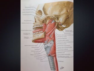
Pharynx
Part 1
Pharynx
Is about 12 cm long
Extending from base of skull to the lower border of the
cricoid cartilage
Opposite the sixth cervical vertebra where it is
continuous with oesophagus
Divided in to 3 parts : nasopharynx,oropharynx and
laryngopharynx
Nasopharynx
Behind the nasal cavity and above the soft palate.
Posterior nasal apertures open into it anterioly
Below it is continuous with oropharynx through
pharyngeal isthmus
The nasopharynx is part of the upper respiratory tract
and does not normally give passage to food or drink
Nasopharynx
Auditory tube opens on the lateral wall of nasopharynx
This opening is bounded above and behind by the tubal
elevation produced by underlying medial end of auditory
tube
A collection of lymphoid tissue,the pharyngeal tonsil or
adenoid is located beneath the mucous membrane
The tonsil may become enlarged in chronic infections of
upper respiratory system leading to mouth breathing
Nasopharynx
The mucous membrane lining the nasopharynx is of
the respiratory type,covered by ciliated cuboidal
epithilium
Receives its sensory and secretomotor nerve supply
through a branch of pterygopalatine gangilion
It resembles the mucous membrane lining the nose
Oropharynx
Extends from soft palate to epiglottis
Oral cavity opens into oropharynx anteriorly through
oropharngeal isthmus ,which is bounded on each side by a
fold of mucous membrane ,the palatoglossal arch
The arch is produced by the underlying palatoglossus
muscle
The lateral wall of oropharynx ,behind the palatoglossal arch
presents a vertical ridge of mucous membrane ,the
palatopharyngeal arch produced by palatopharyngeus
muscle
Oropharynx
Between palatoglossal and palatopharyngeal arches is a
depression ,the tonsillar sinus.
The palatine tonsil ,a mass of lymphoid tissue, is situated beneath
the mucous membrane lining the sinus
Laterally the tonsil is enclosed by a fibrous capsule
The tonsil is very variable in size and is frequently the site of
infection which causes it to become enlarged and painful
(tonsillitis)
The oropharynx is lined by stratified squamous epithilium, nerve
supply is from pharyngeal plexus
Laryngopharynx
Extends from epiglottis to the lower border of the cricoid
cartilage
Laryngopharynx is lined by stratified squamous epithilium and
innervated by vagus through the internal and recurrent
laryngeal branches
On functional nasopharynx regarded as part of nasal cavity
Unlike oropharynx and laryngopharynx it is respiratory in
function ,lined by respiratory mucosa which receives its
sensory innervation from trigiminal nerve
Muscles of pharynx
Three paired of constrictor muscles , meet their
fellows of opposite side in the posterior pharyngeal
wall at the median pharyngeal ligament and raphe
Three other muscles,these are stylopharngeus
,salpinopharyngeus ,palatopharyngeus
Superior constrictor
Arises from lower two thirds of posterior border of the medial
pterygoid plate (including hamulus) and from the posterior end of
mylohyoid line on the medial surface of the mandible
Between hamulus and the mandible the fibres arise from the
pterygomandibular raphe where they interdigitate with fibers of
buccinator
Between the upper border of the muscle and the base of the skull
is a gap through which the cartilaginous part of auditory tube and
levator veli palatini muscle pass
The remainder of the gap is closed by pharyngobasilar fascia
Middle constrictor
Attached anteriorly to stylohyoid ligament, the lesser
Cornu and upper border of greater Cornu of hyoid
bone
The upper fibers diverge upwards,passing superficial
to the lower part of superior constrictor
The lower fibers run horizontally passing deep to
upper part of inferior constrictor
Inferior constrictor
Consists of two parts (thyropharyngeus and cricopharyngeus)
Thyropharyngeus arises from oblique line on the lamina of the
thyroid cartilage and inserted in to pharyngeal raphe
Cricopharyngeus attached anteriorly to the side of the arch of
cricoid cartilage
It encircles the lower most part of pharynx
A weak area called killians dehiscence, is present between
thyropharyngeus and cricopharyngeus giving rise to pharyngeal
pouch
Salpinopharyngeus
Arises from the cartilage of auditory tube
It passes downwards beneath the pharyngeal mucous
membrane and raised to form salpinopharyngeal fold
and blends with constrictor muscle
Stylopharngeus muscle
Arises from medial surfaces of styloid process
It passes between the internal and external carotid
arteries
It crosses the lower border of superior constrictor
muscle and continues downwards inside the middle
constrictor , beneath the pharyngeal mucous
membrane to be inserted into posterior border of
thyroid cartilage and the side wall of the pharynx
Pharyngeal muscles , with exception of
stylopharngeus are innervated by accessory nerve
Stylopharngeus is the only muscle derived from third
pharyngeal arch and supplied by glossopharyngeal
nerve

pharynx part1

  • 1.
  • 2.
    Pharynx Is about 12cm long Extending from base of skull to the lower border of the cricoid cartilage Opposite the sixth cervical vertebra where it is continuous with oesophagus Divided in to 3 parts : nasopharynx,oropharynx and laryngopharynx
  • 4.
    Nasopharynx Behind the nasalcavity and above the soft palate. Posterior nasal apertures open into it anterioly Below it is continuous with oropharynx through pharyngeal isthmus The nasopharynx is part of the upper respiratory tract and does not normally give passage to food or drink
  • 5.
    Nasopharynx Auditory tube openson the lateral wall of nasopharynx This opening is bounded above and behind by the tubal elevation produced by underlying medial end of auditory tube A collection of lymphoid tissue,the pharyngeal tonsil or adenoid is located beneath the mucous membrane The tonsil may become enlarged in chronic infections of upper respiratory system leading to mouth breathing
  • 6.
    Nasopharynx The mucous membranelining the nasopharynx is of the respiratory type,covered by ciliated cuboidal epithilium Receives its sensory and secretomotor nerve supply through a branch of pterygopalatine gangilion It resembles the mucous membrane lining the nose
  • 7.
    Oropharynx Extends from softpalate to epiglottis Oral cavity opens into oropharynx anteriorly through oropharngeal isthmus ,which is bounded on each side by a fold of mucous membrane ,the palatoglossal arch The arch is produced by the underlying palatoglossus muscle The lateral wall of oropharynx ,behind the palatoglossal arch presents a vertical ridge of mucous membrane ,the palatopharyngeal arch produced by palatopharyngeus muscle
  • 8.
    Oropharynx Between palatoglossal andpalatopharyngeal arches is a depression ,the tonsillar sinus. The palatine tonsil ,a mass of lymphoid tissue, is situated beneath the mucous membrane lining the sinus Laterally the tonsil is enclosed by a fibrous capsule The tonsil is very variable in size and is frequently the site of infection which causes it to become enlarged and painful (tonsillitis) The oropharynx is lined by stratified squamous epithilium, nerve supply is from pharyngeal plexus
  • 9.
    Laryngopharynx Extends from epiglottisto the lower border of the cricoid cartilage Laryngopharynx is lined by stratified squamous epithilium and innervated by vagus through the internal and recurrent laryngeal branches On functional nasopharynx regarded as part of nasal cavity Unlike oropharynx and laryngopharynx it is respiratory in function ,lined by respiratory mucosa which receives its sensory innervation from trigiminal nerve
  • 10.
    Muscles of pharynx Threepaired of constrictor muscles , meet their fellows of opposite side in the posterior pharyngeal wall at the median pharyngeal ligament and raphe Three other muscles,these are stylopharngeus ,salpinopharyngeus ,palatopharyngeus
  • 12.
    Superior constrictor Arises fromlower two thirds of posterior border of the medial pterygoid plate (including hamulus) and from the posterior end of mylohyoid line on the medial surface of the mandible Between hamulus and the mandible the fibres arise from the pterygomandibular raphe where they interdigitate with fibers of buccinator Between the upper border of the muscle and the base of the skull is a gap through which the cartilaginous part of auditory tube and levator veli palatini muscle pass The remainder of the gap is closed by pharyngobasilar fascia
  • 13.
    Middle constrictor Attached anteriorlyto stylohyoid ligament, the lesser Cornu and upper border of greater Cornu of hyoid bone The upper fibers diverge upwards,passing superficial to the lower part of superior constrictor The lower fibers run horizontally passing deep to upper part of inferior constrictor
  • 14.
    Inferior constrictor Consists oftwo parts (thyropharyngeus and cricopharyngeus) Thyropharyngeus arises from oblique line on the lamina of the thyroid cartilage and inserted in to pharyngeal raphe Cricopharyngeus attached anteriorly to the side of the arch of cricoid cartilage It encircles the lower most part of pharynx A weak area called killians dehiscence, is present between thyropharyngeus and cricopharyngeus giving rise to pharyngeal pouch
  • 16.
    Salpinopharyngeus Arises from thecartilage of auditory tube It passes downwards beneath the pharyngeal mucous membrane and raised to form salpinopharyngeal fold and blends with constrictor muscle
  • 18.
    Stylopharngeus muscle Arises frommedial surfaces of styloid process It passes between the internal and external carotid arteries It crosses the lower border of superior constrictor muscle and continues downwards inside the middle constrictor , beneath the pharyngeal mucous membrane to be inserted into posterior border of thyroid cartilage and the side wall of the pharynx
  • 19.
    Pharyngeal muscles ,with exception of stylopharngeus are innervated by accessory nerve Stylopharngeus is the only muscle derived from third pharyngeal arch and supplied by glossopharyngeal nerve