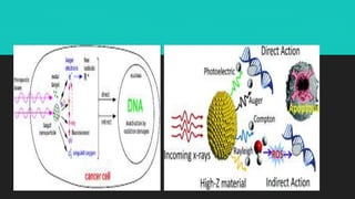
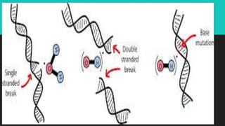
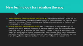
Radiotherapy uses ionizing radiation to treat cancer. There are two main types - external beam radiotherapy which uses radiation from outside the body, and brachytherapy which places radioactive sources inside or near the tumor. The radiation damages cancer cell DNA directly or through free radicals, limiting cell division and causing cell death. Different techniques are used depending on the tumor location and size to deliver precise radiation doses while sparing surrounding healthy tissues.
Management of cacrinoma cervix: Techniques of radiotherapy (2D conventional, 3D Conformal radiotherapy (3DCRT) and IMRT with a review of various contouring guidelines.
Sharing about “A typical day in the life as Radiation Therapy Technologist (RTT)” includes their roles, responsibilities, duties, working protocols, management, working stress, daily challenges in this modern radiotherapy era. As well as a bit information about how to become a RTT in India.
IORT uses a high single-fraction radiation dose (10-30 Gy) is delivered during surgery to a surgically-exposed tumour bed, immediately after a chunk of the tumour has been surgically excised. This slide includes topics like APBI, IOERT, IOHDR.
Here you can know about the teletherapy techniques which is used in radiation therapy. It can also help you to prepare notes on them. You can download it in your PC or laptop to see the gif, it will clear the concept better.
A summary of recent innovations in radiation oncology focussing on the priniciples of different techniques and their application. An overview of clinical results has also been given
A review of advances in Brachytherapy treatment planning and delivery in last decade or so, with main focus on brachytherapy for Prostate cancer, Breast cancer and Cervical cancer
Common Types of Cancer Treatment
Surgery: An operation where doctors cut out tissue with cancer cells. Chemotherapy: Special medicines that shrink or kill cancer cells that we cannot see. Radiation therapy: Using high-energy rays (similar to X-rays) to kill cancer cells.
Radiotherapy /certified fixed orthodontic courses by Indian dental academy Indian dental academy
The Indian Dental Academy is the Leader in continuing dental education , training dentists in all aspects of dentistry and offering a wide range of dental certified courses in different formats.
Indian dental academy provides dental crown & Bridge,rotary endodontics,fixed orthodontics,
Dental implants courses.for details pls visit www.indiandentalacademy.com ,or call
0091-9248678078
The Indian Dental Academy is the Leader in continuing dental education , training dentists in all aspects of dentistry and
offering a wide range of dental certified courses in different formats.
Title: Sense of Smell
Presenter: Dr. Faiza, Assistant Professor of Physiology
Qualifications:
MBBS (Best Graduate, AIMC Lahore)
FCPS Physiology
ICMT, CHPE, DHPE (STMU)
MPH (GC University, Faisalabad)
MBA (Virtual University of Pakistan)
Learning Objectives:
Describe the primary categories of smells and the concept of odor blindness.
Explain the structure and location of the olfactory membrane and mucosa, including the types and roles of cells involved in olfaction.
Describe the pathway and mechanisms of olfactory signal transmission from the olfactory receptors to the brain.
Illustrate the biochemical cascade triggered by odorant binding to olfactory receptors, including the role of G-proteins and second messengers in generating an action potential.
Identify different types of olfactory disorders such as anosmia, hyposmia, hyperosmia, and dysosmia, including their potential causes.
Key Topics:
Olfactory Genes:
3% of the human genome accounts for olfactory genes.
400 genes for odorant receptors.
Olfactory Membrane:
Located in the superior part of the nasal cavity.
Medially: Folds downward along the superior septum.
Laterally: Folds over the superior turbinate and upper surface of the middle turbinate.
Total surface area: 5-10 square centimeters.
Olfactory Mucosa:
Olfactory Cells: Bipolar nerve cells derived from the CNS (100 million), with 4-25 olfactory cilia per cell.
Sustentacular Cells: Produce mucus and maintain ionic and molecular environment.
Basal Cells: Replace worn-out olfactory cells with an average lifespan of 1-2 months.
Bowman’s Gland: Secretes mucus.
Stimulation of Olfactory Cells:
Odorant dissolves in mucus and attaches to receptors on olfactory cilia.
Involves a cascade effect through G-proteins and second messengers, leading to depolarization and action potential generation in the olfactory nerve.
Quality of a Good Odorant:
Small (3-20 Carbon atoms), volatile, water-soluble, and lipid-soluble.
Facilitated by odorant-binding proteins in mucus.
Membrane Potential and Action Potential:
Resting membrane potential: -55mV.
Action potential frequency in the olfactory nerve increases with odorant strength.
Adaptation Towards the Sense of Smell:
Rapid adaptation within the first second, with further slow adaptation.
Psychological adaptation greater than receptor adaptation, involving feedback inhibition from the central nervous system.
Primary Sensations of Smell:
Camphoraceous, Musky, Floral, Pepperminty, Ethereal, Pungent, Putrid.
Odor Detection Threshold:
Examples: Hydrogen sulfide (0.0005 ppm), Methyl-mercaptan (0.002 ppm).
Some toxic substances are odorless at lethal concentrations.
Characteristics of Smell:
Odor blindness for single substances due to lack of appropriate receptor protein.
Behavioral and emotional influences of smell.
Transmission of Olfactory Signals:
From olfactory cells to glomeruli in the olfactory bulb, involving lateral inhibition.
Primitive, less old, and new olfactory systems with different path
TEST BANK for Operations Management, 14th Edition by William J. Stevenson, Ve...kevinkariuki227
TEST BANK for Operations Management, 14th Edition by William J. Stevenson, Verified Chapters 1 - 19, Complete Newest Version.pdf
micro teaching on communication m.sc nursing.pdfAnurag Sharma
Microteaching is a unique model of practice teaching. It is a viable instrument for the. desired change in the teaching behavior or the behavior potential which, in specified types of real. classroom situations, tends to facilitate the achievement of specified types of objectives.
Lung Cancer: Artificial Intelligence, Synergetics, Complex System Analysis, S...Oleg Kshivets
RESULTS: Overall life span (LS) was 2252.1±1742.5 days and cumulative 5-year survival (5YS) reached 73.2%, 10 years – 64.8%, 20 years – 42.5%. 513 LCP lived more than 5 years (LS=3124.6±1525.6 days), 148 LCP – more than 10 years (LS=5054.4±1504.1 days).199 LCP died because of LC (LS=562.7±374.5 days). 5YS of LCP after bi/lobectomies was significantly superior in comparison with LCP after pneumonectomies (78.1% vs.63.7%, P=0.00001 by log-rank test). AT significantly improved 5YS (66.3% vs. 34.8%) (P=0.00000 by log-rank test) only for LCP with N1-2. Cox modeling displayed that 5YS of LCP significantly depended on: phase transition (PT) early-invasive LC in terms of synergetics, PT N0—N12, cell ratio factors (ratio between cancer cells- CC and blood cells subpopulations), G1-3, histology, glucose, AT, blood cell circuit, prothrombin index, heparin tolerance, recalcification time (P=0.000-0.038). Neural networks, genetic algorithm selection and bootstrap simulation revealed relationships between 5YS and PT early-invasive LC (rank=1), PT N0—N12 (rank=2), thrombocytes/CC (3), erythrocytes/CC (4), eosinophils/CC (5), healthy cells/CC (6), lymphocytes/CC (7), segmented neutrophils/CC (8), stick neutrophils/CC (9), monocytes/CC (10); leucocytes/CC (11). Correct prediction of 5YS was 100% by neural networks computing (area under ROC curve=1.0; error=0.0).
CONCLUSIONS: 5YS of LCP after radical procedures significantly depended on: 1) PT early-invasive cancer; 2) PT N0--N12; 3) cell ratio factors; 4) blood cell circuit; 5) biochemical factors; 6) hemostasis system; 7) AT; 8) LC characteristics; 9) LC cell dynamics; 10) surgery type: lobectomy/pneumonectomy; 11) anthropometric data. Optimal diagnosis and treatment strategies for LC are: 1) screening and early detection of LC; 2) availability of experienced thoracic surgeons because of complexity of radical procedures; 3) aggressive en block surgery and adequate lymph node dissection for completeness; 4) precise prediction; 5) adjuvant chemoimmunoradiotherapy for LCP with unfavorable prognosis.
These lecture slides, by Dr Sidra Arshad, offer a quick overview of physiological basis of a normal electrocardiogram.
Learning objectives:
1. Define an electrocardiogram (ECG) and electrocardiography
2. Describe how dipoles generated by the heart produce the waveforms of the ECG
3. Describe the components of a normal electrocardiogram of a typical bipolar leads (limb II)
4. Differentiate between intervals and segments
5. Enlist some common indications for obtaining an ECG
Study Resources:
1. Chapter 11, Guyton and Hall Textbook of Medical Physiology, 14th edition
2. Chapter 9, Human Physiology - From Cells to Systems, Lauralee Sherwood, 9th edition
3. Chapter 29, Ganong’s Review of Medical Physiology, 26th edition
4. Electrocardiogram, StatPearls - https://www.ncbi.nlm.nih.gov/books/NBK549803/
5. ECG in Medical Practice by ABM Abdullah, 4th edition
6. ECG Basics, http://www.nataliescasebook.com/tag/e-c-g-basics
Pulmonary Thromboembolism - etilogy, types, medical- Surgical and nursing man...VarunMahajani
Disruption of blood supply to lung alveoli due to blockage of one or more pulmonary blood vessels is called as Pulmonary thromboembolism. In this presentation we will discuss its causes, types and its management in depth.
Couples presenting to the infertility clinic- Do they really have infertility...Sujoy Dasgupta
Dr Sujoy Dasgupta presented the study on "Couples presenting to the infertility clinic- Do they really have infertility? – The unexplored stories of non-consummation" in the 13th Congress of the Asia Pacific Initiative on Reproduction (ASPIRE 2024) at Manila on 24 May, 2024.
Anti ulcer drugs and their Advance pharmacology ||
Anti-ulcer drugs are medications used to prevent and treat ulcers in the stomach and upper part of the small intestine (duodenal ulcers). These ulcers are often caused by an imbalance between stomach acid and the mucosal lining, which protects the stomach lining.
||Scope: Overview of various classes of anti-ulcer drugs, their mechanisms of action, indications, side effects, and clinical considerations.
Ethanol (CH3CH2OH), or beverage alcohol, is a two-carbon alcohol
that is rapidly distributed in the body and brain. Ethanol alters many
neurochemical systems and has rewarding and addictive properties. It
is the oldest recreational drug and likely contributes to more morbidity,
mortality, and public health costs than all illicit drugs combined. The
5th edition of the Diagnostic and Statistical Manual of Mental Disorders
(DSM-5) integrates alcohol abuse and alcohol dependence into a single
disorder called alcohol use disorder (AUD), with mild, moderate,
and severe subclassifications (American Psychiatric Association, 2013).
In the DSM-5, all types of substance abuse and dependence have been
combined into a single substance use disorder (SUD) on a continuum
from mild to severe. A diagnosis of AUD requires that at least two of
the 11 DSM-5 behaviors be present within a 12-month period (mild
AUD: 2–3 criteria; moderate AUD: 4–5 criteria; severe AUD: 6–11 criteria).
The four main behavioral effects of AUD are impaired control over
drinking, negative social consequences, risky use, and altered physiological
effects (tolerance, withdrawal). This chapter presents an overview
of the prevalence and harmful consequences of AUD in the U.S.,
the systemic nature of the disease, neurocircuitry and stages of AUD,
comorbidities, fetal alcohol spectrum disorders, genetic risk factors, and
pharmacotherapies for AUD.
MANAGEMENT OF ATRIOVENTRICULAR CONDUCTION BLOCK.pdfJim Jacob Roy
Cardiac conduction defects can occur due to various causes.
Atrioventricular conduction blocks ( AV blocks ) are classified into 3 types.
This document describes the acute management of AV block.
Factory Supply Best Quality Pmk Oil CAS 28578–16–7 PMK Powder in Stockrebeccabio
Factory Supply Best Quality Pmk Oil CAS 28578–16–7 PMK Powder in Stock
Telegram: bmksupplier
signal: +85264872720
threema: TUD4A6YC
You can contact me on Telegram or Threema
Communicate promptly and reply
Free of customs clearance, Double Clearance 100% pass delivery to USA, Canada, Spain, Germany, Netherland, Poland, Italy, Sweden, UK, Czech Republic, Australia, Mexico, Russia, Ukraine, Kazakhstan.Door to door service
Hot Selling Organic intermediates
2. Radiotherapy---TYPES
Radiotherapy and chemotherapy are the important modalities of therapy
for human cancers apart from surgery
Primary therapy---radiotherapy in carcinoma cervix and chemotherapy in
gestational trophoblastic neoplasia
Multidisciplinary approach---- is needed for the treatment of some
malignancies to improve the outcome.
Palliation approach ---- incapacitating symptoms when cure may not be
achieved.
3. Radiotherapy---Mechanism
➢ Radiotherapy is a science about use of ionizing radiation (IR) mainly to
treat malignant tumors.
It depends on the ABSORBED DOSE (AD) – energy transmitted to
irradiated tissues (Gr).
➢ The first time of using X-rays was in 1896.
4. Radiobiology of normal tissues
The effects of radiation on tissues are generally of two types:
A. Loss of mature functional cells by apoptosis (programmed cell death). This usually
occurs within 24 hours of radiation.
B. Loss of cellular reproductive capacity. The severity depends upon the total dose of
radiation, length of time over which radiotherapy is delivered and the radiosensitivity of
the particular cell types. Usually lost cells are replaced by proliferation of surviving stem
cells or progenitor cells.
5. Ionizing radiation
(i) Electromagnetic radiation--Electromagnetic radiation:This consists of
quanta of energy and wavelength (photon radiation). They are of two
types—X-rays and gamma rays. These electromagnetic waves travel in
discrete bundles called ‘Photons’.
(ii) Particulate radiation.-- This consists of atomic subparticles such as
electrons, protons and neutrons. Only electrons (β-rays) are used in
radiotherapy
6. Ionizing radiation
Gamma rays are produced spontaneously as a result of decay of the atomic nucleus of
some radio-active isotopes. 60Cobalt or 192Iridium is a source of γ-rays.
„X-rays are produced outside the atomic nucleus. When fast-moving electrons approach
the fields around the nuclei of atoms of a target material (tungsten), they are deflected
from their path. The energy thus emitted in the form of electromagnetic radiation
(photons) is X-rays. Machines such as betatron (circular fashion) and linear accelerator
(linear fashion) can accelerate electrons with high kinetic energy. Therefore, X-rays
generated by these machines are very high in energy
7.
8.
9. Radiotherapy -- mechanism
X-rays and gamma rays are collectively called photons. When photons interact with
matter (tissue), three effects are observed: (i) photoelectric effect, (ii) compton scattering
and (iii) pair production. In human radiation therapy, compton scattering is the major
interaction of photons with tissue .
X-rays and gamma rays have shorter wavelength and high frequency. They have high
kinetic energy. X-rays and gamma rays possess considerable power of tissue penetration
depending on the photon energy and the density of the matter through which they pass.
The photon energy produced from radioactive cobalt is 1.2 million electron volts (Mev).
External photon beam radiation is usually derived from a linear accelerator
10. Radioactive isotopes used in gynecological malignancies
Element isotope Energy (mev) Half-life clinical use
Cesium 137 (137Cs) 0.514 30 yrs intracavitary implants-
temporary
Radium 236 (236Ra) 3.26 1600 yrs historical
Cobalt 60 (60Co) 1.173 1.173 5.3 yrs
iridium 192 (192ir) 0.38 74.2 days interstitial implant
(temporary)
iodine 125 (125i) 0.028 60.2 days interstitial implant
(permanent)
phosphorus 32 (32p) none 14.3 days intracavitary (permanent)
11. Techniques of radiation therapy
Brachytherapy---It gives a very high dose of radiation where the source of
radiation is placed within, or close to the tumor. The application may be (i)
Intracavitary (ii) Interstitial or (iii) Surface (skin). Damage to normal tissues
is less as there is rapid falloff of radiation around the source (inverse
square law).
Intracavitary: The devices for brachytherapy consist of hollow stem
(intrauterine tandem), which is placed within the uterine cavity .Especially
designed devices used for vaginal placements are called vaginal ovoids or
colpostats.
12. Techniques of radiation therapy
Interstitial form of brachytherapy consists of placement of radioactive
sources (needles, wires or seeds) within the tissues.
Commonly used sources are Iridium-192 (192Ir), Cesium-137 (137Cs) and
Cobalt 60 (60Co). Small volume of tumor, as in early cases of vaginal
carcinoma, can be treated with the method. Normal tissues are spared
from radiation injury.
Intraperitoneal instillation is another mode of local therapy--gold or
phosphorus,
13. After loading technique:
It is a modern development of brachytherapy to prevent radiation
complications to the personnel. A mock insertion of applicators is
performed and an X-ray is taken to note their exact position.
After loading technique may be manual or by remote control. Later on,
live radioactive sources are introduced by remote control in identical
manner. Remote after loading system uses Selectron (137Cs) or high dose
Selectron (60Co). Remote control systems allow complete protection of
staff from radiation exposure
14. Techniques of radiation therapy
Brachytherapy can be either low dose rate (LDR) or a high dose rate (HDR) system. LDR
require hospital admission and deliver dose at about 50-100 cGy/hour. HDR systems are
commonly done as outpatient basis. The dose rate delivered is at 100 cGy/minute.
Advantages: (a) Localized high radiation dose to a small tumor volume with high local
control. Radiation dose in the surrounding normal tissues is less as there is sharp fall-off
according to inverse square law
Disadvantages: (a) Large tumors are usually unsuitable unless used following EBRT and/or
chemotherapy. (b) Risks of exposure to medical and nursing personnel due to gamma
rays.
15. Brachytherapy technique
Small radioactive sources, mainly radium sulphate is mixed with some inert powder and
packed in small needles or tubes. These are used for interstitial, intracavitary or surface
applications. Radiation sources for intracavitary radiation are Radium (226Ra), Cesium
(137Cs) or Cobalt (60Co). The container is made up of platinum, gold or alloy steel to
absorb alpha and beta particles and allowing the gamma rays to sterilize the cancer cells.
In carcinoma cervix, the tandems are inserted in the uterine cavity and the ovoids and
colpostats are placed in the vaginal vault under anesthesia
16. Brachytherapy technique
In Paris and Manchester techniques, the source strength is smaller but exposure time is increased.
The vaginal source is away from the cervix. They are used with either preloaded or afterloaded
special applicators.
One treatment period in Paris technique is 96–200 hours as compared to Stockholm technique
where each application is 24–28 hours in duration .
Manchester system, which is a modification of the Paris technique, delivers constant isodose at
different depths, regardless of the size of the uterus and vagina. In Stockholm technique large high
intensity source with less exposure time is given, but the vaginal source is closer to the cervix
22. External Beam Radiotherapy (EBRT)
EBRT or teletherapy is the treatment with beams of ionizing radiation produced from a
source external to the patient.
Superficial tumors may be treated with X-rays of low energy in the range of 80-300 KV.
Deeper-seated tumors are usually treated using mega-voltage photons. Cobalt 60 is the
common teletherapy source for EBRT, the other one is cesium 137.
External radiation therapy is used to treat large volumes (tumor, lymph nodes,
parametrium) .
It is designed to deliver a uniform radiation dose to the tumor volume without ‘hot’ (excess
dose) or ‘cold’ (under dose) spots. Accurate tumor localization and volume measurement
are essential. Greater the tumor volume, higher the radiation dose required
23. Instillation of Radioisotopes into the
Peritoneal or Pleural Cavity
Radioactive isotopes of either gold or phosphorus, linked to carrier
colloids, are commonly used in ovarian cancer. This can give radiation only
to a depth of 4-8 mm. Radioactive chromic phosphate (32P) emits pure β-
rays and has got longer half-life (14.3 days) and deeper penetration (8
mm) power compared to radio gold (198Au). Small volume of tumor in the
peritoneal or pleural cavity is treated with solution of radioisotopes.
24. Measurement of Radiation Radiation
absorption dose (Gray)
is the unit used to measure the amount of energy absorbed per unit mass
of tissue. One gray (Gy) is equivalent to 1 Joule/kg which is equivalent to
100 rads. Currently, the term centigray (cGy) is used.
One cGy is equivalent to one rad. Amount of radiation the patient
receives is calculated by dosimetry. Homogeneous irradiation of tissues is
desirable . Primary tumor should receive high dose.
Brachytherapy and teletherapy should be combined to provide adequate
irradiation to the primary tumor as well as the pelvic lymph nodes and the
parametrium
25. Biological effects of radiation (radiobiology)
(1) Direct action: Where the radiation is absorbed, it causes damage to
DNA directly. This is the predominant mechanism of action of particulate
radiation (neutrons).
(2) Indirect action: Where the radiation interact with other substances (H2
O) in the cell to produce free radicals (OH– ) which in turn damage the
DNA.
Radiation, depending on the dose and time of exposure may cause (a)
gene mutation (b) abnormal cell mitosis and (c) derangement of
reproductive ability of the cell—“progeria”.
26. The target for radiation injury is DNA
Ultimately, there is limited cell mitosis and mitotic cell death. There is
cytoplasmic vacuolation and fragmentation.
Ionizing radiation also produces damage to nuclear and plasma
membranes. This effect of ionizing radiation is common for both the
normal and neoplastic tissues, encountered in the radiation path.
Radiation complications are mainly due to interaction with the normal
tissues .When the radiation effect to a cell is sublethal, cellular DNA may
undergo repair and the cell survives. Lethal effect kills the cell.
27. Radiation dose
According to the “inverse square law” there is reduction of radiation at a
distance from the source in brachytherapy. This protects the normal
tissues
28. Advantages of primary radiotherapy- ca cervix
Wider applicability in all stages of carcinoma cervix.
Survival rate 85%, comparable with that of surgery in early stages.
Less primary mortality and morbidity.
Individualization of dose distributions/requirement possible.
29. Contraindication of radiotherapy
Associated PID—acute or chronic, pelvic kidney.
Associated myoma, prolapse (procidentia), ovarian tumor or genital
fistula.
Young patient (to preserve ovarian function).
Vaginal stenosis — placement of radiation source is inadequate.
Cases with adenocarcinoma or adenosquamous carcinoma — surgery is
preferred.
30. Radiation reactions
EARLY
1. anorexia, nausea, vomiting, lassitude or even fever
2. diarrhea (radiation enteritis)
3. leukopenia and thrombocytopenia, anemia
4. intestinal reaction such as enteritis, colitis, proctitis
5. urinary—cystitis, pyelitis, hematuria
6. Skin reaction such as peeling often found in moist area of the vulva. this is
almost absent in megavoltage radiotherapy
31. Radiation reactions
late (due to vasculitis and fibrosis):
atrophic changes of vulval skin and vaginal stricture
Radiation fibrosis
pathological fracture due to osteoporosis
stricture, bleeding per rectum, perforation, obstruction
malabsorption syndrome with megaloblastic anemia
Proctosigmoiditis
radiation menopause
32. Radio sensitivity
means the response of the tumor to irradiation. Radiosensitivity is measured in terms of loss of cellular
proliferative capacity due to the damage to DNA. Accumulation of sublethal injury following repeat
radiation leads to ultimate DNA damage and cell death.
Radiosensitivity depends on several factors:
Tissue hypoxia—higher the hypoxic fraction of cells, the less (2-3 times) is the radiation response.
Hypoxic cells are more resistant to radiation compared to toxic cells.
Proportion of mitotic (clonogenic) cells— clonogenic cells are more radiosensitive.
Cell cycle—mitotic cells (M phase) and G2 cells are more radiosensitive compared to late S-phase cells
Tumor specificity—certain tumors (dysgerminomas) are more radiosensitive than the others.
Tumor volume—smaller the tumor volume → lesser the hypoxic cells → less the radiation dose better the
radiation response.
33. Radio sensitivity
Lesser the photon wavelength more is the penetrating power and energy
of ionizing radiation. Supervoltage and megavoltage radiation (60Co,
137Cs, 226Ra, betatron, linear accelerator) have the following advantages
over the orthovoltage one.
They have higher energy of radiation, less skin injury, less lateral
scattering and more tissue penetration at a greater depth. They are
suitable for the deep seated tumors (e.g. carcinomas of the cervix and
endometrium).
34. Fractionation
Is the division of a total dose of external beam radiotherapy into small (daily) doses.
Thus it spares normal tissue damage preferentially.
External beam radiotherapy is usually fractionated and is given once daily for five
times a week. A dose of 180-220 cGy per fraction is used. This is based on the
ability of the cells to accumulate and repair the sublethal injury.
Tumor tissue takes longer time to recover from radiation damage compared to
normal tissue. Fractionation allows normal tissue (intestinal mucosa, bone marrow)
to repair sublethal injury (sparing effect). On the other hand irradiation results in
accumulation of sublethal damage and ultimate loss of reproductive capacity in
tumor tissue.
35. Radiopotentiators and hypoxic cell sensitizers
Chemotherapeutic agents
Cisplatin, paclitaxel ,Gemcitabine, doxorubicin
Others
Metronidazole, misonidazole ,tumor necrosis factor (tnf), interferon, acyclovir
Intraoperative radiation of large fraction of 1500-2500 cGy are delivered directly to the area
selected. Periaortic node irradiation (biopsy proven) at the time of staging laparotomy is
possible.
Hyperthermia is found helpful as an active anti-neoplastic agent and a significant
radiosensitizer.
36. New technology for radiation therapy
Three-dimensional conformal radiation therapy (3D CRT) uses imaging modalities (CT, MRI and PET
scaning). Beam placement using a CT simulation is used. 3D conformal therapy can shape the beam
to conform to the target. This can help to arrange the beams to maximize dose to the tumor and
minimize dose to normal tissues.
Intensity modulated radiation therapy (IMRT): IMRT uses the power of computers to shape and
perform thousands of iterations of planning to maximize the tumor dose and to minimize normal
tissue dose. Both 3D CRT and IMRT use small collimator “leaves” to shape the beam finely. These
“leaves” are mobile and can vary the beam intensity. It allows irregular shapes (tumor) to be treated
and has the benefit of reduced radiation to normal tissues (bowel, bladder). Tomotherapy and cone-
beam CT may allow more precise localization of beam and verification of dose delivered.
37. New technology for radiation therapy
Stereotactic radiotherapy and Gamma-Knife radiation are similar to IMRT
and 3D CRT to allow precise high dose delivery of external radiation.
Steoreatactic radiation uses a modification of linear accelerator