Definitions
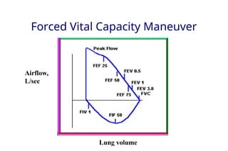
FVC –Forced Vital Capacity
Volume of air exhaled after a maximal inspiration to total lung capacity. This
volume is expressed in Liters
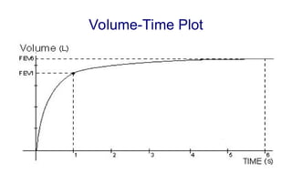
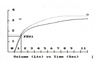
FEV1 – Forced Expiratory Volume in 1 second
Volume of air exhaled in the first second of expiration.
This volume is expressed in Liters
FEF 25-75%
Mean expiratory flow during the middle half of the FVC maneuver; reflects
flow through later emptying airways, not necessarily the small airways
FEV1/FVC – Ratio (%)
Volume of air expired in the first second, expressed as a percent of FVC
6.
Performance of FVCmaneuver
Patient assumes the position (typically standing)
• Puts nose clip on
• Inhales maximally
• Puts mouthpiece in mouth and closes lips around mouthpiece
(open circuit)
• Exhales as hard and fast and long as possible
• Repeat instructions if necessary – effective coaching is essential
• Give simple instructions
• Repeat minimum of three times (check for repeatability)
7.
Special Considerations inPediatric
Patients
Ability to perform spirometry dependent on
developmental age of child, personality, and interest.
Patients need a calm, relaxed environment and
good coaching. Patience is key.
Be creative
Use incentives
Even with the best of environments and coaching, a
child may not be able to perform spirometry.
8.
ATS Acceptable Criteria
WithinManeuver
Free from artifacts, such as
• Cough during the first second of exhalation
• Glottis closure that influences the measurement
• Early termination or cut-off
• Effort that is not maximal throughout
• Leak
• Obstructed mouthpiece
Good starts
• Extrapolated volume < 5% of FVC or 0.15 L, whichever is greater
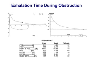
Satisfactory exhalation
• Duration of ≥ 6 s (3 s for children < 10) or a plateau in the volume–
time curve or
• If the subject cannot or should not continue to exhale
9.
ATS Acceptable Criteria
WithinManeuver
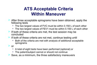
After three acceptable spirograms have been obtained, apply the
following tests
• The two largest values of FVC must be within 0.150 L of each other
• The two largest values of FEV1 must be within 0.150 L of each other
If both of these criteria are met, the test session may be
concluded
If both of these criteria are not met, continue testing until
• Both of the criteria are met with analysis of additional acceptable
spirograms
or
• A total of eight tests have been performed (optional) or
• The patient/subject cannot or should not continue
Save, as a minimum, the three satisfactory maneuvers
10.
Spirometry Interpretation: Sowhat
constitutes normal?
Normal values vary and depend on:
• Height
• Age
• Gender
• Ethnicity
11.
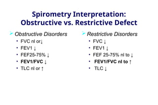
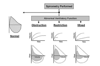
Spirometry Interpretation:
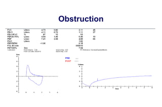
Obstructive vs.Restrictive Defect
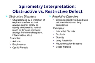
Obstructive Disorders
• Characterized by a limitation of
expiratory airflow so that
airways cannot empty as
rapidly compared to normal
(such as through narrowed
airways from bronchospasm,
inflammation, etc.)
Examples:
• Asthma
• Emphysema
• Cystic Fibrosis
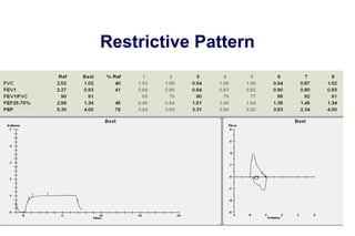
Restrictive Disorders
• Characterized by reduced lung
volumes/decreased lung
compliance
Examples:
• Interstitial Fibrosis
• Scoliosis
• Obesity
• Lung Resection
• Neuromuscular diseases
• Cystic Fibrosis
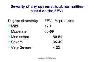
Severity of anyspirometric abnormalities
based on the FEV1
Degree of severity FEV1 % predicted
Mild >70
Moderate 60-69
Mod severe 50-59
Severe 35-49
Very Severe < 35
based on ATS/ERS criteria
14.
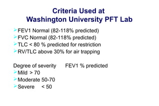
Criteria Used at
WashingtonUniversity PFT Lab
FEV1 Normal (82-118% predicted)
FVC Normal (82-118% predicted)
TLC < 80 % predicted for restriction
RV/TLC above 30% for air trapping
Degree of severity FEV1 % predicted
Mild > 70
Moderate 50-70
Severe < 50
16.
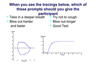
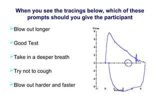
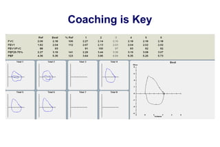
When you seethe tracings below, which of
these prompts should you give the
participant
Take in a deeper breath
Blow out harder
and faster
Try not to cough
Blow out longer
Good Test
17.
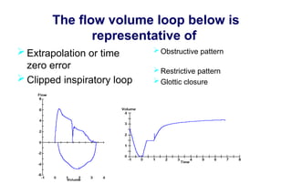
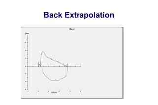
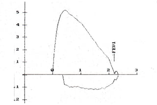
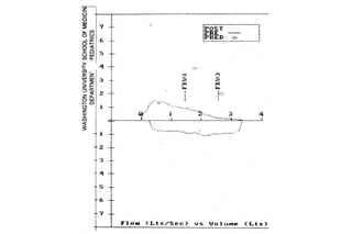
The flow volumeloop below is
representative of
Extrapolation or time
zero error
Clipped inspiratory loop
Obstructive pattern
Restrictive pattern
Glottic closure
18.
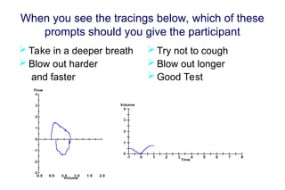
When you seethe tracings below, which of these
prompts should you give the participant
Blow out longer
Good Test
Take in a deeper breath
Try not to cough
Blow out harder and faster
19.
When you seethe tracings below, which of these
prompts should you give the participant
Take in a deeper breath
Blow out harder
and faster
Try not to cough
Blow out longer
Good Test
20.
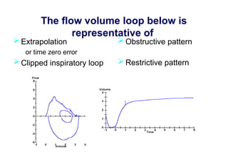
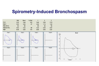
The flow volumeloop below is
representative of
Extrapolation
or time zero error
Clipped inspiratory loop
Obstructive pattern
Restrictive pattern
Glottic closure
FEF 25-75%
What isit?
What does it measure?
Is it a measure of small airways?
37.
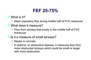
FEF 25-75%
Whatis it?
• Mean expiratory flow during middle half of FVC maneuver
What does it measure?
• Flow from airways that empty in the middle half of FVC
maneuver
Is it a measure of small airways?
• Maybe in normals
• In asthma, or obstructive disease, it measures flow from
more obstructed airways which could be small or larger
with more obstruction
Dysanapsis
Green, Mead,Turner. Variability of maximum expiratory
flow-volume curves. J Appl Physiol 1974 37:67-74
• Variability in flows among healthy adults not altered when
flows were corrected for vital capacity
• Lung static recoil and bronchomotor tone contributed little
to variability
Concluded that variability in flows between individuals
due to differences in airway size independent of
lung/parenchyma size
Differences may have embryological basis, reflecting
disproportionate but physiologically normal growth within
an organ
40.
Dysanapsis
Mead. Dysanapsis innormal lungs assessed by the
relationship between maximal flow, static recoil, and vital
capacity. Am Rev Respir Dis 1980 121:339-342
• “There is no association whatsoever between
airway diameter and lung size.”
• There are differences between men and women
(men 17% larger than women) and between boys
and men (boys in late teens similar to girls,
suggesting that growth in males occurs late)
41.
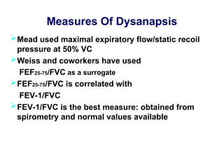
Measures Of Dysanapsis
Meadused maximal expiratory flow/static recoil
pressure at 50% VC
Weiss and coworkers have used
FEF25-75/FVC as a surrogate
FEF25-75/FVC is correlated with
FEV-1/FVC
FEV-1/FVC is the best measure: obtained from
spirometry and normal values available
42.
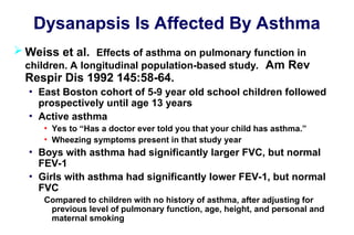
Dysanapsis Is AffectedBy Asthma
Weiss et al. Effects of asthma on pulmonary function in
children. A longitudinal population-based study. Am Rev
Respir Dis 1992 145:58-64.
• East Boston cohort of 5-9 year old school children followed
prospectively until age 13 years
• Active asthma
• Yes to “Has a doctor ever told you that your child has asthma.”
• Wheezing symptoms present in that study year
• Boys with asthma had significantly larger FVC, but normal
FEV-1
• Girls with asthma had significantly lower FEV-1, but normal
FVC
Compared to children with no history of asthma, after adjusting for
previous level of pulmonary function, age, height, and personal and
maternal smoking
43.
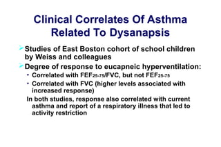
Clinical Correlates OfAsthma
Related To Dysanapsis
Studies of East Boston cohort of school children
by Weiss and colleagues
Degree of response to eucapneic hyperventilation:
• Correlated with FEF25-75/FVC, but not FEF25-75
• Correlated with FVC (higher levels associated with
increased response)
In both studies, response also correlated with current
asthma and report of a respiratory illness that led to
activity restriction
44.
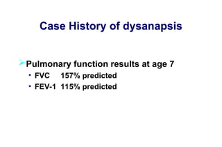
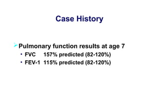
Case History ofdysanapsis
Pulmonary function results at age 7
• FVC 157% predicted
• FEV-1 115% predicted
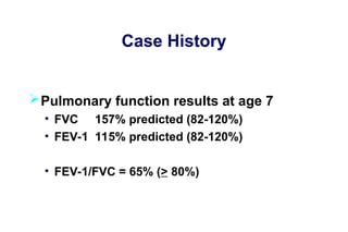
Case History
Pulmonary functionresults at age 7
• FVC 157% predicted (82-120%)
• FEV-1 115% predicted (82-120%)
• FEV-1/FVC = 65% (> 80%)
47.
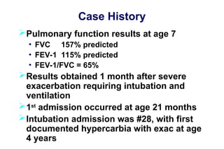
Case History
Pulmonary functionresults at age 7
• FVC 157% predicted
• FEV-1 115% predicted
• FEV-1/FVC = 65%
Results obtained 1 month after severe
exacerbation requiring intubation and
ventilation
1st
admission occurred at age 21 months
Intubation admission was #28, with first
documented hypercarbia with exac at age
4 years
Conclusions
Spirometry is:
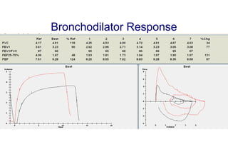
• Usefulin asthma diagnosis and management
• Useful in diagnosis of conditions that can present with
wheezing, or airway noise that can be hard to
distinguish from wheezing
• Requires considerable expertise, particularly in children
FEF25-75% does not measure small airways, but
instead airways more obstructed that empty later
in exhalation
Editor's Notes
#10 Height varies directly with vc
VC increases with age up to age 20 years then becomes inversely proportion to age
Women usually with lower vc than men