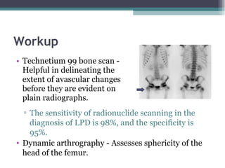
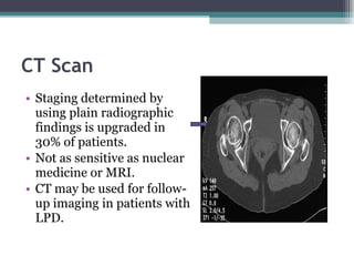
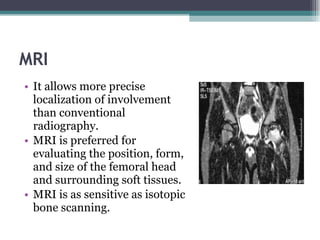
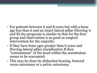
Legg Calve Perthes Disease
History FIRST DESCRIBED BY  LEGG  AND  WALDENSTORM   IN 1909 AND BY  PERTHES  AND CALVE  IN 1910
Definition Legg-Calvé-Perthes disease (LCPD) is the name given to idiopathic osteonecrosis of the capital femoral epiphysis in a child.
Epidemiology Disorder of the hip in young children Usually ages 4-8yo As early as 2yo, as late as teens Boys:Girls= 4-5:1 Bilateral 10-12% No evidence of inheritance
Prevalence :
Etiology Unknown Past theories: infection, inflammation, trauma, congenital Most current theories involve vascular compromise Sanches 1973: “second infarction theory”
Causes Proposed theories . Excessive femoral antiversion. Synovitis. Generalized skeletal disorder. Arterial anomalies.
 
Pathogenesis Histologic changes described by 1913 Secondary ossification center= covered by cartilage of 3 zones: Superficial Epiphyseal Thin cartilage zone Capillaries penetrate thin zone from below
Epiphyseal cartilage in LCP disease: Superficial zone is normal but thickened Middle zone has 1) areas of extreme hypercellularity in clusters and 2) areas of loose fibrocartilaginous matrix Superficial and middle layers nourished by synovial fluid Deep layer relies on blood supply
Physeal plate : cleft formation, amorphis debris, blood extravasation Metaphyseal region : normal bone separated by cartilaginous matrix Epiphyseal changes can be seen also in greater trochanter, acetabulum
Blood Supply
Radiographic Stages Four Waldenstrom stages: 1) Initial stage 2) Fragmentation stage 3) Reossification stage 4) Healed stage
Initial Stage Early radiographic signs: Failure of femoral ossific nucleus to grow Widening of medial joint space “ Crescent sign” Irregular physeal plate Blurry/ radiolucent metaphysis
Fragmentation Stage Bony epiphysis begins to fragment Areas of increased lucency and density Evidence of repair aspects of disease
Reossification Stage Normal bone density returns Alterations in shape of femoral head and neck evident
Healed Stage Left with residual deformity from disease and repair process Differs from AVN following Fx or dislocation
Group I
Group II
Group III
Group IV
3 groups: A) no lateral pillar involvment B) >50% lat height intact C) <50% lat height intact Lateral Pillar Classification
Salter-Thompson Classification Simplification of Catterall Based on status of lateral margin of capital femoral epiphysis Group A (Catterall I & II equivalent) Group B (Catterall III & IV equivalent)
Clinical Features Stature usually shorter than peers Quadriceps and gluteal muscle wasting is common, Trandelenburg test positive (drop of the hip on the unsupported side) Acute phase; range of motion at the hip joint is limited due to muscle spasms Progressively; limited internal rotation and abduction is likely due to impingement lesions (hence the Roll test, guarding on affected side) Later stage; global reduction in all ranges of motion assoc. with pain, indicating joint arthritis
Age- 4 to 10 years, with peak incidence at 7 Gender- Boys (5:1 ratio) but it tends to be more severe in girls Height Passive smoking or maternal smoking at pregnancy ADHD? Increased physical activity Family Hx of; skeletal dysplasias or thrombotic disease Ethnicity; more common in Whites, Eskimos, Japanese Social Hx- associated with low socio-economic status Risk Factors
Differential Diagnosis
Workup Technetium 99 bone scan - Helpful in delineating the extent of avascular changes before they are evident on plain radiographs. The sensitivity of radionuclide scanning in the diagnosis of LPD is 98%, and the specificity is 95%.  Dynamic arthrography - Assesses sphericity of the head of the femur.
Ultrasonography may provide significant diagnostic clues to differentiate early Perthes' from transient synovitis.  T Futami, Y Kasahara, S Suzuki, S Ushikubo and T Tsuchiya  Journal of Bone and Joint Surgery - British Volume, Vol 73-B, Issue 4, 635-639  Ultrasonography in transient synvitis and early Perthes’ disease
CT Scan Staging determined by using plain radiographic findings is upgraded in 30% of patients. Not as sensitive as nuclear medicine or MRI. CT may be used for follow-up imaging in patients with LPD.
MRI It allows more precise localization of involvement than conventional radiography. MRI is preferred for evaluating the position, form, and size of the femoral head and surrounding soft tissues.  MRI is as sensitive as isotopic bone scanning.
Outcome variables Age Extent of involvement Duration  Remodeling potential Premature physeal closure Type of treatment Stage of disease at treatment.
Treatment Options Overall goal of treatment Reduce hip irritability and pain Restore/maintain hip mobility Prevent femoral head from extruding or collapsing  “CONTAINMENT ” Regain  spherical shape of femoral head
Below 6 years and Herring A/B Mainstay of treatment would be to OBSERVE with 6-12 month reassessment. Patients in this age group need bed rest and anti inflammatory medication at most. NO evidence that abduction splints or surgery beneficial Prognosis is good for the majority
Non Surgical treatment NSAIDS Traction Casts and braces (Scottish Rite Orthosis)
Above 6 and Herring class B Containment of the head within the acetabulum is warranted This is achieved by; Abduction bracing Femoral varus osteotomy Pelvic ostotomy
Age between 6-8 and Herring class C Results of intervention have been equivocal. Above 9 years Often have Herring class B or C Prognosis is poor Early containment is key,  by  pelvic osteotomy and internal fixation
Osteotomies
Summary For patients less that 6 years old the prognosis is good for the majority.  If they are stiff or painful they respond to bed rest, traction and pain relieving anti-inflammatory medication.  There is no evidence that abduction splints or surgical intervention is warranted in the majority of these younger patients.
For patients between 6 and 8 years but with a bone age less than 6 and an intact lateral pillar (Herring A and B) the prognosis is similar to that for the first group and observation is as good as surgical intervention for the majority.  If they have bone ages greater than 6 years and Herring lateral pillar classification B then &quot;containment&quot; of the head within the acetabulum seems to be warranted.  This may be done by abduction bracing, femoral varus osteotomy or a pelvic osteotomy.
If they are between 6 and 8 and are in lateral pillar group C then the result of intervention are equivocal.  Children presenting with Perthes disease at age 9 or older often have lateral pillar B or C and a poor prognosis.  The trend is towards early containment of these hips although stiffness can be a problem following early pelvic (Salter's) osteotomy.
Follow-up Initially, close follow-up is required to determine the extent of necrosis. Once the healing phase has been entered, follow-up can be every 6 months. Long-term follow-up is necessary to determine the final outcome.
Complications   Femoral  Shortening stiffness Malrotation Limp Positive trendelenburg  Pelvic Lenghtening Stiffness Chondrolysis Failure of containment
Prognosis   The younger the age of onset of LCPD, the better the prognosis. Children older than 10 years have a very high risk of developing osteoarthritis. Most patients have a favorable outcome. Prognosis is proportional to the degree of radiologic involvement.

Legg+Calve+Perthes+Disease

  • 1.
  • 2.
    History FIRST DESCRIBEDBY LEGG AND WALDENSTORM IN 1909 AND BY PERTHES AND CALVE IN 1910
  • 3.
    Definition Legg-Calvé-Perthes disease(LCPD) is the name given to idiopathic osteonecrosis of the capital femoral epiphysis in a child.
  • 4.
    Epidemiology Disorder ofthe hip in young children Usually ages 4-8yo As early as 2yo, as late as teens Boys:Girls= 4-5:1 Bilateral 10-12% No evidence of inheritance
  • 5.
  • 6.
    Etiology Unknown Pasttheories: infection, inflammation, trauma, congenital Most current theories involve vascular compromise Sanches 1973: “second infarction theory”
  • 7.
    Causes Proposed theories. Excessive femoral antiversion. Synovitis. Generalized skeletal disorder. Arterial anomalies.
  • 8.
  • 9.
    Pathogenesis Histologic changesdescribed by 1913 Secondary ossification center= covered by cartilage of 3 zones: Superficial Epiphyseal Thin cartilage zone Capillaries penetrate thin zone from below
  • 10.
    Epiphyseal cartilage inLCP disease: Superficial zone is normal but thickened Middle zone has 1) areas of extreme hypercellularity in clusters and 2) areas of loose fibrocartilaginous matrix Superficial and middle layers nourished by synovial fluid Deep layer relies on blood supply
  • 11.
    Physeal plate :cleft formation, amorphis debris, blood extravasation Metaphyseal region : normal bone separated by cartilaginous matrix Epiphyseal changes can be seen also in greater trochanter, acetabulum
  • 12.
  • 13.
    Radiographic Stages FourWaldenstrom stages: 1) Initial stage 2) Fragmentation stage 3) Reossification stage 4) Healed stage
  • 14.
    Initial Stage Earlyradiographic signs: Failure of femoral ossific nucleus to grow Widening of medial joint space “ Crescent sign” Irregular physeal plate Blurry/ radiolucent metaphysis
  • 15.
    Fragmentation Stage Bonyepiphysis begins to fragment Areas of increased lucency and density Evidence of repair aspects of disease
  • 16.
    Reossification Stage Normalbone density returns Alterations in shape of femoral head and neck evident
  • 17.
    Healed Stage Leftwith residual deformity from disease and repair process Differs from AVN following Fx or dislocation
  • 18.
  • 19.
  • 20.
  • 21.
  • 22.
    3 groups: A)no lateral pillar involvment B) >50% lat height intact C) <50% lat height intact Lateral Pillar Classification
  • 23.
    Salter-Thompson Classification Simplificationof Catterall Based on status of lateral margin of capital femoral epiphysis Group A (Catterall I & II equivalent) Group B (Catterall III & IV equivalent)
  • 24.
    Clinical Features Statureusually shorter than peers Quadriceps and gluteal muscle wasting is common, Trandelenburg test positive (drop of the hip on the unsupported side) Acute phase; range of motion at the hip joint is limited due to muscle spasms Progressively; limited internal rotation and abduction is likely due to impingement lesions (hence the Roll test, guarding on affected side) Later stage; global reduction in all ranges of motion assoc. with pain, indicating joint arthritis
  • 25.
    Age- 4 to10 years, with peak incidence at 7 Gender- Boys (5:1 ratio) but it tends to be more severe in girls Height Passive smoking or maternal smoking at pregnancy ADHD? Increased physical activity Family Hx of; skeletal dysplasias or thrombotic disease Ethnicity; more common in Whites, Eskimos, Japanese Social Hx- associated with low socio-economic status Risk Factors
  • 26.
  • 27.
    Workup Technetium 99bone scan - Helpful in delineating the extent of avascular changes before they are evident on plain radiographs. The sensitivity of radionuclide scanning in the diagnosis of LPD is 98%, and the specificity is 95%. Dynamic arthrography - Assesses sphericity of the head of the femur.
  • 28.
    Ultrasonography may providesignificant diagnostic clues to differentiate early Perthes' from transient synovitis. T Futami, Y Kasahara, S Suzuki, S Ushikubo and T Tsuchiya Journal of Bone and Joint Surgery - British Volume, Vol 73-B, Issue 4, 635-639 Ultrasonography in transient synvitis and early Perthes’ disease
  • 29.
    CT Scan Stagingdetermined by using plain radiographic findings is upgraded in 30% of patients. Not as sensitive as nuclear medicine or MRI. CT may be used for follow-up imaging in patients with LPD.
  • 30.
    MRI It allowsmore precise localization of involvement than conventional radiography. MRI is preferred for evaluating the position, form, and size of the femoral head and surrounding soft tissues. MRI is as sensitive as isotopic bone scanning.
  • 31.
    Outcome variables AgeExtent of involvement Duration Remodeling potential Premature physeal closure Type of treatment Stage of disease at treatment.
  • 32.
    Treatment Options Overallgoal of treatment Reduce hip irritability and pain Restore/maintain hip mobility Prevent femoral head from extruding or collapsing “CONTAINMENT ” Regain spherical shape of femoral head
  • 33.
    Below 6 yearsand Herring A/B Mainstay of treatment would be to OBSERVE with 6-12 month reassessment. Patients in this age group need bed rest and anti inflammatory medication at most. NO evidence that abduction splints or surgery beneficial Prognosis is good for the majority
  • 34.
    Non Surgical treatmentNSAIDS Traction Casts and braces (Scottish Rite Orthosis)
  • 35.
    Above 6 andHerring class B Containment of the head within the acetabulum is warranted This is achieved by; Abduction bracing Femoral varus osteotomy Pelvic ostotomy
  • 36.
    Age between 6-8and Herring class C Results of intervention have been equivocal. Above 9 years Often have Herring class B or C Prognosis is poor Early containment is key, by pelvic osteotomy and internal fixation
  • 37.
  • 38.
    Summary For patientsless that 6 years old the prognosis is good for the majority. If they are stiff or painful they respond to bed rest, traction and pain relieving anti-inflammatory medication. There is no evidence that abduction splints or surgical intervention is warranted in the majority of these younger patients.
  • 39.
    For patients between6 and 8 years but with a bone age less than 6 and an intact lateral pillar (Herring A and B) the prognosis is similar to that for the first group and observation is as good as surgical intervention for the majority. If they have bone ages greater than 6 years and Herring lateral pillar classification B then &quot;containment&quot; of the head within the acetabulum seems to be warranted. This may be done by abduction bracing, femoral varus osteotomy or a pelvic osteotomy.
  • 40.
    If they arebetween 6 and 8 and are in lateral pillar group C then the result of intervention are equivocal. Children presenting with Perthes disease at age 9 or older often have lateral pillar B or C and a poor prognosis. The trend is towards early containment of these hips although stiffness can be a problem following early pelvic (Salter's) osteotomy.
  • 41.
    Follow-up Initially, closefollow-up is required to determine the extent of necrosis. Once the healing phase has been entered, follow-up can be every 6 months. Long-term follow-up is necessary to determine the final outcome.
  • 42.
    Complications Femoral Shortening stiffness Malrotation Limp Positive trendelenburg Pelvic Lenghtening Stiffness Chondrolysis Failure of containment
  • 43.
    Prognosis The younger the age of onset of LCPD, the better the prognosis. Children older than 10 years have a very high risk of developing osteoarthritis. Most patients have a favorable outcome. Prognosis is proportional to the degree of radiologic involvement.

Editor's Notes

  • #5 -incidence of positive family hx ranges from 1.6% to 20%, but no hard evidence of predisposition -is more common in certain geographic areas (urban&gt;rural)=nutritional?, later born children, strong association with ADHD (33%)
  • #7 -Phemister- thought it was infectious but cx neg -Axhausen- thought bacillary embolism with weak infection which healed quickly -1975 Matsoukas showed association with prenatal rubella -1973 Sanches, infarcted animal femoral heads, unable to produce typical histologic picture of LCPdz with one infarction, could do it with a second. Supported by Inoue using human histologic material
  • #10 -few human specimens have been studied, each showing only a stage of the dz and usually from sample of just one part of the involved head. Histologically not well illucidated
  • #11 -changes in zone 2 are abnormal, have different histochemical and US properties vs normal, also see small 2ndary ossification centers directly on the abnormal cartilage matrix -synovial fluid nourishes 2 superficial layers, continue to proliferate -deep layer affected by ischemic process
  • #12 -physeal plate: thinner than normal, irregular cell columns and cartilage masses -metaphysis: cartilage does not ossify, proliferates with bone, causes tongues of cartilage extending into metaphysis -skeletal surveys shows contour irregularities in 48% of normal contralateral capital epiphysis, suggesting it is a generalized disorder, more appropriately named a syndrome
  • #15 -growth failure due to lack of blood supply -affected femoral ossific nucleus appears radiodense (relative osteopenia of surrounding bone vs. increased mass in that area?) -affected femoral head appears smaller vs. other side -wide med joint space due to: synovitis? Decreased head volume from necrosis and collapse? Due to increased blood flow to soft tissues (eg. Lig teres) causing lateral displacement? Most likely due to epiphyseal cartilage hypertrophy (x-ray phenomenon) -crescent sign= subchondral radiolucent zone, likely results from a subchondral stress fracture and the extent of this zone determines the extent of the necrotic fragment
  • #16 -increased radiodensity due to new bone forming on old bone
  • #18 -AVN process after fx/dislocation does not undergo fragmentation
  • #19 -25% of anterocentral head involved -no sequestrum, no subchondral fx’s, normal metaphysis
  • #20 -50% of anterolateral region involved -evidence of sequestrum/ subchondral (anterior) fx, med/lat pillars intact
  • #21 -75% of head involved -large sequestrum, lat pillar (column) involved, sclerotic junction btwn normal/abnormal -subchondral fx line extends into post ½ of epiphysis
  • #22 -whole head involved, widespread epiphyseal collapse -diffuse or central metaphyseal lesion -Posterior remodeling of ephiphysis -poor prognosis