The document describes a new surgical approach called the Paley-Pentagon osteotomy to correct complex deformities of the tibial plafond. Three cases are presented where the osteotomy was used to realign the ankle joint. All cases resulted in improved radiographic alignment and stability of the ankle joint as well as improved foot and ankle function and decreased pain. However, ankle range of motion was decreased. The osteotomy addresses intra-articular deformities through a single subtractive osteotomy, avoiding more destructive joint procedures.
Evolution of orthognathic surgery /certified fixed orthodontic courses by Ind...Indian dental academy
The Indian Dental Academy is the Leader in continuing dental education , training dentists in all aspects of dentistry and offering a wide range of dental certified courses in different formats.
The Indian Dental Academy is the Leader in continuing dental education , training dentists in all aspects of dentistry and
offering a wide range of dental certified courses in different formats.for more details please visit
www.indiandentalacademy.com
Changes on Maxillary Sinus and Pharyngeal Airway Space after orthognathic sur...Turgut Novruzlu
Presentation about impact of Bimaxillary orthognathic surgery on Maxillary Sinuses and PAS. Evaluation with CBCT. Article investigation. 2018 Research, rejected null hypothesis that orthognathic surgeries does not affect dimensions maxillary sinuses and PAS. BSSO. Bilateral Split Sagittal Osteotomy, LeFort1
maxillary osteotomies are the surgical procedure to correct dentofacial deformities of upper jaw. It includes Le Fort I, II & III, and subapical osteotomies.
Repositioning and fixation of simple, non displaced mandibular angle fractures by means of minimum exposure of the fracture site and fixation by wiring osteosynthesis.
Indian Dental Academy: will be one of the most relevant and exciting training center with best faculty and flexible training programs for dental professionals who wish to advance in their dental practice,Offers certified courses in Dental implants,Orthodontics,Endodontics,Cosmetic Dentistry, Prosthetic Dentistry, Periodontics and General Dentistry.
comminuted fracture of left patellar with displacement case presentationJOEL RAJAN U
A patella fracture is a break of the kneecap. Symptoms include pain, swelling, and bruising to the front of the knee. A person may also be unable to walk. Complications may include injury to the tibia, femur, or knee ligaments. It typically results from a hard blow to the front of the knee or falling on the knee.
Severe
patellofemoral arthritis secondary to patellofemoral
malalignment
treated by Fulkerson osteotomy plus tricortical
bone graft. A retrospective cohort of 45 knees.
Union Rate of Tibiotalocalcaneal Nail with Internal or External Bone Stimulat...
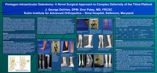
Pentagon Intraarticular Osteotomy: A Novel Surgical Approach to Complex Deformity of the Tibial Plafond
1. Case 2: 5 y/o ♂ with Ollier’s disease Case 1: 14 y/o ♂ with tibial hemimelia Pentagon Intraarticular Osteotomy: A Novel Surgical Approach to Complex Deformity of the Tibial Plafond J. George DeVries, DPM; Dror Paley, MD, FRCSC Rubin Institute for Advanced Orthopedics – Sinai Hospital, Baltimore, Maryland Introduction : Proximal migration of the fibula leads to lateral talar shift and subsequent ankle joint degeneration 1,2 . In children, the physis of the distal fibula should be at the level of the ankle joint line. Chronic proximal migration with lateral talar shift leads to a V-shaped joint surface of the plafond and a space between the medial malleolus and the talus. The talus comes to rest upon the proximally migrated lateral malleolus. The purpose of this study is to describe a new intra-articular osteotomy to realign the ankle joint and foot. Methods : Three Paley-Pentagon osteotomies were performed in the past two years. Patients were followed at an average of 12.3 +/- 7.6 months. Subjective and objective findings were recorded from chart review. Surgical Procedure : The proximal cut of the osteotomy is made perpendicular to the tibia. The two distal cuts are made parallel to their respective parts of the tibial plafond. A middle split is made into the plafond and wedged open to make the medial and lateral parts of the plafond parallel. The distance between the proximal cut and the lateral cut is the amount of shortening of the fibula relative to the ankle joint. The segment of bone removed from the tibia looks like a pentagon. Literature Review : Ankle joint pathology has been dealt with by using joint destructive procedures such as ankle arthrodesis or arthroplasty if there is significant arthrosis, 3,4 or by corrective supramalleolar osteotomies if there is remaining healthy joint space. 2,5 Supramalleolar osteotomies are unable to address intra-articular pathology. Intra-articular osteotomies have been described for the hip and knee joints. 2 Recently, Bluman and Chiodo suggest a lateral distal tibial opening osteotomy may be used to restore intra-articular congruity of the ankle, and describe a technique to do so through a fibular door with bone grafting and internal fixation, but do not report on their results. 6 This is the first report of an intra-articular supramalleolar osteotomy to reconstruct the ankle joint. Results : All cases resulted in improved radiographic angles, stability of the ankle joint, and function of the foot and ankle. Patients were satisfied with the outcomes of the procedure. Case 1 had required a brace prior to the osteotomy, but not afterward. Case 3 is currently involved in cheerleading. There was a decrease in pain in all cases; however there was also a decrease in ankle ROM. Discussion : This comprehensive osteotomy is capable of addressing multiple complex deformities at the ankle and within the joint in a single surgery. In the short term follow-up of these patients there has been improved function and decreased deformity without performing a joint destructive procedure. References : 1) Yablon IG, Heller FG, Shouse L. The fey role of the lateral malleolus in displaced fractures of the ankle. JBJS-Am. 59:169-73, 1977. 2) Paley D. Principles of Deformity Correction 1st Ed. Springer-Verlag Berlin, Germany. 2002. 3) Holt ES, Hansen ST, Mayo KA, Sangeorzan BJ. Ankle arthrodesis using internal screw fixation. Clin Orthop Relat Res. 268:21-8, 1991. 4) Knecht SI, Estin M, Callaghan JJ, Zimmerman MB, Alliman KJ, Alvine FG, Saltzman CL. The Agility total ankle arthroplasty. Seven to sixteen-year follow-up. J Bone Joint Surg Am. 86-A:1161-71, 2004. 5) Harstall R, Lehmann O, Krause F, Weber M. Supramalleolar lateral closing wedge osteotomy for the treatment of varus ankle arthrosis. Foot Ankle Int. 28:542-8, 2007. 6) Bluman EM, Chiodo CP. Valgus ankle deformity and arthritis. Foot Ankle Clin N Am. 13:443-470, 2008. . Pre-operative clinical picture of hindfoot valgus Pre-operative radiographs show intra-articular deformity of the tibial plafond Intraoperative radiograph demonstrating subtractive nature of the osteotomy. The medial portion will be rotated inferiorly to re-establish the medial malleolus Post-operative clinical picture of corrected hindfoot valgus, taken prior to leg lengthening Final ankle radiographs taken at 19 months postoperatively shows improved ankle architecture and stability, with bony healing of all osteotomies Pre-operative clinical picture of hindfoot valgus Pre-operative radiographs show intra-articular deformity of the tibial plafond as well as proximal malalignment Intraoperative radiograph shows subtractive nature of the osteotomy. Again, the medial portion will be rotated inferiorly to re-establish the medial malleolus Postoperative clinical picture of corrected hindfoot valgus. Notice genu valgum which was later corrected Final radiographs taken at 12 months, prior to lengthening and deformity correction of left femur and tibia. Case 3: 13 y/o ♀ with history of chainsaw trauma to ankle Preoperative radiograph and CT scan shows the intra-articular nature of the deformity. Notice the fibular nonunion. Intraoperative radiograph after all fragments are reduced prior to fixation Radiograph at 6 months postoperatively showing restoration of calcaneal valgus and hardware placement Illustration demonstrating the basic principle behind the pentagon osteotomy 6 scar irritation 95 72 90 80 3: KP 12 superficial wound slough 80 90 N/A N/A 2: ER 19 none 70 85 76 95 1: JC F/U months Complications Post-op LDTA Post-op ADTA Pre-op LDTA Pre-op ADTA Pt