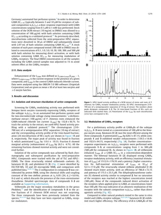
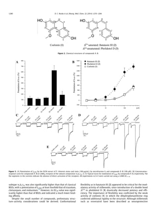
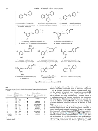
Identification of dihydrostilbenes in Pholidota chinensis as a new
scaffold for GABAA receptor modulators
Diana C. Rueda a
, Angela Schöffmann b
, Maria De Mieri a
, Melanie Raith a
, Evelyn A. Jähne a
,
Steffen Hering b
, Matthias Hamburger a,⇑
a
Division of Pharmaceutical Biology, University of Basel, Klingelbergstrasse 50, CH-4056 Basel, Switzerland
b
Institute of Pharmacology and Toxicology, University of Vienna, Althanstrasse 14, A-1090 Vienna, Austria
a r t i c l e i n f o
Article history:
Received 3 October 2013
Revised 20 December 2013
Accepted 3 January 2014
Available online 10 January 2014
Keywords:
Pholidota chinensis
HPLC-based activity profiling
GABAA receptor modulators
Dihydrostilbenes
a b s t r a c t
A dichloromethane extract of stems and roots of Pholidota chinensis (Orchidaceae) enhanced
GABA-induced chloride currents (IGABA) by 132.75 ± 36.69% when tested at 100 lg/mL in a two-micro-
electrode voltage clamp assay, on Xenopus laevis oocytes expressing recombinant a1b2c2S GABAA recep-
tors. By means of an HPLC-based activity profiling approach, the three structurally related stilbenoids
coelonin (1), batatasin III (2), and pholidotol D (3) were identified in the active fractions of the extract.
Dihydrostilbene 2 enhanced IGABA by 1512.19 ± 176.47% at 300 lM, with an EC50 of 52.51 ± 16.96 lM,
while compounds 1 and 3 showed much lower activity. The relevance of conformational flexibility for
receptor modulation by stilbenoids was confirmed with a series of 13 commercially available stilbenes
and their corresponding semisynthetic dihydro derivatives. Dihydrostilbenes showed higher activity in
the oocyte assay than their corresponding stilbenes. The dihydro derivatives of tetramethoxy-piceatannol
(12) and pterostilbene (20) were the most active among these derivatives, but they showed lower effi-
ciencies than compound 2. Batatasin III (2) showed high efficiency but no significant subunit specificity
when tested on the receptor subtypes a1b2c2s, a2b2c2s, a3b2c2s, a4b2c2s, a5b2c2s, a1b1c2s, and a1b3c2s.
Dihydrostilbenes represent a new scaffold for GABAA receptor modulators.
Ó 2014 Elsevier Ltd. All rights reserved.
1. Introduction
GABAA receptors are ligand-gated chloride channels physiolog-
ically activated by GABA, the major inhibitory neurotransmitter in
the brain. Structurally, they are heteropentameric assemblies
forming a central chloride-selective channel. Up to now 19 differ-
ent subunits (a1–6, b1–3, c1–3, d, e, h, p, q1–3) have been identi-
fied. Depending on the nature, stoichiometry, and arrangement of
these subunits, the receptor subtypes exhibit distinct pharmaco-
logical profiles providing the potential for rational drug therapy
in several disorders related with impaired GABAergic function,
such as epilepsy, insomnia, anxiety, and mood disorders.1–3
GABAA
receptors are the target for many clinically important drugs such as
benzodiazepines (BDZs), barbiturates, neuroactive steroids, anes-
thetics, and certain other CNS depressants. Due to their lack of
subunit specificity, these drugs show a number of adverse side ef-
fects. Hence, there is a need for subtype-selective drugs devoid of
the side effects of the classical BDZs. Despite the availability of
experimental subunit-specific GABAergic drugs for more than a
decade, no subtype-selective GABAA receptor modulators have
been introduced into clinical practice.3,4
In a search for new natural product-derived GABAA receptor
modulators, we screened a library of 880 fungal and plant extracts
in an automated functional two-microelectrode voltage clamp as-
say on Xenopus oocytes5
transiently expressing GABAA receptors
of the a1b2c2S subtype, the most abundant subunit combination
in the human brain.2
In this screening the dichloromethane extract
of stems and roots of Pholidota chinensis LINDL (Orchidaceae)
showed promising activity.
Orchidaceae is the largest family of flowering plants, with
around 25,000 species in over 800 genera. The family shows world-
wide distribution, with greatest diversity in tropical and subtropi-
cal climate zones. Apart from their ornamental value, many orchids
have been used as medicinal plants. In traditional Chinese medi-
cine we find the earliest written records for medicinal uses of orch-
ids.6–8
In Chinese folk medicine, the whole plant of P. chinensis (shi
xian tao) has long been used in the treatment of diverse conditions,
such as hypertension, headache, gastroenteritis, and bronchitis.
Previous pharmacological studies on P. chinensis reported sedative
and anticonvulsant activities.9–12
The genus Pholidota comprises
approximately 30 species distributed from tropical Asia to tropical
0968-0896/$ - see front matter Ó 2014 Elsevier Ltd. All rights reserved.
http://dx.doi.org/10.1016/j.bmc.2014.01.008
Abbreviations: BDZs, benzodiazepines; GABA, gamma-aminobutyric acid;
GABAARs, gamma-aminobutyric acid type A receptors.
⇑ Corresponding author. Tel.: +41 61 2671425; fax: +41 61 2671474.
E-mail address: matthias.hamburger@unibas.ch (M. Hamburger).
Bioorganic & Medicinal Chemistry 22 (2014) 1276–1284
Contents lists available at ScienceDirect
Bioorganic & Medicinal Chemistry
journal homepage: www.elsevier.com/locate/bmc
Australia. Phytochemical studies on the genus showed the pres-
ence of triterpenes, steroids, lignans, benzoxepines, and stilbe-
noids.10
Stilbenoids exhibit a limited but heterogeneous
distribution in the plant kingdom, and have been most widely re-
ported from the Orchidaceae family. Dihydrostilbenes and 9,10-
dihydrophenanthrenes have been previously identified in the
genus Pholidota.13–17
In this study, batatasin III (2) isolated from a DCM extract of
P. chinensis was identified as a positive GABAA receptor modulator
by means of HPLC-based activity profiling,18
a miniaturized
approach for the rapid identification of new bioactive natural com-
pounds,19–22
that we have been successfully applying in the discov-
ery of GABAA receptor ligands of natural origin.23–27
The subunit
selectivity of 2 was assessed at GABAA receptor subtypes a2b2c2s,
a3b2c2s, a4b2c2s, a5b2c2s, a1b1c2s, and a1b3c2s. Furthermore,
dihydrostilbenes were established as a new scaffold for GABAA
receptor modulators, by comparison of the performance of a series
of commercially available stilbenes and their semisynthetic
dihydro derivatives on the oocyte assay.
2. Experimental
2.1. General procedures
1D and 2D NMR spectra were measured at room temperature
on a Bruker Avance III 500 MHz spectrometer (Bruker BioSpin,
Fällanden, Switzerland) operating at 500.13 MHz. Spectra were re-
corded at 291.2 K with a 1 mm TXI probe with z-gradient. The fol-
lowing settings were used: 128 scans for 1
H spectra; 8 scans for
1
H1
H-COSY spectra (cosygpqf pulse program); 32 scans and 256
increments for HSQC experiments (hsqcedetgp pulse program); 64
scans and 128 increments for HMBC spectra (hmbcgp pulse pro-
gram). Spectra were analyzed by TopSpin 3.0 software (Bruker).
High resolution mass spectra (HPLC–ESITOFMS) were recorded in
positive mode, m/z range 100–800, on a Bruker microTOF ESIMS
system (Bruker Daltonics, Bremen, Germany) connected via a
T-splitter (1:10) to an Agilent HP 1100 system consisting of a deg-
asser, a binary mixing pump, autosampler, column oven, and a
diode array detector (G1315B) (Agilent Technologies, Waldbronn,
Germany). Nitrogen was used as a nebulizing gas at a pressure of
2.0 bar, and as drying gas at a flow rate of 9.0 L/min (dry gas tem-
perature 240 °C). Capillary voltage was set at 45,000 V; hexapole at
230.0 Vpp. Instrument calibration was done with a reference solu-
tion of sodium formate 0.1% in 2-propanol/water (1:1) containing
5 mM NaOH. Data acquisition and processing was performed with
Bruker Daltonics Hystar 3.0 software. Semi-preparative HPLC sep-
arations for activity profiling and purification were performed with
an Agilent HP 1100 series system consisting of a quaternary pump,
autosampler, column oven, and diode array detector (G1315B).
Waters SunFire™ C18 (3.5 lm, 3.0 Â 150 mm) and SunFire™ Prep
C18 (5 lm, 10 Â 150 mm) columns were used for analytical and
semi-preparative HPLC analysis, respectively (Waters, Wexford,
Ireland). Parallel evaporation of semi-preparative HPLC fractions
was performed with a Genevac EZ-2 plus vacuum centrifuge (Gen-
evac Ltd, Ipswich, United Kingdom). Flash chromatography was
performed with pre-packed Buchi SepacoreÒ
silica flash cartridges
(40–63 lm, 40 Â 150 mm) on a Buchi SepacoreÒ
system consisting
of two C-605 pumps, a C-620 control unit, and a C-660 fraction col-
lector (Buchi, Flawil, Switzerland). Sample introduction was car-
ried out with a Buchi Prep Elut adapter filled with the sample
adsorbed onto silica gel. The separation was monitored by TLC. Pre-
parative HPLC separations were performed with a Waters SunFire
Prep C18 OBD column (5 lM, 30 Â 150 mm) connected to a
Shimadzu LC-8A preparative HPLC with SPD-M10A VP diode array
detector. HPLC-grade MeOH (Scharlau Chemie S.A.), acetonitrile
(Scharlau Chemie S.A.) and water were used for HPLC separations.
For analytical separations, HPLC solvents contained 0.1% of HCO2H.
NMR spectra were recorded in methanol-d4 (Armar Chemicals). For
extraction and flash chromatography, distilled technical grade sol-
vents were used. Silica gel (63–200 lm, Merck) was used for open
column chromatography.
2.2. Plant material
Shi Xian Tao (dried stems/roots of P. chinensis Lindl.) was pur-
chased in 2008 from a local herbal market in Kunming and authen-
ticated by Dr. X. Yang (previously Pharmaceutical Biology,
University of Basel, now Kunming Institute of Botany, Chinese
Academy of Science, Kunming, PR China). A voucher specimen
(463) is deposited at the Division of Pharmaceutical Biology, Uni-
versity of Basel.
2.3. Extraction
The plant material was frozen with liquid nitrogen and ground
with a ZM1 ultracentrifugal mill (Retsch). The DCM extract for
screening and HPLC-based activity profiling was prepared with
an ASE 200 extraction system with solvent module (Dionex, Sun-
nyvale CA). Three extraction cycles (5 min each) were performed,
at an extraction pressure of 120 bar and a temperature of 70 °C.
For preparative isolation, 293 g of ground plant material was mac-
erated with DCM (4 Â 1 L, 3 h each, permanent magnetic stirring).
The solvent was evaporated at reduced pressure to yield 10.3 g of
extract. The extracts were stored at 2–8 °C until use.
2.4. Microfractionation for activity profiling
Time-based microfractionation for GABAA receptor activity pro-
filing was performed as previously described,23,27,28
with minor
modifications: separation was done on a semi-preparative HPLC
column with MeOH (solvent A) and water (solvent B), using a gra-
dient from 50% A to 80% A in 30 min, followed by 80% A to 100% A
in 2 min. The flow rate was 4 mL/min, and 10 mg of extract (in
200 lL of DMSO) were injected. A total of 24 microfractions of
90 s each were collected. After parallel evaporation of solvents,
the dry films were redissolved in 1 mL of methanol, and aliquots
of 0.5 mL were dispensed in two vials, dried under N2 gas, and sub-
mitted to bioassay.
2.5. Isolation
An aliquot of the DCM extract (450 mg) was dissolved in CHCl3
and adsorbed onto silica gel (3 g), prior to packing into a Buchi Prep
Elut adapter. Separation was performed on a SepacoreÒ
silica gel
cartridge eluted with an n-hexane (solvent A) and EtOAc (solvent
B) gradient: 0% B to 30% B in 90 min, followed by 30% B to 50% B
in 30 min, and 50% B to 80% B in 30 min. The flow rate was set at
15 mL/min. Fractions of 15 mL were collected and later combined
into 18 fractions (1–18) on the basis of a TLC analysis (detection
at 254, 366, and at daylight after staining with anisaldehyde–sulfu-
ric acid reagent). Fractions 1–18 were submitted to analytical
HPLC–PDA–ESIMS with MeOH (solvent C) and water (solvent D),
using a gradient from 50% C to 100% C in 30 min, hold for
15 min. The flow rate was 0.4 mL/min, and 10 lg of each fraction
(in 10 lL of DMSO) were injected. Fractions 13 and 14 were found
to contain the compounds of interest and were submitted to semi-
preparative HPLC using solvents C and D as eluents. A gradient of
50% C to 60% C in 30 min was used for fraction 13, and isocratic
conditions (50% C, 30 min) for fraction 14. The flow rate was
4 mL/min. Stock solutions in DMSO (50 mg/mL) were prepared
and repeatedly injected in portions of 50–100 lL. A portion
D. C. Rueda et al. / Bioorg. Med. Chem. 22 (2014) 1276–1284 1277
(17 mg) of fraction 13 (25 mg) afforded compounds 1 (2.3 mg) and
2 (6.5 mg). Compound 3 (2 mg) was isolated from 10 mg of fraction
14 (16 mg).
Compounds 1–3 were identified by comparison of their physio-
chemical data (NMR, ESI-TOFMS, and UV–vis) with published val-
ues.14,29–31
The purity was >95% (purity check by 1
H NMR).
2.5.1. Coelonin (1)
1
H NMR (methanol-d4, 500.13 MHz) dH (ppm): 8.13 (1H, d,
J = 8.4 Hz, H-5), 6.62 (1H, dd J = 8.3 and 2.7 Hz, H-6), 6.61 (1H, d,
J = 2.6 Hz, H-8), 6.30 (1H, d, J = 2.5 Hz, H-3), 6.26 (1H, d,
J = 2.5 Hz, H-1), 3.67 (3H, s, 4-OCH3), 2.59 (4H, s, H-9 and H-10);
13
C shifts (derived from multiplicity-edited HSQC and HMBC spec-
tra), dC (ppm): 158.3 (C, C-4), 155.4 (C, C-2), 154.8 (C, C-7), 139.8 (C,
C-8a), 138.7 (C, C-10a), 128.6 (CH, C-5), 125.2 (C, C-4b), 114.8 (C,
C-4a), 113.8 (CH, C-8), 112.2 (CH, C-6), 104.8 (CH, C-1), 100.1
(CH, C-3), 54.2 (4-OCH3), 30.8 (CH2, C-10) 30.1 (CH2, C-9).
HR-ESIMS m/z 243.1016 [M+H]+
(calcd for C15H15O3, 243.1016).
2.5.2. Batatasin III (2)
1
H NMR (methanol-d4, 500.13 MHz) dH (ppm): 7.03 (1H, dd,
J = 7.9 and 7.5 Hz, H-50
), 6.64 (3H, m, H-20
, H-40
, and H-60
), 6.29
(1H, br s, H-2), 6.23 (2H, m, H-4 and H-6), 3.63 (3H, s, 5-OCH3),
2.75 (4H, m, H-a and H-b); 13
C shifts (derived from multiplicity-
edited HSQC and HMBC spectra), dC (ppm): 160.9 (C, C-5), 157.6
(C, C-3), 156.7 (C, C-30
), 144.9 (C, C-1), 143.3 (C, C-10
), 129.0 (CH,
C-50
), 119.8 (CH, C-60
), 115.3 (CH, C-20
), 112.4 (CH, C-40
), 108.1
(CH, C-2), 105.5 (CH, C-6), 98.7 (CH, C-4), 54.3 (CH3, 5-OCH3),
37.6 (CH2, C-b), 37.0 (CH2, C-a). HR-ESIMS m/z 245.1176 [M+H]+
(calcd for C15H17O3, 245.1172).
2.5.3. Pholidotol D (3)
1
H NMR (methanol-d4, 500.13 MHz) dH (ppm): 7.17 (1H, dd,
J = 7.9 and 7.8 Hz, H-50
), 7.00–6.95 (4H, m, H-20
, H-60
, H-a and H-
b), 6.69 (1H, dd, J = 8.2 and 2.2 Hz, H-40
), 6.58 (2H, m, H-2 and H-
6), 6.31 (1H, t, J = 2 Hz, H-4), 3.76 (3H, s, 5-OCH3); 13
C shifts (de-
rived from multiplicity-edited HSQC and HMBC spectra), dC
(ppm): 160.8 (C, C-5), 157.7 (C, C-3), 156.0 (C, C-30
), 139.7 (C, C-
1), 138.5 (C, C-10
), 129.4 (CH, C-50
), 128.6 (CH, C-b), 128.4 (CH, C-
a), 117.8 (CH, C-60
), 114.4 (CH, C-40
), 112.4 (CH, C-20
), 105.8 (CH,
C-2), 103.4 (CH, C-6), 100.3 (CH, C-4), 54.4 (CH3, 5-OCH3). HR-
ESIMS m/z 243.1017 [M+H]+
(calcd for C15H15O3, 243.1016).
Further purification of compound 2 for subunit specificity tests
was achieved by separating a portion of the extract (7.3 g) by open
column chromatography (6 Â 69 cm, 700 g of silica gel), using a
step gradient of n-hexane–EtOAc (100:0, 95:5, 90:10, 85:15,
80:20, 75:25, 70:30, 65:35, 60:40, 55:45, 50:50, 40:60, 30:70,
20:80, 10:90, 0:100, 1 L each) and washing in the end with MeOH
100% (1.5 L). The flow rate was ca. 50 mL/min. The effluent was
combined to 15 fractions (1–15) based on TLC patterns. After
HPLC–PDA–MS analysis, fractions 7 and 8 were selected for isola-
tion of compound 2 by preparative HPLC, with acetonitrile (solvent
A) and water (solvent B), using a gradient from 40% A to 50% A in
30 min, followed by 50% A to 100% A in 5 min, hold for 10 min.
The flow rate was 20 mL/min. Stock solutions in THF (100 mg/
mL) were prepared and repeatedly injected in portions of 300–
400 lL. The separation of fractions 7 (129 mg) and 8 (132 mg),
yielded compound 10.8 mg of 2.
2.6. Synthesis of dihydrostilbenes
2.6.1. Stilbenes
Compounds 4–7, 9, 11, 13, 15, 17, and 19 were purchased from
TCI Europe N.V. Compounds 21 and 25 were purchased from Sig-
ma–Aldrich Co. Compound 23 was purchased from Santa Cruz Bio-
technology, Inc.
2.6.2. General procedure
Dihydro derivatives of compounds 7, 9, 11, 13, 15, 17, 19, 21,
and 23 were prepared by hydrogenation of corresponding stilb-
enes. A standard protocol was followed,32
with minor modifica-
tions. Solutions of each stilbene (10 mg) in absolute EtOH (5 ml)
were stirred under H2 for 3 h in the presence of 10% Pd/C. The reac-
tion mixtures were filtered over Celite to remove the catalyst, and
evaporated to dryness. The resulting residues were purified by
flash column chromatography, using a hexane/EtOAc gradient, to
afford target compounds 8, 10, 12, 14, 16, 18, 20, 22, and 24,
respectively, in yields of 85–95%. The spectroscopic data of com-
pounds were in agreement with the literature, except for com-
pound 24, for which no report was found (1
H NMR spectrum is
provided as Supporting information).32–41
2.6.3. trans-2-Fluoro-40
-methoxy-dihydrostilbene (24)
1
H NMR (chloroform-d4, 500.13 MHz) dH (ppm): 7.26–7.08 (4H,
m), 7.08–7.98 (2H, m), 6.90–6.80 (2H, m), 3.81 (3H, s), 3.05–2.84
(4H, m). HRESI-MS m/z 253.1589 [M+Na]+
(calcd formula weight
for C15H15FO, 230.2774).
2.7. Expression of GABAA receptors
Stage V–VI oocytes from Xenopus laevis were prepared, and
cRNA injected as previously described.23
Female Xenopus laevis
(NASCO, Fort Atkinson, WI) were anesthetized by exposing them
for 15 min to a 0.2% MS-222 (methanesulfonate salt of 3-amino-
benzoic acid ethyl, Sigma) solution before surgically removing
parts of the ovaries. Follicle membranes from isolated oocytes were
enzymatically digested with 2 mg/mL collagenase from Clostridium
histolyticum (Type 1A, Sigma). Synthesis of capped runoff poly(A+)
cRNA transcripts was obtained from linearized cDNA templates
(pCMV vector). Directly after enzymatic isolation, the oocytes were
injected with 50 nL of DEPC-treated water (Sigma) containing dif-
ferent cRNAs at a concentration of approximately 300–3000 pg/nL
per subunit. The amount of injected cRNA mixture was determined
by means of a NanoDrop ND-1000 (Kisker Biotech). To ensure
expression of the gamma subunit in a1b2c2S receptors, rat cRNAs
were mixed in a 1:1:10 ratio. Oocytes were then stored at 18 °C
in ND96 solution containing 1% of penicillin–streptomycin solution
(Sigma–Aldrich). Voltage clamp measurements were performed
between days 1 and 5 after cRNA injection.
2.8. Positive control
Diazepam (7-chloro-1,3-dihydro-1-methyl-5-phenyl-2H-1,4-
benzodiazepin-2-one, Sigma, purity not less than 98%) was used
as positive control. At 1 lM diazepam enhanced IGABA up to
231.3 ± 22.6% (n = 3). See also Figure S1, Supporting information.
2.9. Two-microelectrode voltage clamp studies
Electrophysiological experiments were performed by the two-
microelectrode voltage clamp method making use of a TURBO
TEC 03X amplifier (npi electronic GmbH) at a holding potential
of À70 mV and pCLAMP 10 data acquisition software (Molecular
Devices). Currents were low-pass-filtered at 1 kHz and sampled
at 3 kHz. The bath solution contained 90 mM NaCl, 1 mM KCl,
1 mM MgCl2, 1 mM CaCl2, and 5 mM HEPES (pH 7.4). Electrode fill-
ing solution contained 2 M KCl. Oocytes with maximal current
amplitudes >3 lA were discarded to exclude voltage clamp errors.
2.10. Fast solution exchange during IGABA recordings
Test solutions (100 lL) were applied to the oocytes at a speed of
300 lL/s by means of the ScreeningTool (npi electronic, Tamm,
1278 D. C. Rueda et al. / Bioorg. Med. Chem. 22 (2014) 1276–1284
Germany) automated fast perfusion system.5
In order to determine
GABA EC3–10 (typically between 3 and 10 lM for receptors of sub-
unit composition a1b2c2s), a dose–response experiment with GABA
concentrations ranging from 0.1 lM to 1 mM was performed. Stock
solution of the DCM extract (10 mg/mL in DMSO) was diluted to a
concentration of 100 lg/mL with bath solution containing GABA
EC3–10 according to a validated protocol.23
As previously described,
microfractions collected from the semi-preparative HPLC separa-
tions were dissolved in 30 lL of DMSO and subsequently mixed
with 2.97 mL of bath solution containing GABA EC3–10.23
A stock
solution of each pure compound tested (100 mM in DMSO) was di-
luted to concentrations of 0.1, 1.0, 3.0, 10, 30, 100, 300, and 500 lM
with bath solution for measuring direct activation, or with bath
solution containing GABA EC3–10 for measuring modulation of
GABAA receptors. The final DMSO concentration in all the samples
including the GABA control samples was adjusted to 1% to avoid
solvent effect at the GABAA receptor.
2.11. Data analysis
Enhancement of the IGABA was defined as I(GABA+Comp)/IGABA À 1,
where I(GABA+Comp) is the current response in the presence of a given
compound, and IGABA is the control GABA-induced chloride current.
Data were analyzed using the ORIGIN 7.0 SR0 software (OriginLab
Corporation) and are given as mean ± SE of at least two oocytes and
P2 oocyte batches.
3. Results and discussion
3.1. Isolation and structure elucidation of active compounds
Screening for GABAA modulating activity was performed with
Xenopus laevis oocytes transiently expressing GABAA receptors of
the subtype a1b2c2s. In an automated fast-perfusion system used
for two-microelectrode voltage clamp measurements,5
a dichloro-
methane extract (100 lg/mL) of P. chinensis roots enhanced the
GABA-induced chloride ion current (IGABA) by 132.8 ± 36.7%. To
track the activity in the extract, we used HPLC-based activity pro-
filing with a validated protocol.23
The chromatogram (210–
700 nm) of a semipreparative HPLC separation (10 mg of extract)
and the corresponding activity profile of the time-based fraction-
ation (24 microfractions of 90 s each) are shown in Figure 1B and
A, respectively. The major peak of activity was found in fraction
9, which potentiated IGABA by 119.1 ± 19.1%. Fraction 8 showed
marginal activity (enhancement of IGABA by 26.5 ± 4.7%). All the
remaining fractions showed minimal activity and were not consid-
ered further.
Isolation of the active compounds was achieved by flash chro-
matography and subsequent purification by semi-preparative
HPLC. Compounds were tracked with the aid of TLC and HPLC–
ESIMS. The three structurally related stilbenoids coelonin (1),
batatasin III (2), and pholidotol D (3) (Fig. 2) were identified by
ESI-TOF-MS, 1D and 2D microprobe NMR, and comparison with
published data.14,29–31
The Z configuration in compound 3 was cor-
roborated by proton NMR, using the chemical shifts and coupling
constant of the two olefinic protons at dH 6.95 (2H, d, J = 6.0 Hz,
H-b and a), which discards the presence of the trans-stereoisomer
thunalbene. Detailed spectroscopic data of compounds 1–3 are
available as Supporting information.
Stilbenoids are the major secondary metabolites in the genus
Pholidota,10
and the identification of compounds 1–3 in the ac-
tive fractions of P. chinensis DCM extract was not surprising.
The three compounds have been previously isolated from the
species,10,12,14
but they have not been reported as GABAA recep-
tor modulators.
3.2. Modulation of GABAA receptors
For a preliminary activity profile at GABAARs of the subtype
a1b2c2s, 1–3 were tested at a concentration of 100 lM in the Xeno-
pus oocyte assay. Batatasin III (2) was the most efficient among the
three compounds. It potentiated IGABA by 628.3 ± 87.1%, while com-
pounds 1 and 3 exhibited weaker enhancements (139.5 ± 14.4%
and 192.0 ± 64.1%, respectively) (Fig. 3A). Further concentration–
response experiments on a1b2c2s receptors were performed with
compounds 1–3, at concentrations ranging from 1 to 300 lM
(500 lM for compound 3). As shown in Figure 3B, all stilbenoids
enhanced IGABA at a GABA EC3–10 in a concentration-dependent
manner. The bibenzyl batatasin III (2) displayed strong GABAA
receptor modulatory activity, with an efficiency (maximal stimula-
tion of IGABA) of 1512.9 ± 176.5% and a potency (higher concentra-
tion for half-maximal stimulation of IGABA, or EC50) of
52.5 ± 17.0 lM. The structurally related stilbene pholitodol D (3)
showed much lower activity, with an efficiency of 786.8 ± 72.1%
and potency of 175.5 ± 25.5 lM. The dihydrophenanthrene coelo-
nin (1) showed activity similar to compound 2, but no saturation
of the receptors was reached at the highest concentration tested
(300 lM). None of the compounds induced direct activation of
the receptors when applied prior to GABA, at concentrations lower
than 100 lM. This was indicative of an allosteric modulation of the
receptor with the subunit composition a1b2c2s, rather than direct
agonistic activity (Fig. 3C).
Compared to other natural products tested in the same in vitro
model and GABAA receptor subtype,24,27,28,33
batatasin III (2) exhib-
ited much higher efficiency. The efficiency of 2 in GABAARs of the
Figure 1. HPLC-based activity profiling of a DCM extract of stems and roots of P.
chinensis, for GABAA receptor modulatory activity. (B) HPLC chromatogram (210–
700 nm) of a semipreparative separation of 10 mg of extract. The numbers above
peaks designate compounds 1–3. The 24 time-based fractions of 90 s each are
indicated with dashed lines. (A) Potentiation of the IGABA by each microfraction
(error bars correspond to SE).
D. C. Rueda et al. / Bioorg. Med. Chem. 22 (2014) 1276–1284 1279
subtype a1b2c2s was also significantly higher than that of classical
BDZs, with a potentiation of IGABA at least fourfold that of triazolam,
clotiazepam, and midazolam.34
However, its EC50 value was signif-
icantly higher than that of BDZs and indicated a much lower bind-
ing affinity.
Despite the small number of compounds, preliminary struc-
ture–activity considerations could be derived. Conformational
flexibility as in batatasin III (2) appeared to be critical for the mod-
ulatory activity of stilbenoids, since introduction of a double bond
Db,a
in pholidotol D (3) drastically decreased potency and effi-
ciency. The importance of flexibility was confirmed by the weak
activity of coelonin (1) in which the dihydrophenanthrene ring
conferred additional rigidity to the structure. Although stilbenoids
such as resveratrol have been described as neuroprotective
Figure 3. (A) Potentiation of IGABA by the DCM extract of P. chinensis stems and roots (100 lg/mL), by microfraction 9, and compounds 1–3 (100 lM). (B) Concentration–
response curve for compounds 1–3 on GABAA receptors of the subunit composition a1b2c2S. (C–E) Typical traces for modulation of IGABA by compounds 1–3, respectively. The
flat segments in the currents indicate the absence of direct activation of the receptors. All experiments in A–E were carried out using a GABA EC3–10.
Figure 2. Chemical structures of compounds 1–3.
1280 D. C. Rueda et al. / Bioorg. Med. Chem. 22 (2014) 1276–1284
agents,17,35–38
none of them has been reported as GABAA receptor
ligand so far. Batatasin III (2) is thus the first representative of a
new scaffold for GABAA receptor modulators. It is noteworthy that
compounds with biosynthetically related scaffolds such as flavo-
noids,25,39
coumarins,24
and lignans40
have been previously shown
to possess GABAA receptor modulatory properties.
3.3. GABAA receptor subtype selectivity
Batatasin III (2) was tested for potential a subunit specificity, by
replacing the a1 subunit in the receptor subtype a1b2c2s with a2,
a3, a4, and a5. Likewise, b subunit specificity was evaluated by
replacing b2 with b1 and b3. Concentration-dependent IGABA modu-
lation of compound 2 was evaluated on receptor subtypes a2b2c2s,
a3b2c2s, a4b2c2s, a5b2c2s, a1b1c2s, and a1b3c2s (Table 1).
As shown in Figure 4 and summarized in Table 1, compound 2
did not exhibit subtype specificity, as reflected by comparable EC50
values with all receptor subtypes studied (p > 0.05). The order of
potency of batatasin III (2) in receptor composed by different a
subunits was a4b2c2s > a5b2c2s > a1b2c2s > a3b2c2s > a2b2c2s. The
lower potency on a2b2c2s receptors compared to a4b2c2s was sta-
tistically significant, while there were no significant differences
in efficiency among the other a-containing receptor subtypes. On
GABAA receptors comprising different b subunits, almost no differ-
ences in potency and efficiency were observed. Thus, batatasin III
(2) was a positive allosteric modulator of GABAARs, devoid of sig-
nificant subtype specificity.
3.4. GABAAR modulatory activity of dihydrostilbenes
Flexibility appeared to be a critical factor for the GABAAR mod-
ulatory activity of stilbenoids. To confirm the influence of the dou-
ble bond Db,a
, 13 commercially available stilbenoids and their
corresponding dihydro derivatives (compounds 4–25; Fig. 5) were
tested in the Xenopus oocyte assay. Compounds were initially
tested at a concentration of 100 lM on GABAARs of the subtype
a1b2c2s. As expected, dihydrostilbenes showed higher activity than
the corresponding stilbenes (Table 2, Fig. 6A). These differences in
the activity of stilbenes and their dihydro derivatives were statisti-
cally significant in almost every case, with the exception of the
pairs 4 and 5/6, 9/10, 13/14, and 23/24 (p > 0.05).
Among the stilbenes, tetramethoxy-piceatannol (11), resvera-
trol (13), pterostilbene (19), and resveratrol triacetate (21), dis-
played the highest activity, potentiating IGABA between 100% and
200%. Their corresponding dihydro derivatives showed the highest
activity among dihydrostilbenes, but only compounds 12 and 20
showed efficiencies comparable to that of batatasin III (2)
(544.5 ± 104.4% and 660.6 ± 100.2% respectively). A comparison
of the activity of the dihydrostilbenes at 100 lM revealed that
the bibenzyl scaffold alone (6) does not possess any GABAAR mod-
ulatory activity. In general, substituents at C-3 and C-5 (12, 14, 16,
18, 20, and 22) resulted in an enhancement of the activity. Increas-
ing the lipophilicity by replacing the hydroxy groups at C-3 and C-5
with bulkier oxygenated functions (12, 20, and 22) enhanced the
Figure 4. (A) a-Subunit dependency of batatasin III (2), depicted as concentration–response curves, with GABAA receptors of the subunit compositions a1b2c2s, a2b2c2s,
a3b2c2s, a4b2c2s, and a5b2c2s. (B) b-Subunit dependency of batatasin III (2), depicted as concentration–response curves with GABAA receptors of the subunit compositions
a1b1c2s, a1b2c2s, and a1b3c2s. (C and D) Typical traces for modulation of IGABA by compound 2, in receptors with different a and b subunit composition, respectively. All
experiments were performed using a GABA EC3–10.
Table 1
Potencies and efficiencies of batatasin III (2) for GABAA receptors of different subunit
compositions
Subtype EC50 (lM) Max. potentiation of IGABA
(EC3–10) (Imax) (%)
Hill coeff. (nH) na
a1b2c2s 52.5 ± 17.0 1512.9 ± 176.5 1.4 ± 0.3 5
a2b2c2s 80.8 ± 22.1 1026.5 ± 139.2 1.2 ± 0.1 6
a3b2c2s 67.3 ± 18.6 1694.2 ± 229.0 1.2 ± 0.1 5
a4b2c2s 26.2 ± 3.6 1588.2 ± 97.5 1.5 ± 0.1 6
a5b2c2s 46.7 ± 9.0 1375.7 ± 76.5 1.3 ± 0.1 5
a1b1c2s 66.7 ± 21.0 1251.3 ± 157.0 1.8 ± 0.4 5
a1b3c2s 67.2 ± 10.5 1252.9 ± 79.9 1.4 ± 0.1 5
a
Number of experiments.
D. C. Rueda et al. / Bioorg. Med. Chem. 22 (2014) 1276–1284 1281
activity of dihydrostilbenes. The role of substituents in ring B was
less clear within this compounds series. In the case of compounds
12 and 20, different substitution patterns in ring B did not influ-
ence the activity. In contrast, when comparing compounds 14,
16, and 18, addition of a hydroxy group in C-40
or C-60
led to a sig-
nificant decrease of activity. Introduction of a halogen atom as in
25 induced slight negative receptor modulation, and substitution
at C-4 (compound 10) decreased activity. Since we had only one
pair of cis and trans isomers (4 and 5, both inactive at 100 lM),
the role of geometric isomerism could not be assessed in more
detail.
The dihydro derivatives of tetramethoxy-piceatannol and ptero-
stilbene (compounds 12 and 20, respectively) were submitted to
further concentration–response experiments on a1b2c2s receptors.
Both compounds enhanced IGABA at a GABA EC3–10 in a concentra-
tion-dependent manner (Fig. 6B). Compounds 12 and 20 had lower
efficiency than the natural dihydrostilbene 2 (Table 3), with max-
imal stimulations of IGABA of 870.7 ± 106.8% and 694.2 ± 86.0%,
respectively. In terms of potency, 20 was comparable to 2 (EC50
54.5 ± 13.4 lM), whereas 12 was twice as potent (EC50 20.2 ±
Figure 5. Chemical structures of compounds 4–25.
Table 2
Potentiation of IGABA in a1b2c2s receptors by compounds 4–25, at a test concentration
of 100 lM
Stilbenes Dihydrostilbenes
Compound Max. potentiation
of IGABA
na
Compound Max. potentiation
of IGABA
na
4 12.6 ± 9.2 3 6 8.3 ± 22.9 3
5 À11.3 ± 12.3 3
7 À7.3 ± 0.1 3 8 51.6 ± 1.1 3
9 À24.8 ± 4.8 3 10 À20.9 ± 3.7 3
11 101.3 ± 0.9 3 12 544.5 ± 140.4 3
13 121.9 ± 21.8 3 14 162.2 ± 17.5 3
15 À35.4 ± 9.8 3 16 86.3 ± 9.9 3
17 À19.7 ± 3.9 3 18 44.1 ± 19.7 3
19 212.4 ± 10.9 3 20 660.6 ± 100.2 3
21 122.8 ± 18.6 3 22 227.7 ± 1.3 2
23 À22.9 ± 7.5 3 24 À16.8 ± 7.9 3
Diazepamb
(1 lM)
231.3 ± 22.6 3 25 À12.9 ± 0.4 3
a
Number of experiments.
b
Positive control.
1282 D. C. Rueda et al. / Bioorg. Med. Chem. 22 (2014) 1276–1284
6.4 lM). This suggests that increased lipophilicity of ring B may
have a positive effect on the potency of dihydrostilbenes. However,
further studies with a larger series of compounds are needed for
confirmation. None of the compounds induced direct activation
of the receptors when applied prior to GABA, at concentrations
lower than 100 lM (Fig. 6C).
Stilbenoids have attracted significant attention in recent years
due to their wide range of useful properties, including applications
in optics, biochemistry, and chemotherapy.13,41
The stilbenoid
scaffold can be considered as a priviledged structure.42,43
However,
there have been no reports on GABAA receptor modulatory activity
of stilbenoids up to now, despite a significant number of publica-
tions on biological activities of natural stilbenoids, and in particu-
lar, on resveratrol. Dihydrostilbenes such as 2 may thus be an
interesting starting point for the synthesis of new GABAA receptor
modulators.
4. Conclusions
With the aid of an HPLC-based profiling approach, we identified
batatasin III (2) as the major compound responsible for GABAAR
modulatory activity of the dichloromethane extract of P. chinensis.
This dihydrostilbene showed allosteric modulation in a1b2c2s
GABAA receptors with a higher efficiency than any other natural
products tested up to now, but its EC50 value was significantly
higher than that of BDZs. Dihydrostilbenes represent a new scaf-
fold for GABAA receptor modulators.
The conformational flexibility of dihydrostilbenoids appeared
critical for GABAAR modulatory properties. For a further explora-
tion of this scaffold, conformationally restricted derivatives should
be synthesized in order to explore in more detail the optimal ori-
entation of the aromatic rings and substituents.
Acknowledgements
Financial support was provided by the Swiss National Science
Foundation through project 205320_126888 (M.H.). D.C.R. thanks
the Swiss Federal Commission for Scholarships for Foreign Stu-
dents (FCS) and the Department of Education of Canton Basel
(Erziehungsdepartement des Kantons Basel-Stadt) for fellowships
granted in 2011 and 2012, respectively.
Supplementary data
Supplementary data associated with this article can be found, in
the online version, at http://dx.doi.org/10.1016/j.bmc.2014.01.008.
References and notes
1. Olsen, R. W.; Sieghart, W. Pharmacol. Rev. 2008, 60, 243.
2. Rudolph, U.; Knoflach, F. Nat. Rev. Drug Disc. 2011, 10, 685.
3. D’Hulst, C.; Atack, J. R.; Kooy, R. F. Drug Discovery Today 2009, 14, 866.
4. Tan, K. R.; Rudolph, U.; Lüscher, C. Trends Neurosci. 2011, 34, 188.
5. Baburin, I.; Beyl, S.; Hering, S. Pflug. Arch. Eur. J. Physiol. 2006, 453, 117.
6. Pérez Gutiérrez, R. M. J. Med. Plants Res. 2010, 4, 592.
7. Williams, R.; Martin, S.; Hu, J.-F.; Garo, E.; Rice, S.; Norman, V.; Lawrence, J.;
Hough, G.; Goering, M.; O’Neil-Johnson, M.; Eldridge, G.; Starks, C. Planta Med.
2011, 78, 160.
8. Bulpitt, C. J.; Li, Y.; Bulpitt, P. F.; Wang, J. J. R. Soc. Med. 2007, 100, 558.
9. Wang, J.; Matsuzaki, K.; Kitanaka, S. Chem. Pharm. Bull. (Tokyo) 2006, 54, 1216.
10. Bandi, A. K. R.; Lee, D.-U. Chem. Biodiversity 2011, 8, 1400.
11. Wu, B.; Qu, H.; Cheng, Y. Chem. Biodiversity 1803, 2008, 5.
Figure 6. (A) Potentiation of IGABA by compounds 4–25 (100 lM). (B) Potentiation of IGABA by compounds 2, 12 and 20. Concentration–response curves are shown for GABAA
receptors of the subunit composition a1b2c2S. (C and D) Typical traces for modulation of IGABA by compounds 12 and 20, respectively. The inward currents induced in the
absence of GABA (C and D) indicate direct activation of the receptors. All experiments shown in A–D were performed using a GABA EC3–10.
Table 3
Potencies and efficiencies of compounds 2, 3, 12 and 20 for a1b2c2s GABAA receptors
Compound EC50 (lM) Max. potentiation of IGABA
(EC3–10) (Imax) (%)
Hill coeff.
(nH)
na
2 52.5 ± 17.0 1512.9 ± 176.5 1.2 ± 0.1 5
3 175.5 ± 25.5 786.8 ± 72.1 1.5 ± 0.2 5
12 20.2 ± 6.4 870.7 ± 106.8 2.3 ± 1.0 4
20 54.5 ± 13.4 694.2 ± 86.0 1.6 ± 0.2 4
a
Number of experiments.
D. C. Rueda et al. / Bioorg. Med. Chem. 22 (2014) 1276–1284 1283
12. Yao, S.; Tang, C.-P.; Li, X.-Q.; Ye, Y. Helv. Chim. Acta 2008, 91, 2122.
13. Rivière, C.; Pawlus, A. D.; Mérillon, J.-M. Nat. Prod. Rep. 2012, 29, 1317.
14. Majumder, P. L.; Roychowdhury, M.; Chakraborty, S. Phytochemistry 1998, 49,
2375.
15. Kovacs, A.; Vasas, A.; Hohmann, J. Phytochemistry 2008, 69, 1084.
16. Shen, T.; Wang, X.-N.; Lou, H.-X. Nat. Prod. Rep. 2009, 26, 916.
17. Greger, H. J. Nat. Prod. 2012, 75, 2261.
18. Potterat, O.; Hamburger, M. Curr. Org. Chem. 2006, 10, 899.
19. Danz, H.; Stoyanova, S.; Wippich, P.; Brattstrom, A.; Hamburger, M. Planta Med.
2001, 67, 411.
20. Dittmann, K.; Gerhauser, C.; Klimo, K.; Hamburger, M. Planta Med. 2004, 70,
909.
21. Adams, M.; Christen, M.; Plitzko, I.; Zimmermann, S.; Brun, R.; Kaiser, M.;
Hamburger, M. J. Nat. Prod. 2010, 73, 897.
22. Adams, M.; Zimmermann, S.; Kaiser, M.; Brun, R.; Hamburger, M. Nat. Prod.
Commun. 2009, 4, 1377.
23. Kim, H. J.; Baburin, I.; Khom, S.; Hering, S.; Hamburger, M. Planta Med. 2008, 74,
521.
24. Li, Y.; Plitzko, I.; Zaugg, J.; Hering, S.; Hamburger, M. J. Nat. Prod. 2010, 73, 768.
25. Yang, X.; Baburin, I.; Plitzko, I.; Hering, S.; Hamburger, M. Mol. Diversity 2011,
15, 361.
26. Zaugg, J.; Khom, S.; Eigenmann, D.; Baburin, I.; Hamburger, M.; Hering, S. J. Nat.
Prod. 2011, 74, 1764.
27. Zaugg, J.; Eickmeier, E.; Rueda, D. C.; Hering, S.; Hamburger, M. Fitoterapia
2011, 82, 434.
28. Zaugg, J.; Baburin, I.; Strommer, B.; Kim, H. J.; Hering, S.; Hamburger, M. J. Nat.
Prod. 2010, 73, 185.
29. Wang, J.; Wang, L.; Kitanaka, S. J. Nat. Med. 2007, 61, 381.
30. Majumder, P.; Laha, S.; Datta, N. Phytochemistry 1982, 21, 478.
31. Hashimoto, T.; Hasegawa, K.; Yamaguchi, H.; Saito, M.; Ishimoto, S.
Phytochemistry 1974, 13, 2849.
32. Oh, K. B.; Kim, S. H.; Lee, J.; Cho, W. J.; Lee, T.; Kim, S. J. Med. Chem. 2004, 47,
2418.
33. Zaugg, J.; Eickmeier, E.; Ebrahimi, S. N.; Baburin, I.; Hering, S.; Hamburger, M. J.
Nat. Prod. 2011, 74, 1437.
34. Khom, S.; Baburin, I.; Timin, E. N.; Hohaus, A.; Sieghart, W.; Hering, S. Mol.
Pharmacol. 2006, 69, 640.
35. Richard, T.; Pawlus, A. D.; Iglésias, M.-L.; Pedrot, E.; Waffo-Teguo, P.; Mérillon,
J.-M.; Monti, J.-P. Ann. N.Y. Acad. Sci. 2011, 1215, 103.
36. Vingtdeux, V.; Dreses-Werringloer, U.; Zhao, H.; Davies, P.; Marambaud, P. BMC
Neurosci. 2008, 9, S6.
37. Sun, A. Y.; Wang, Q.; Simonyi, A.; Sun, G. Y. Mol. Neurobiol. 2010, 41, 375.
38. Zhang, F.; Liu, J.; Shi, J. S. Eur. J. Pharmacol. 2010, 636, 1.
39. Hanrahan, J. R.; Chebib, M.; Johnston, G. A. Br. J. Pharmacol. 2011, 163, 234.
40. Zaugg, J.; Ebrahimi, S. N.; Smiesko, M.; Baburin, I.; Hering, S.; Hamburger, M.
Phytochemistry 2011, 72, 2385.
41. Gray, E. E.; Rabenold, L. E.; Goess, B. C. Tetrahedron Lett. 2011, 52, 6177.
42. Welsch, M. E.; Snyder, S. A.; Stockwell, B. R. Curr. Opin. Chem. Biol. 2010, 14, 1.
43. Breinbauer, R.; Vetter, I. R.; Waldmann, H. Angew. Chem., Int. Ed. 2002, 41, 2878.
1284 D. C. Rueda et al. / Bioorg. Med. Chem. 22 (2014) 1276–1284

Paper 3_2

  • 1.
    Identification of dihydrostilbenesin Pholidota chinensis as a new scaffold for GABAA receptor modulators Diana C. Rueda a , Angela Schöffmann b , Maria De Mieri a , Melanie Raith a , Evelyn A. Jähne a , Steffen Hering b , Matthias Hamburger a,⇑ a Division of Pharmaceutical Biology, University of Basel, Klingelbergstrasse 50, CH-4056 Basel, Switzerland b Institute of Pharmacology and Toxicology, University of Vienna, Althanstrasse 14, A-1090 Vienna, Austria a r t i c l e i n f o Article history: Received 3 October 2013 Revised 20 December 2013 Accepted 3 January 2014 Available online 10 January 2014 Keywords: Pholidota chinensis HPLC-based activity profiling GABAA receptor modulators Dihydrostilbenes a b s t r a c t A dichloromethane extract of stems and roots of Pholidota chinensis (Orchidaceae) enhanced GABA-induced chloride currents (IGABA) by 132.75 ± 36.69% when tested at 100 lg/mL in a two-micro- electrode voltage clamp assay, on Xenopus laevis oocytes expressing recombinant a1b2c2S GABAA recep- tors. By means of an HPLC-based activity profiling approach, the three structurally related stilbenoids coelonin (1), batatasin III (2), and pholidotol D (3) were identified in the active fractions of the extract. Dihydrostilbene 2 enhanced IGABA by 1512.19 ± 176.47% at 300 lM, with an EC50 of 52.51 ± 16.96 lM, while compounds 1 and 3 showed much lower activity. The relevance of conformational flexibility for receptor modulation by stilbenoids was confirmed with a series of 13 commercially available stilbenes and their corresponding semisynthetic dihydro derivatives. Dihydrostilbenes showed higher activity in the oocyte assay than their corresponding stilbenes. The dihydro derivatives of tetramethoxy-piceatannol (12) and pterostilbene (20) were the most active among these derivatives, but they showed lower effi- ciencies than compound 2. Batatasin III (2) showed high efficiency but no significant subunit specificity when tested on the receptor subtypes a1b2c2s, a2b2c2s, a3b2c2s, a4b2c2s, a5b2c2s, a1b1c2s, and a1b3c2s. Dihydrostilbenes represent a new scaffold for GABAA receptor modulators. Ó 2014 Elsevier Ltd. All rights reserved. 1. Introduction GABAA receptors are ligand-gated chloride channels physiolog- ically activated by GABA, the major inhibitory neurotransmitter in the brain. Structurally, they are heteropentameric assemblies forming a central chloride-selective channel. Up to now 19 differ- ent subunits (a1–6, b1–3, c1–3, d, e, h, p, q1–3) have been identi- fied. Depending on the nature, stoichiometry, and arrangement of these subunits, the receptor subtypes exhibit distinct pharmaco- logical profiles providing the potential for rational drug therapy in several disorders related with impaired GABAergic function, such as epilepsy, insomnia, anxiety, and mood disorders.1–3 GABAA receptors are the target for many clinically important drugs such as benzodiazepines (BDZs), barbiturates, neuroactive steroids, anes- thetics, and certain other CNS depressants. Due to their lack of subunit specificity, these drugs show a number of adverse side ef- fects. Hence, there is a need for subtype-selective drugs devoid of the side effects of the classical BDZs. Despite the availability of experimental subunit-specific GABAergic drugs for more than a decade, no subtype-selective GABAA receptor modulators have been introduced into clinical practice.3,4 In a search for new natural product-derived GABAA receptor modulators, we screened a library of 880 fungal and plant extracts in an automated functional two-microelectrode voltage clamp as- say on Xenopus oocytes5 transiently expressing GABAA receptors of the a1b2c2S subtype, the most abundant subunit combination in the human brain.2 In this screening the dichloromethane extract of stems and roots of Pholidota chinensis LINDL (Orchidaceae) showed promising activity. Orchidaceae is the largest family of flowering plants, with around 25,000 species in over 800 genera. The family shows world- wide distribution, with greatest diversity in tropical and subtropi- cal climate zones. Apart from their ornamental value, many orchids have been used as medicinal plants. In traditional Chinese medi- cine we find the earliest written records for medicinal uses of orch- ids.6–8 In Chinese folk medicine, the whole plant of P. chinensis (shi xian tao) has long been used in the treatment of diverse conditions, such as hypertension, headache, gastroenteritis, and bronchitis. Previous pharmacological studies on P. chinensis reported sedative and anticonvulsant activities.9–12 The genus Pholidota comprises approximately 30 species distributed from tropical Asia to tropical 0968-0896/$ - see front matter Ó 2014 Elsevier Ltd. All rights reserved. http://dx.doi.org/10.1016/j.bmc.2014.01.008 Abbreviations: BDZs, benzodiazepines; GABA, gamma-aminobutyric acid; GABAARs, gamma-aminobutyric acid type A receptors. ⇑ Corresponding author. Tel.: +41 61 2671425; fax: +41 61 2671474. E-mail address: matthias.hamburger@unibas.ch (M. Hamburger). Bioorganic & Medicinal Chemistry 22 (2014) 1276–1284 Contents lists available at ScienceDirect Bioorganic & Medicinal Chemistry journal homepage: www.elsevier.com/locate/bmc
  • 2.
    Australia. Phytochemical studieson the genus showed the pres- ence of triterpenes, steroids, lignans, benzoxepines, and stilbe- noids.10 Stilbenoids exhibit a limited but heterogeneous distribution in the plant kingdom, and have been most widely re- ported from the Orchidaceae family. Dihydrostilbenes and 9,10- dihydrophenanthrenes have been previously identified in the genus Pholidota.13–17 In this study, batatasin III (2) isolated from a DCM extract of P. chinensis was identified as a positive GABAA receptor modulator by means of HPLC-based activity profiling,18 a miniaturized approach for the rapid identification of new bioactive natural com- pounds,19–22 that we have been successfully applying in the discov- ery of GABAA receptor ligands of natural origin.23–27 The subunit selectivity of 2 was assessed at GABAA receptor subtypes a2b2c2s, a3b2c2s, a4b2c2s, a5b2c2s, a1b1c2s, and a1b3c2s. Furthermore, dihydrostilbenes were established as a new scaffold for GABAA receptor modulators, by comparison of the performance of a series of commercially available stilbenes and their semisynthetic dihydro derivatives on the oocyte assay. 2. Experimental 2.1. General procedures 1D and 2D NMR spectra were measured at room temperature on a Bruker Avance III 500 MHz spectrometer (Bruker BioSpin, Fällanden, Switzerland) operating at 500.13 MHz. Spectra were re- corded at 291.2 K with a 1 mm TXI probe with z-gradient. The fol- lowing settings were used: 128 scans for 1 H spectra; 8 scans for 1 H1 H-COSY spectra (cosygpqf pulse program); 32 scans and 256 increments for HSQC experiments (hsqcedetgp pulse program); 64 scans and 128 increments for HMBC spectra (hmbcgp pulse pro- gram). Spectra were analyzed by TopSpin 3.0 software (Bruker). High resolution mass spectra (HPLC–ESITOFMS) were recorded in positive mode, m/z range 100–800, on a Bruker microTOF ESIMS system (Bruker Daltonics, Bremen, Germany) connected via a T-splitter (1:10) to an Agilent HP 1100 system consisting of a deg- asser, a binary mixing pump, autosampler, column oven, and a diode array detector (G1315B) (Agilent Technologies, Waldbronn, Germany). Nitrogen was used as a nebulizing gas at a pressure of 2.0 bar, and as drying gas at a flow rate of 9.0 L/min (dry gas tem- perature 240 °C). Capillary voltage was set at 45,000 V; hexapole at 230.0 Vpp. Instrument calibration was done with a reference solu- tion of sodium formate 0.1% in 2-propanol/water (1:1) containing 5 mM NaOH. Data acquisition and processing was performed with Bruker Daltonics Hystar 3.0 software. Semi-preparative HPLC sep- arations for activity profiling and purification were performed with an Agilent HP 1100 series system consisting of a quaternary pump, autosampler, column oven, and diode array detector (G1315B). Waters SunFire™ C18 (3.5 lm, 3.0 Â 150 mm) and SunFire™ Prep C18 (5 lm, 10 Â 150 mm) columns were used for analytical and semi-preparative HPLC analysis, respectively (Waters, Wexford, Ireland). Parallel evaporation of semi-preparative HPLC fractions was performed with a Genevac EZ-2 plus vacuum centrifuge (Gen- evac Ltd, Ipswich, United Kingdom). Flash chromatography was performed with pre-packed Buchi SepacoreÒ silica flash cartridges (40–63 lm, 40 Â 150 mm) on a Buchi SepacoreÒ system consisting of two C-605 pumps, a C-620 control unit, and a C-660 fraction col- lector (Buchi, Flawil, Switzerland). Sample introduction was car- ried out with a Buchi Prep Elut adapter filled with the sample adsorbed onto silica gel. The separation was monitored by TLC. Pre- parative HPLC separations were performed with a Waters SunFire Prep C18 OBD column (5 lM, 30 Â 150 mm) connected to a Shimadzu LC-8A preparative HPLC with SPD-M10A VP diode array detector. HPLC-grade MeOH (Scharlau Chemie S.A.), acetonitrile (Scharlau Chemie S.A.) and water were used for HPLC separations. For analytical separations, HPLC solvents contained 0.1% of HCO2H. NMR spectra were recorded in methanol-d4 (Armar Chemicals). For extraction and flash chromatography, distilled technical grade sol- vents were used. Silica gel (63–200 lm, Merck) was used for open column chromatography. 2.2. Plant material Shi Xian Tao (dried stems/roots of P. chinensis Lindl.) was pur- chased in 2008 from a local herbal market in Kunming and authen- ticated by Dr. X. Yang (previously Pharmaceutical Biology, University of Basel, now Kunming Institute of Botany, Chinese Academy of Science, Kunming, PR China). A voucher specimen (463) is deposited at the Division of Pharmaceutical Biology, Uni- versity of Basel. 2.3. Extraction The plant material was frozen with liquid nitrogen and ground with a ZM1 ultracentrifugal mill (Retsch). The DCM extract for screening and HPLC-based activity profiling was prepared with an ASE 200 extraction system with solvent module (Dionex, Sun- nyvale CA). Three extraction cycles (5 min each) were performed, at an extraction pressure of 120 bar and a temperature of 70 °C. For preparative isolation, 293 g of ground plant material was mac- erated with DCM (4 Â 1 L, 3 h each, permanent magnetic stirring). The solvent was evaporated at reduced pressure to yield 10.3 g of extract. The extracts were stored at 2–8 °C until use. 2.4. Microfractionation for activity profiling Time-based microfractionation for GABAA receptor activity pro- filing was performed as previously described,23,27,28 with minor modifications: separation was done on a semi-preparative HPLC column with MeOH (solvent A) and water (solvent B), using a gra- dient from 50% A to 80% A in 30 min, followed by 80% A to 100% A in 2 min. The flow rate was 4 mL/min, and 10 mg of extract (in 200 lL of DMSO) were injected. A total of 24 microfractions of 90 s each were collected. After parallel evaporation of solvents, the dry films were redissolved in 1 mL of methanol, and aliquots of 0.5 mL were dispensed in two vials, dried under N2 gas, and sub- mitted to bioassay. 2.5. Isolation An aliquot of the DCM extract (450 mg) was dissolved in CHCl3 and adsorbed onto silica gel (3 g), prior to packing into a Buchi Prep Elut adapter. Separation was performed on a SepacoreÒ silica gel cartridge eluted with an n-hexane (solvent A) and EtOAc (solvent B) gradient: 0% B to 30% B in 90 min, followed by 30% B to 50% B in 30 min, and 50% B to 80% B in 30 min. The flow rate was set at 15 mL/min. Fractions of 15 mL were collected and later combined into 18 fractions (1–18) on the basis of a TLC analysis (detection at 254, 366, and at daylight after staining with anisaldehyde–sulfu- ric acid reagent). Fractions 1–18 were submitted to analytical HPLC–PDA–ESIMS with MeOH (solvent C) and water (solvent D), using a gradient from 50% C to 100% C in 30 min, hold for 15 min. The flow rate was 0.4 mL/min, and 10 lg of each fraction (in 10 lL of DMSO) were injected. Fractions 13 and 14 were found to contain the compounds of interest and were submitted to semi- preparative HPLC using solvents C and D as eluents. A gradient of 50% C to 60% C in 30 min was used for fraction 13, and isocratic conditions (50% C, 30 min) for fraction 14. The flow rate was 4 mL/min. Stock solutions in DMSO (50 mg/mL) were prepared and repeatedly injected in portions of 50–100 lL. A portion D. C. Rueda et al. / Bioorg. Med. Chem. 22 (2014) 1276–1284 1277
  • 3.
    (17 mg) offraction 13 (25 mg) afforded compounds 1 (2.3 mg) and 2 (6.5 mg). Compound 3 (2 mg) was isolated from 10 mg of fraction 14 (16 mg). Compounds 1–3 were identified by comparison of their physio- chemical data (NMR, ESI-TOFMS, and UV–vis) with published val- ues.14,29–31 The purity was >95% (purity check by 1 H NMR). 2.5.1. Coelonin (1) 1 H NMR (methanol-d4, 500.13 MHz) dH (ppm): 8.13 (1H, d, J = 8.4 Hz, H-5), 6.62 (1H, dd J = 8.3 and 2.7 Hz, H-6), 6.61 (1H, d, J = 2.6 Hz, H-8), 6.30 (1H, d, J = 2.5 Hz, H-3), 6.26 (1H, d, J = 2.5 Hz, H-1), 3.67 (3H, s, 4-OCH3), 2.59 (4H, s, H-9 and H-10); 13 C shifts (derived from multiplicity-edited HSQC and HMBC spec- tra), dC (ppm): 158.3 (C, C-4), 155.4 (C, C-2), 154.8 (C, C-7), 139.8 (C, C-8a), 138.7 (C, C-10a), 128.6 (CH, C-5), 125.2 (C, C-4b), 114.8 (C, C-4a), 113.8 (CH, C-8), 112.2 (CH, C-6), 104.8 (CH, C-1), 100.1 (CH, C-3), 54.2 (4-OCH3), 30.8 (CH2, C-10) 30.1 (CH2, C-9). HR-ESIMS m/z 243.1016 [M+H]+ (calcd for C15H15O3, 243.1016). 2.5.2. Batatasin III (2) 1 H NMR (methanol-d4, 500.13 MHz) dH (ppm): 7.03 (1H, dd, J = 7.9 and 7.5 Hz, H-50 ), 6.64 (3H, m, H-20 , H-40 , and H-60 ), 6.29 (1H, br s, H-2), 6.23 (2H, m, H-4 and H-6), 3.63 (3H, s, 5-OCH3), 2.75 (4H, m, H-a and H-b); 13 C shifts (derived from multiplicity- edited HSQC and HMBC spectra), dC (ppm): 160.9 (C, C-5), 157.6 (C, C-3), 156.7 (C, C-30 ), 144.9 (C, C-1), 143.3 (C, C-10 ), 129.0 (CH, C-50 ), 119.8 (CH, C-60 ), 115.3 (CH, C-20 ), 112.4 (CH, C-40 ), 108.1 (CH, C-2), 105.5 (CH, C-6), 98.7 (CH, C-4), 54.3 (CH3, 5-OCH3), 37.6 (CH2, C-b), 37.0 (CH2, C-a). HR-ESIMS m/z 245.1176 [M+H]+ (calcd for C15H17O3, 245.1172). 2.5.3. Pholidotol D (3) 1 H NMR (methanol-d4, 500.13 MHz) dH (ppm): 7.17 (1H, dd, J = 7.9 and 7.8 Hz, H-50 ), 7.00–6.95 (4H, m, H-20 , H-60 , H-a and H- b), 6.69 (1H, dd, J = 8.2 and 2.2 Hz, H-40 ), 6.58 (2H, m, H-2 and H- 6), 6.31 (1H, t, J = 2 Hz, H-4), 3.76 (3H, s, 5-OCH3); 13 C shifts (de- rived from multiplicity-edited HSQC and HMBC spectra), dC (ppm): 160.8 (C, C-5), 157.7 (C, C-3), 156.0 (C, C-30 ), 139.7 (C, C- 1), 138.5 (C, C-10 ), 129.4 (CH, C-50 ), 128.6 (CH, C-b), 128.4 (CH, C- a), 117.8 (CH, C-60 ), 114.4 (CH, C-40 ), 112.4 (CH, C-20 ), 105.8 (CH, C-2), 103.4 (CH, C-6), 100.3 (CH, C-4), 54.4 (CH3, 5-OCH3). HR- ESIMS m/z 243.1017 [M+H]+ (calcd for C15H15O3, 243.1016). Further purification of compound 2 for subunit specificity tests was achieved by separating a portion of the extract (7.3 g) by open column chromatography (6 Â 69 cm, 700 g of silica gel), using a step gradient of n-hexane–EtOAc (100:0, 95:5, 90:10, 85:15, 80:20, 75:25, 70:30, 65:35, 60:40, 55:45, 50:50, 40:60, 30:70, 20:80, 10:90, 0:100, 1 L each) and washing in the end with MeOH 100% (1.5 L). The flow rate was ca. 50 mL/min. The effluent was combined to 15 fractions (1–15) based on TLC patterns. After HPLC–PDA–MS analysis, fractions 7 and 8 were selected for isola- tion of compound 2 by preparative HPLC, with acetonitrile (solvent A) and water (solvent B), using a gradient from 40% A to 50% A in 30 min, followed by 50% A to 100% A in 5 min, hold for 10 min. The flow rate was 20 mL/min. Stock solutions in THF (100 mg/ mL) were prepared and repeatedly injected in portions of 300– 400 lL. The separation of fractions 7 (129 mg) and 8 (132 mg), yielded compound 10.8 mg of 2. 2.6. Synthesis of dihydrostilbenes 2.6.1. Stilbenes Compounds 4–7, 9, 11, 13, 15, 17, and 19 were purchased from TCI Europe N.V. Compounds 21 and 25 were purchased from Sig- ma–Aldrich Co. Compound 23 was purchased from Santa Cruz Bio- technology, Inc. 2.6.2. General procedure Dihydro derivatives of compounds 7, 9, 11, 13, 15, 17, 19, 21, and 23 were prepared by hydrogenation of corresponding stilb- enes. A standard protocol was followed,32 with minor modifica- tions. Solutions of each stilbene (10 mg) in absolute EtOH (5 ml) were stirred under H2 for 3 h in the presence of 10% Pd/C. The reac- tion mixtures were filtered over Celite to remove the catalyst, and evaporated to dryness. The resulting residues were purified by flash column chromatography, using a hexane/EtOAc gradient, to afford target compounds 8, 10, 12, 14, 16, 18, 20, 22, and 24, respectively, in yields of 85–95%. The spectroscopic data of com- pounds were in agreement with the literature, except for com- pound 24, for which no report was found (1 H NMR spectrum is provided as Supporting information).32–41 2.6.3. trans-2-Fluoro-40 -methoxy-dihydrostilbene (24) 1 H NMR (chloroform-d4, 500.13 MHz) dH (ppm): 7.26–7.08 (4H, m), 7.08–7.98 (2H, m), 6.90–6.80 (2H, m), 3.81 (3H, s), 3.05–2.84 (4H, m). HRESI-MS m/z 253.1589 [M+Na]+ (calcd formula weight for C15H15FO, 230.2774). 2.7. Expression of GABAA receptors Stage V–VI oocytes from Xenopus laevis were prepared, and cRNA injected as previously described.23 Female Xenopus laevis (NASCO, Fort Atkinson, WI) were anesthetized by exposing them for 15 min to a 0.2% MS-222 (methanesulfonate salt of 3-amino- benzoic acid ethyl, Sigma) solution before surgically removing parts of the ovaries. Follicle membranes from isolated oocytes were enzymatically digested with 2 mg/mL collagenase from Clostridium histolyticum (Type 1A, Sigma). Synthesis of capped runoff poly(A+) cRNA transcripts was obtained from linearized cDNA templates (pCMV vector). Directly after enzymatic isolation, the oocytes were injected with 50 nL of DEPC-treated water (Sigma) containing dif- ferent cRNAs at a concentration of approximately 300–3000 pg/nL per subunit. The amount of injected cRNA mixture was determined by means of a NanoDrop ND-1000 (Kisker Biotech). To ensure expression of the gamma subunit in a1b2c2S receptors, rat cRNAs were mixed in a 1:1:10 ratio. Oocytes were then stored at 18 °C in ND96 solution containing 1% of penicillin–streptomycin solution (Sigma–Aldrich). Voltage clamp measurements were performed between days 1 and 5 after cRNA injection. 2.8. Positive control Diazepam (7-chloro-1,3-dihydro-1-methyl-5-phenyl-2H-1,4- benzodiazepin-2-one, Sigma, purity not less than 98%) was used as positive control. At 1 lM diazepam enhanced IGABA up to 231.3 ± 22.6% (n = 3). See also Figure S1, Supporting information. 2.9. Two-microelectrode voltage clamp studies Electrophysiological experiments were performed by the two- microelectrode voltage clamp method making use of a TURBO TEC 03X amplifier (npi electronic GmbH) at a holding potential of À70 mV and pCLAMP 10 data acquisition software (Molecular Devices). Currents were low-pass-filtered at 1 kHz and sampled at 3 kHz. The bath solution contained 90 mM NaCl, 1 mM KCl, 1 mM MgCl2, 1 mM CaCl2, and 5 mM HEPES (pH 7.4). Electrode fill- ing solution contained 2 M KCl. Oocytes with maximal current amplitudes >3 lA were discarded to exclude voltage clamp errors. 2.10. Fast solution exchange during IGABA recordings Test solutions (100 lL) were applied to the oocytes at a speed of 300 lL/s by means of the ScreeningTool (npi electronic, Tamm, 1278 D. C. Rueda et al. / Bioorg. Med. Chem. 22 (2014) 1276–1284
  • 4.
    Germany) automated fastperfusion system.5 In order to determine GABA EC3–10 (typically between 3 and 10 lM for receptors of sub- unit composition a1b2c2s), a dose–response experiment with GABA concentrations ranging from 0.1 lM to 1 mM was performed. Stock solution of the DCM extract (10 mg/mL in DMSO) was diluted to a concentration of 100 lg/mL with bath solution containing GABA EC3–10 according to a validated protocol.23 As previously described, microfractions collected from the semi-preparative HPLC separa- tions were dissolved in 30 lL of DMSO and subsequently mixed with 2.97 mL of bath solution containing GABA EC3–10.23 A stock solution of each pure compound tested (100 mM in DMSO) was di- luted to concentrations of 0.1, 1.0, 3.0, 10, 30, 100, 300, and 500 lM with bath solution for measuring direct activation, or with bath solution containing GABA EC3–10 for measuring modulation of GABAA receptors. The final DMSO concentration in all the samples including the GABA control samples was adjusted to 1% to avoid solvent effect at the GABAA receptor. 2.11. Data analysis Enhancement of the IGABA was defined as I(GABA+Comp)/IGABA À 1, where I(GABA+Comp) is the current response in the presence of a given compound, and IGABA is the control GABA-induced chloride current. Data were analyzed using the ORIGIN 7.0 SR0 software (OriginLab Corporation) and are given as mean ± SE of at least two oocytes and P2 oocyte batches. 3. Results and discussion 3.1. Isolation and structure elucidation of active compounds Screening for GABAA modulating activity was performed with Xenopus laevis oocytes transiently expressing GABAA receptors of the subtype a1b2c2s. In an automated fast-perfusion system used for two-microelectrode voltage clamp measurements,5 a dichloro- methane extract (100 lg/mL) of P. chinensis roots enhanced the GABA-induced chloride ion current (IGABA) by 132.8 ± 36.7%. To track the activity in the extract, we used HPLC-based activity pro- filing with a validated protocol.23 The chromatogram (210– 700 nm) of a semipreparative HPLC separation (10 mg of extract) and the corresponding activity profile of the time-based fraction- ation (24 microfractions of 90 s each) are shown in Figure 1B and A, respectively. The major peak of activity was found in fraction 9, which potentiated IGABA by 119.1 ± 19.1%. Fraction 8 showed marginal activity (enhancement of IGABA by 26.5 ± 4.7%). All the remaining fractions showed minimal activity and were not consid- ered further. Isolation of the active compounds was achieved by flash chro- matography and subsequent purification by semi-preparative HPLC. Compounds were tracked with the aid of TLC and HPLC– ESIMS. The three structurally related stilbenoids coelonin (1), batatasin III (2), and pholidotol D (3) (Fig. 2) were identified by ESI-TOF-MS, 1D and 2D microprobe NMR, and comparison with published data.14,29–31 The Z configuration in compound 3 was cor- roborated by proton NMR, using the chemical shifts and coupling constant of the two olefinic protons at dH 6.95 (2H, d, J = 6.0 Hz, H-b and a), which discards the presence of the trans-stereoisomer thunalbene. Detailed spectroscopic data of compounds 1–3 are available as Supporting information. Stilbenoids are the major secondary metabolites in the genus Pholidota,10 and the identification of compounds 1–3 in the ac- tive fractions of P. chinensis DCM extract was not surprising. The three compounds have been previously isolated from the species,10,12,14 but they have not been reported as GABAA recep- tor modulators. 3.2. Modulation of GABAA receptors For a preliminary activity profile at GABAARs of the subtype a1b2c2s, 1–3 were tested at a concentration of 100 lM in the Xeno- pus oocyte assay. Batatasin III (2) was the most efficient among the three compounds. It potentiated IGABA by 628.3 ± 87.1%, while com- pounds 1 and 3 exhibited weaker enhancements (139.5 ± 14.4% and 192.0 ± 64.1%, respectively) (Fig. 3A). Further concentration– response experiments on a1b2c2s receptors were performed with compounds 1–3, at concentrations ranging from 1 to 300 lM (500 lM for compound 3). As shown in Figure 3B, all stilbenoids enhanced IGABA at a GABA EC3–10 in a concentration-dependent manner. The bibenzyl batatasin III (2) displayed strong GABAA receptor modulatory activity, with an efficiency (maximal stimula- tion of IGABA) of 1512.9 ± 176.5% and a potency (higher concentra- tion for half-maximal stimulation of IGABA, or EC50) of 52.5 ± 17.0 lM. The structurally related stilbene pholitodol D (3) showed much lower activity, with an efficiency of 786.8 ± 72.1% and potency of 175.5 ± 25.5 lM. The dihydrophenanthrene coelo- nin (1) showed activity similar to compound 2, but no saturation of the receptors was reached at the highest concentration tested (300 lM). None of the compounds induced direct activation of the receptors when applied prior to GABA, at concentrations lower than 100 lM. This was indicative of an allosteric modulation of the receptor with the subunit composition a1b2c2s, rather than direct agonistic activity (Fig. 3C). Compared to other natural products tested in the same in vitro model and GABAA receptor subtype,24,27,28,33 batatasin III (2) exhib- ited much higher efficiency. The efficiency of 2 in GABAARs of the Figure 1. HPLC-based activity profiling of a DCM extract of stems and roots of P. chinensis, for GABAA receptor modulatory activity. (B) HPLC chromatogram (210– 700 nm) of a semipreparative separation of 10 mg of extract. The numbers above peaks designate compounds 1–3. The 24 time-based fractions of 90 s each are indicated with dashed lines. (A) Potentiation of the IGABA by each microfraction (error bars correspond to SE). D. C. Rueda et al. / Bioorg. Med. Chem. 22 (2014) 1276–1284 1279
  • 5.
    subtype a1b2c2s wasalso significantly higher than that of classical BDZs, with a potentiation of IGABA at least fourfold that of triazolam, clotiazepam, and midazolam.34 However, its EC50 value was signif- icantly higher than that of BDZs and indicated a much lower bind- ing affinity. Despite the small number of compounds, preliminary struc- ture–activity considerations could be derived. Conformational flexibility as in batatasin III (2) appeared to be critical for the mod- ulatory activity of stilbenoids, since introduction of a double bond Db,a in pholidotol D (3) drastically decreased potency and effi- ciency. The importance of flexibility was confirmed by the weak activity of coelonin (1) in which the dihydrophenanthrene ring conferred additional rigidity to the structure. Although stilbenoids such as resveratrol have been described as neuroprotective Figure 3. (A) Potentiation of IGABA by the DCM extract of P. chinensis stems and roots (100 lg/mL), by microfraction 9, and compounds 1–3 (100 lM). (B) Concentration– response curve for compounds 1–3 on GABAA receptors of the subunit composition a1b2c2S. (C–E) Typical traces for modulation of IGABA by compounds 1–3, respectively. The flat segments in the currents indicate the absence of direct activation of the receptors. All experiments in A–E were carried out using a GABA EC3–10. Figure 2. Chemical structures of compounds 1–3. 1280 D. C. Rueda et al. / Bioorg. Med. Chem. 22 (2014) 1276–1284
  • 6.
    agents,17,35–38 none of themhas been reported as GABAA receptor ligand so far. Batatasin III (2) is thus the first representative of a new scaffold for GABAA receptor modulators. It is noteworthy that compounds with biosynthetically related scaffolds such as flavo- noids,25,39 coumarins,24 and lignans40 have been previously shown to possess GABAA receptor modulatory properties. 3.3. GABAA receptor subtype selectivity Batatasin III (2) was tested for potential a subunit specificity, by replacing the a1 subunit in the receptor subtype a1b2c2s with a2, a3, a4, and a5. Likewise, b subunit specificity was evaluated by replacing b2 with b1 and b3. Concentration-dependent IGABA modu- lation of compound 2 was evaluated on receptor subtypes a2b2c2s, a3b2c2s, a4b2c2s, a5b2c2s, a1b1c2s, and a1b3c2s (Table 1). As shown in Figure 4 and summarized in Table 1, compound 2 did not exhibit subtype specificity, as reflected by comparable EC50 values with all receptor subtypes studied (p > 0.05). The order of potency of batatasin III (2) in receptor composed by different a subunits was a4b2c2s > a5b2c2s > a1b2c2s > a3b2c2s > a2b2c2s. The lower potency on a2b2c2s receptors compared to a4b2c2s was sta- tistically significant, while there were no significant differences in efficiency among the other a-containing receptor subtypes. On GABAA receptors comprising different b subunits, almost no differ- ences in potency and efficiency were observed. Thus, batatasin III (2) was a positive allosteric modulator of GABAARs, devoid of sig- nificant subtype specificity. 3.4. GABAAR modulatory activity of dihydrostilbenes Flexibility appeared to be a critical factor for the GABAAR mod- ulatory activity of stilbenoids. To confirm the influence of the dou- ble bond Db,a , 13 commercially available stilbenoids and their corresponding dihydro derivatives (compounds 4–25; Fig. 5) were tested in the Xenopus oocyte assay. Compounds were initially tested at a concentration of 100 lM on GABAARs of the subtype a1b2c2s. As expected, dihydrostilbenes showed higher activity than the corresponding stilbenes (Table 2, Fig. 6A). These differences in the activity of stilbenes and their dihydro derivatives were statisti- cally significant in almost every case, with the exception of the pairs 4 and 5/6, 9/10, 13/14, and 23/24 (p > 0.05). Among the stilbenes, tetramethoxy-piceatannol (11), resvera- trol (13), pterostilbene (19), and resveratrol triacetate (21), dis- played the highest activity, potentiating IGABA between 100% and 200%. Their corresponding dihydro derivatives showed the highest activity among dihydrostilbenes, but only compounds 12 and 20 showed efficiencies comparable to that of batatasin III (2) (544.5 ± 104.4% and 660.6 ± 100.2% respectively). A comparison of the activity of the dihydrostilbenes at 100 lM revealed that the bibenzyl scaffold alone (6) does not possess any GABAAR mod- ulatory activity. In general, substituents at C-3 and C-5 (12, 14, 16, 18, 20, and 22) resulted in an enhancement of the activity. Increas- ing the lipophilicity by replacing the hydroxy groups at C-3 and C-5 with bulkier oxygenated functions (12, 20, and 22) enhanced the Figure 4. (A) a-Subunit dependency of batatasin III (2), depicted as concentration–response curves, with GABAA receptors of the subunit compositions a1b2c2s, a2b2c2s, a3b2c2s, a4b2c2s, and a5b2c2s. (B) b-Subunit dependency of batatasin III (2), depicted as concentration–response curves with GABAA receptors of the subunit compositions a1b1c2s, a1b2c2s, and a1b3c2s. (C and D) Typical traces for modulation of IGABA by compound 2, in receptors with different a and b subunit composition, respectively. All experiments were performed using a GABA EC3–10. Table 1 Potencies and efficiencies of batatasin III (2) for GABAA receptors of different subunit compositions Subtype EC50 (lM) Max. potentiation of IGABA (EC3–10) (Imax) (%) Hill coeff. (nH) na a1b2c2s 52.5 ± 17.0 1512.9 ± 176.5 1.4 ± 0.3 5 a2b2c2s 80.8 ± 22.1 1026.5 ± 139.2 1.2 ± 0.1 6 a3b2c2s 67.3 ± 18.6 1694.2 ± 229.0 1.2 ± 0.1 5 a4b2c2s 26.2 ± 3.6 1588.2 ± 97.5 1.5 ± 0.1 6 a5b2c2s 46.7 ± 9.0 1375.7 ± 76.5 1.3 ± 0.1 5 a1b1c2s 66.7 ± 21.0 1251.3 ± 157.0 1.8 ± 0.4 5 a1b3c2s 67.2 ± 10.5 1252.9 ± 79.9 1.4 ± 0.1 5 a Number of experiments. D. C. Rueda et al. / Bioorg. Med. Chem. 22 (2014) 1276–1284 1281
  • 7.
    activity of dihydrostilbenes.The role of substituents in ring B was less clear within this compounds series. In the case of compounds 12 and 20, different substitution patterns in ring B did not influ- ence the activity. In contrast, when comparing compounds 14, 16, and 18, addition of a hydroxy group in C-40 or C-60 led to a sig- nificant decrease of activity. Introduction of a halogen atom as in 25 induced slight negative receptor modulation, and substitution at C-4 (compound 10) decreased activity. Since we had only one pair of cis and trans isomers (4 and 5, both inactive at 100 lM), the role of geometric isomerism could not be assessed in more detail. The dihydro derivatives of tetramethoxy-piceatannol and ptero- stilbene (compounds 12 and 20, respectively) were submitted to further concentration–response experiments on a1b2c2s receptors. Both compounds enhanced IGABA at a GABA EC3–10 in a concentra- tion-dependent manner (Fig. 6B). Compounds 12 and 20 had lower efficiency than the natural dihydrostilbene 2 (Table 3), with max- imal stimulations of IGABA of 870.7 ± 106.8% and 694.2 ± 86.0%, respectively. In terms of potency, 20 was comparable to 2 (EC50 54.5 ± 13.4 lM), whereas 12 was twice as potent (EC50 20.2 ± Figure 5. Chemical structures of compounds 4–25. Table 2 Potentiation of IGABA in a1b2c2s receptors by compounds 4–25, at a test concentration of 100 lM Stilbenes Dihydrostilbenes Compound Max. potentiation of IGABA na Compound Max. potentiation of IGABA na 4 12.6 ± 9.2 3 6 8.3 ± 22.9 3 5 À11.3 ± 12.3 3 7 À7.3 ± 0.1 3 8 51.6 ± 1.1 3 9 À24.8 ± 4.8 3 10 À20.9 ± 3.7 3 11 101.3 ± 0.9 3 12 544.5 ± 140.4 3 13 121.9 ± 21.8 3 14 162.2 ± 17.5 3 15 À35.4 ± 9.8 3 16 86.3 ± 9.9 3 17 À19.7 ± 3.9 3 18 44.1 ± 19.7 3 19 212.4 ± 10.9 3 20 660.6 ± 100.2 3 21 122.8 ± 18.6 3 22 227.7 ± 1.3 2 23 À22.9 ± 7.5 3 24 À16.8 ± 7.9 3 Diazepamb (1 lM) 231.3 ± 22.6 3 25 À12.9 ± 0.4 3 a Number of experiments. b Positive control. 1282 D. C. Rueda et al. / Bioorg. Med. Chem. 22 (2014) 1276–1284
  • 8.
    6.4 lM). Thissuggests that increased lipophilicity of ring B may have a positive effect on the potency of dihydrostilbenes. However, further studies with a larger series of compounds are needed for confirmation. None of the compounds induced direct activation of the receptors when applied prior to GABA, at concentrations lower than 100 lM (Fig. 6C). Stilbenoids have attracted significant attention in recent years due to their wide range of useful properties, including applications in optics, biochemistry, and chemotherapy.13,41 The stilbenoid scaffold can be considered as a priviledged structure.42,43 However, there have been no reports on GABAA receptor modulatory activity of stilbenoids up to now, despite a significant number of publica- tions on biological activities of natural stilbenoids, and in particu- lar, on resveratrol. Dihydrostilbenes such as 2 may thus be an interesting starting point for the synthesis of new GABAA receptor modulators. 4. Conclusions With the aid of an HPLC-based profiling approach, we identified batatasin III (2) as the major compound responsible for GABAAR modulatory activity of the dichloromethane extract of P. chinensis. This dihydrostilbene showed allosteric modulation in a1b2c2s GABAA receptors with a higher efficiency than any other natural products tested up to now, but its EC50 value was significantly higher than that of BDZs. Dihydrostilbenes represent a new scaf- fold for GABAA receptor modulators. The conformational flexibility of dihydrostilbenoids appeared critical for GABAAR modulatory properties. For a further explora- tion of this scaffold, conformationally restricted derivatives should be synthesized in order to explore in more detail the optimal ori- entation of the aromatic rings and substituents. Acknowledgements Financial support was provided by the Swiss National Science Foundation through project 205320_126888 (M.H.). D.C.R. thanks the Swiss Federal Commission for Scholarships for Foreign Stu- dents (FCS) and the Department of Education of Canton Basel (Erziehungsdepartement des Kantons Basel-Stadt) for fellowships granted in 2011 and 2012, respectively. Supplementary data Supplementary data associated with this article can be found, in the online version, at http://dx.doi.org/10.1016/j.bmc.2014.01.008. References and notes 1. Olsen, R. W.; Sieghart, W. Pharmacol. Rev. 2008, 60, 243. 2. Rudolph, U.; Knoflach, F. Nat. Rev. Drug Disc. 2011, 10, 685. 3. D’Hulst, C.; Atack, J. R.; Kooy, R. F. Drug Discovery Today 2009, 14, 866. 4. Tan, K. R.; Rudolph, U.; Lüscher, C. Trends Neurosci. 2011, 34, 188. 5. Baburin, I.; Beyl, S.; Hering, S. Pflug. Arch. Eur. J. Physiol. 2006, 453, 117. 6. Pérez Gutiérrez, R. M. J. Med. Plants Res. 2010, 4, 592. 7. Williams, R.; Martin, S.; Hu, J.-F.; Garo, E.; Rice, S.; Norman, V.; Lawrence, J.; Hough, G.; Goering, M.; O’Neil-Johnson, M.; Eldridge, G.; Starks, C. Planta Med. 2011, 78, 160. 8. Bulpitt, C. J.; Li, Y.; Bulpitt, P. F.; Wang, J. J. R. Soc. Med. 2007, 100, 558. 9. Wang, J.; Matsuzaki, K.; Kitanaka, S. Chem. Pharm. Bull. (Tokyo) 2006, 54, 1216. 10. Bandi, A. K. R.; Lee, D.-U. Chem. Biodiversity 2011, 8, 1400. 11. Wu, B.; Qu, H.; Cheng, Y. Chem. Biodiversity 1803, 2008, 5. Figure 6. (A) Potentiation of IGABA by compounds 4–25 (100 lM). (B) Potentiation of IGABA by compounds 2, 12 and 20. Concentration–response curves are shown for GABAA receptors of the subunit composition a1b2c2S. (C and D) Typical traces for modulation of IGABA by compounds 12 and 20, respectively. The inward currents induced in the absence of GABA (C and D) indicate direct activation of the receptors. All experiments shown in A–D were performed using a GABA EC3–10. Table 3 Potencies and efficiencies of compounds 2, 3, 12 and 20 for a1b2c2s GABAA receptors Compound EC50 (lM) Max. potentiation of IGABA (EC3–10) (Imax) (%) Hill coeff. (nH) na 2 52.5 ± 17.0 1512.9 ± 176.5 1.2 ± 0.1 5 3 175.5 ± 25.5 786.8 ± 72.1 1.5 ± 0.2 5 12 20.2 ± 6.4 870.7 ± 106.8 2.3 ± 1.0 4 20 54.5 ± 13.4 694.2 ± 86.0 1.6 ± 0.2 4 a Number of experiments. D. C. Rueda et al. / Bioorg. Med. Chem. 22 (2014) 1276–1284 1283
  • 9.
    12. Yao, S.;Tang, C.-P.; Li, X.-Q.; Ye, Y. Helv. Chim. Acta 2008, 91, 2122. 13. Rivière, C.; Pawlus, A. D.; Mérillon, J.-M. Nat. Prod. Rep. 2012, 29, 1317. 14. Majumder, P. L.; Roychowdhury, M.; Chakraborty, S. Phytochemistry 1998, 49, 2375. 15. Kovacs, A.; Vasas, A.; Hohmann, J. Phytochemistry 2008, 69, 1084. 16. Shen, T.; Wang, X.-N.; Lou, H.-X. Nat. Prod. Rep. 2009, 26, 916. 17. Greger, H. J. Nat. Prod. 2012, 75, 2261. 18. Potterat, O.; Hamburger, M. Curr. Org. Chem. 2006, 10, 899. 19. Danz, H.; Stoyanova, S.; Wippich, P.; Brattstrom, A.; Hamburger, M. Planta Med. 2001, 67, 411. 20. Dittmann, K.; Gerhauser, C.; Klimo, K.; Hamburger, M. Planta Med. 2004, 70, 909. 21. Adams, M.; Christen, M.; Plitzko, I.; Zimmermann, S.; Brun, R.; Kaiser, M.; Hamburger, M. J. Nat. Prod. 2010, 73, 897. 22. Adams, M.; Zimmermann, S.; Kaiser, M.; Brun, R.; Hamburger, M. Nat. Prod. Commun. 2009, 4, 1377. 23. Kim, H. J.; Baburin, I.; Khom, S.; Hering, S.; Hamburger, M. Planta Med. 2008, 74, 521. 24. Li, Y.; Plitzko, I.; Zaugg, J.; Hering, S.; Hamburger, M. J. Nat. Prod. 2010, 73, 768. 25. Yang, X.; Baburin, I.; Plitzko, I.; Hering, S.; Hamburger, M. Mol. Diversity 2011, 15, 361. 26. Zaugg, J.; Khom, S.; Eigenmann, D.; Baburin, I.; Hamburger, M.; Hering, S. J. Nat. Prod. 2011, 74, 1764. 27. Zaugg, J.; Eickmeier, E.; Rueda, D. C.; Hering, S.; Hamburger, M. Fitoterapia 2011, 82, 434. 28. Zaugg, J.; Baburin, I.; Strommer, B.; Kim, H. J.; Hering, S.; Hamburger, M. J. Nat. Prod. 2010, 73, 185. 29. Wang, J.; Wang, L.; Kitanaka, S. J. Nat. Med. 2007, 61, 381. 30. Majumder, P.; Laha, S.; Datta, N. Phytochemistry 1982, 21, 478. 31. Hashimoto, T.; Hasegawa, K.; Yamaguchi, H.; Saito, M.; Ishimoto, S. Phytochemistry 1974, 13, 2849. 32. Oh, K. B.; Kim, S. H.; Lee, J.; Cho, W. J.; Lee, T.; Kim, S. J. Med. Chem. 2004, 47, 2418. 33. Zaugg, J.; Eickmeier, E.; Ebrahimi, S. N.; Baburin, I.; Hering, S.; Hamburger, M. J. Nat. Prod. 2011, 74, 1437. 34. Khom, S.; Baburin, I.; Timin, E. N.; Hohaus, A.; Sieghart, W.; Hering, S. Mol. Pharmacol. 2006, 69, 640. 35. Richard, T.; Pawlus, A. D.; Iglésias, M.-L.; Pedrot, E.; Waffo-Teguo, P.; Mérillon, J.-M.; Monti, J.-P. Ann. N.Y. Acad. Sci. 2011, 1215, 103. 36. Vingtdeux, V.; Dreses-Werringloer, U.; Zhao, H.; Davies, P.; Marambaud, P. BMC Neurosci. 2008, 9, S6. 37. Sun, A. Y.; Wang, Q.; Simonyi, A.; Sun, G. Y. Mol. Neurobiol. 2010, 41, 375. 38. Zhang, F.; Liu, J.; Shi, J. S. Eur. J. Pharmacol. 2010, 636, 1. 39. Hanrahan, J. R.; Chebib, M.; Johnston, G. A. Br. J. Pharmacol. 2011, 163, 234. 40. Zaugg, J.; Ebrahimi, S. N.; Smiesko, M.; Baburin, I.; Hering, S.; Hamburger, M. Phytochemistry 2011, 72, 2385. 41. Gray, E. E.; Rabenold, L. E.; Goess, B. C. Tetrahedron Lett. 2011, 52, 6177. 42. Welsch, M. E.; Snyder, S. A.; Stockwell, B. R. Curr. Opin. Chem. Biol. 2010, 14, 1. 43. Breinbauer, R.; Vetter, I. R.; Waldmann, H. Angew. Chem., Int. Ed. 2002, 41, 2878. 1284 D. C. Rueda et al. / Bioorg. Med. Chem. 22 (2014) 1276–1284