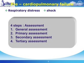
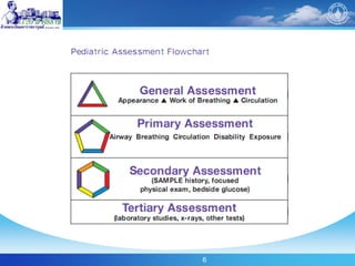
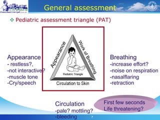
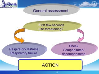
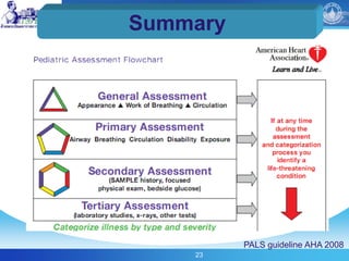
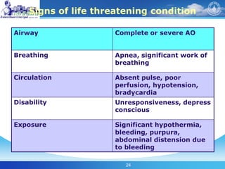
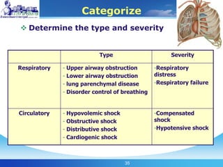
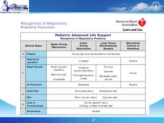
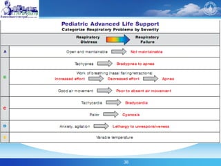
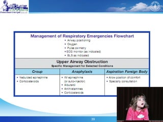
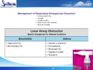
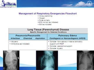
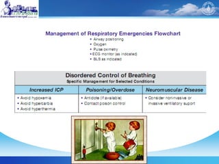
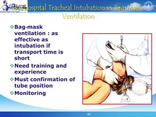
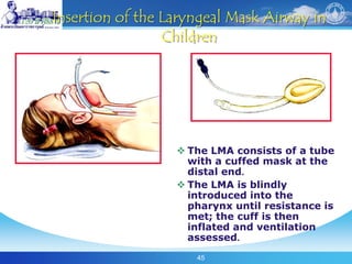
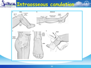
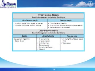
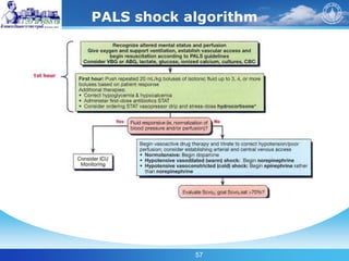
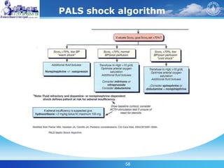
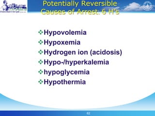
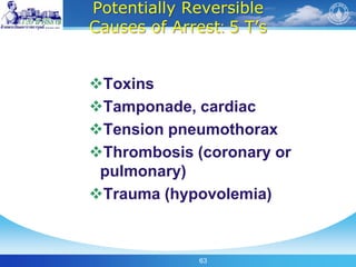
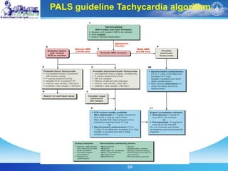
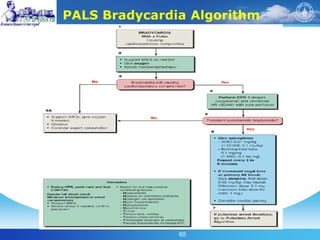
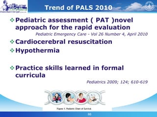
1
From PALS guideline 2005, 2006, 2009 AHA :
Emergency Medicine Conference : Future of Pre-hospital and Emergency Care
Illustrated by Chodchanok Vijarnsorn MD.
Division of Pediatric Cardiology, Department of Pediatrics,
Faculty of Medicine, Siriraj Hospital
21/6/2010
2
Agenda
Two parts
Call fast
Look-listen-feel and airway maintain and
check pulse
Chest compression (new guideline)
3
Etiologies:
Out of hospital cardiac arrest :
Respiratory failure & Shock
By stand
Basic life support alone
In hospital cardiac arrest
Multiple etiologies
Poor outcome
Effective CPR better survival *
4
Many etiologies
Cardiopulmonary failure
Cardiopulmonary arrest
Death Cardiopulmonary recovery
Impaired Unimpaired
neurologic neurologic
recovery recovery
Respiratory failure Shock
5
Pre – cardiopulmonary failure
 Respiratory distress  shock
4 steps : Assessment
1. General assessment
2. Primary assessment
3. Secondary assessment
4. Tertiary assessment
6
7
General assessment
 Pediatric assessment triangle (PAT)
Appearance
- restless?,
-not interactive?
-muscle tone
-Cry/speech
Breathing
-increase effort?
-noise on respiration
-nasalflaring
-retraction
Circulation
-pale? mottling?
-bleeding
First few seconds
Life threatening?
8
First few seconds
Life threatening?
General assessment
Respiratory distress
Respiratory failure
Shock
Compensated/
decompensated
ACTION
9
Primary assessment
 Primary assessment : ABCDE
 - A : airway
 - B : breathing
 - C : circulation
 - D : disability
 - E : exposure
 ( PE, look listen feel, include V/S &
oxygen saturation)
ACTION
10
A : Airway
Chest movement
Breath sound
Feel : air passes through nose and
mouth
Upper airway : clear/ maintainable,
not maintainable
Increase respiratory effort, inspiratory force/absent?
Snoring, stridor?
Retraction?
11
Breathing
RR
Respiratory effort
Tidal volume
Airway and lung sound
Pulse oximetry
 94% = adequate oxygenation
 < 94% airway intervention
 < 90% in 100% oxygen ( non
rebreathing mask  advanced
intervention : assisted ventilation
12
13
Circulatory
 Cardiovascular
function
- skin color : mottling
- HR
- BP
-Pulse
(peripheral/central)
- capillary refill
 End organ
- brain perfusion
- skin perfusion
- renal perfusion (
urine output)
14
Definition of hypotension
 Term (0-28 day)…………… < 60 mmHg
 Infant (1-12 mo)………….. < 70 mmHg
 Children 1-10 y-o (5th P). < 70 + 2 (age yr)
 Children > 10 y-o…………..< 90 mmHg
15
Capillary refill
Normal < 2 seconds
Prolonged capillary refill > 2 sec
In case : shock, hypothermia, severe
dehydration
Warm shock :
capillary refill < 2 sec due to peripheral vasodilatation
16
Pulse check : central pulse
Use femoral / brachial
pulse : < 1 year-old
17
Disability
AVPU pediatric response scale
Glasglow coma scales
Pupillary response to light
AVPU
Alert
Voice
Painful
Unresponsiveness
18
Exposure
Trauma
Burn
Child abuse
Skin lesion
19
Action
General management for all patients
Airway position
Oxygen
Pulse oxymetry
EKG monitor as indicated
BLS as indicated
20
Secondary assessment
3. Secondary assessment
- SAMPLE
- S : Signs and symptoms
- A : Allergies
- M : medication
- P : past medical history
- L : last meal
- E : events leading to
presentation
ACTION
21
Tertiary assessment
 Laboratory : ABG, VBG, Hb, SVO2 sat,
HCO3, lactate,
 Radiography : CXR, echocardiography
 Exhale CO2, PEFR, CVP
 Emphasize : Anytime you identify a life
threatening condition, initiate appropriate
care immediately
22
Assess
Categorize
Decide
Action
If you recognize a life threatening condition at any time,
immediately begin life saving intervention and
activate the emergency response system
23
Summary
PALS guideline AHA 2008
24
Signs of life threatening condition
Airway Complete or severe AO
Breathing Apnea, significant work of
breathing
Circulation Absent pulse, poor
perfusion, hypotension,
bradycardia
Disability Unresponsiveness, depress
conscious
Exposure Significant hypothermia,
bleeding, purpura,
abdominal distension due
to bleeding
25
Life saving intervention
 ABC/CPR
 100% oxygen
 Assisted ventilation :
bag mask, ETT
 Cardiac and
respiratory monitoring
: EKG, pulse oximetry
 Intravenous / I/O
 Bolus isotonic
crystalloid
 Lab study : DTX, ABG
 Drugs
 Electrical therapy
ACTION
26
New recommendation : Bag & mask
ventilation :
E-C clamp
Give 2 breath chest move?
(12-20 breath/min for child)
27
28
PALS and neonatal update
Good PALS begin with good BLS
Lay person (1 choice) : 30:2 (8 yr)
HCP : 1 rescue : 30:2
HCP : 2 rescue : 15:2 (teenage)
Child chest compression > 1 or 2 hands
29
Chest compression
Nipple line for child
Below nipple line in infant
30
31
32
Coronary Perfusion Pressure Improves With Sequential
Compressions
CPP at 5:1 ratio
CPP at 15:2 ratio
Survival with 15:2
33
“Continue CPR as much as
possible except rhythm check”
34
Key change in BLS
Effective rescue breath and visualization
of chest rising
Fully recoil chest
Single shock for VF
(2 J/kg mono-bi phasic continue CPR,
rhythm check only at 2 min)
AED 1-8 years old
35
Categorize
 Determine the type and severity
Type Severity
Respiratory - Upper airway obstruction
- Lower airway obstruction
- lung parenchymal disease
- Disorder control of breathing
-Respiratory
distress
-Respiratory failure
Circulatory - Hypovolemic shock
- Obstructive shock
- Distributive shock
- Cardiogenic shock
-Compensated
shock
-Hypotensive shock
36
Recognition of respiratory
distress and failure
37
38
39
40
41
42
43
Prehospital Tracheal Intubation vs Bag-Mask
Ventilation
Bag-mask
ventilation : as
effective as
intubation if
transport time is
short
Need training and
experience
Must confirmation of
tube position
Monitoring
44
Use of Cuffed Endotracheal Tubes
In-hospital setting, a cuffed ETT : as
safe as an uncuffed tube for infants
(except the newborn) and children
Keep cuff inflation pressure <20 cm
H2O
Cuffed ETT size (mm) = (age (yr) /4) + 3
Uncuff size (mm): (age (yr) /4) + 4
Depth : age (yr)/2 + 12
45
Insertion of the Laryngeal Mask Airway in
Children
 The LMA consists of a tube
with a cuffed mask at the
distal end.
 The LMA is blindly
introduced into the
pharynx until resistance is
met; the cuff is then
inflated and ventilation
assessed.
46
Verification of Endotracheal Tube Placement
bilateral chest movement and listen
for equal breath sounds over both
lung fields
gastric insufflation sounds
exhaled CO2
pulse oximeter
direct laryngoscopy
chest x-ray
47
Colorimetric Exhaled CO2 Detector
Colorimetric
exhaled CO2
detector device
changes color (from
purple to yellow)
with detection of
exhaled CO2
“additional”
confirmation with
clinical assessment
48
Recognition of shock
in pediatric patient
PALS update 2008-2009
49
Myocardial contractility
preload
afterload
Stroke volume
Heart rate
Cardiac output
Tissue perfusion
Blood pressure
ปัจจัยที่มีผลต่อ tissue perfusion
CaO2, Hb
50
Etiology of shock
 1.hypovolemic shock
 - severe dehydration, blood loss, burn, sepsis
 2.Cardiogenic shock
 -congenital heart disease, acquire heart disease,
myocarditis, arrhythmia
 3.Distributive shock
 -anaphylaxis, sepsis, spinal shock
 4. Obstructive shock
 - cardiac tamponade, tension pneumothorax
51
52
Recognition of shock flow chart
53
54
55
Intraosseous canulation
56
57
PALS shock algorithm
58
PALS shock algorithm
59
60
Medications : Maintain CO postresuscitation Stabilization
61
62
Potentially Reversible
Causes of Arrest: 6 H’s
Hypovolemia
Hypoxemia
Hydrogen ion (acidosis)
Hypo-/hyperkalemia
hypoglycemia
Hypothermia
63
Potentially Reversible
Causes of Arrest: 5 T’s
Toxins
Tamponade, cardiac
Tension pneumothorax
Thrombosis (coronary or
pulmonary)
Trauma (hypovolemia)
64
PALS Tachycardia Algorithm
PALS guideline Tachycardia algorithm
65
PALS Bradycardia Algorithm
66
Trend of PALS 2010
Pediatric assessment ( PAT )novel
approach for the rapid evaluation
Pediatric Emergency Care - Vol 26 Number 4, April 2010
Cardiocerebral resuscitation
Hypothermia
Practice skills learned in formal
curricula
Pediatrics 2009; 124; 610-619
67

Pals%20update%202005%20to%202010 chodchanok

  • 1.
    1 From PALS guideline2005, 2006, 2009 AHA : Emergency Medicine Conference : Future of Pre-hospital and Emergency Care Illustrated by Chodchanok Vijarnsorn MD. Division of Pediatric Cardiology, Department of Pediatrics, Faculty of Medicine, Siriraj Hospital 21/6/2010
  • 2.
    2 Agenda Two parts Call fast Look-listen-feeland airway maintain and check pulse Chest compression (new guideline)
  • 3.
    3 Etiologies: Out of hospitalcardiac arrest : Respiratory failure & Shock By stand Basic life support alone In hospital cardiac arrest Multiple etiologies Poor outcome Effective CPR better survival *
  • 4.
    4 Many etiologies Cardiopulmonary failure Cardiopulmonaryarrest Death Cardiopulmonary recovery Impaired Unimpaired neurologic neurologic recovery recovery Respiratory failure Shock
  • 5.
    5 Pre – cardiopulmonaryfailure  Respiratory distress  shock 4 steps : Assessment 1. General assessment 2. Primary assessment 3. Secondary assessment 4. Tertiary assessment
  • 6.
  • 7.
    7 General assessment  Pediatricassessment triangle (PAT) Appearance - restless?, -not interactive? -muscle tone -Cry/speech Breathing -increase effort? -noise on respiration -nasalflaring -retraction Circulation -pale? mottling? -bleeding First few seconds Life threatening?
  • 8.
    8 First few seconds Lifethreatening? General assessment Respiratory distress Respiratory failure Shock Compensated/ decompensated ACTION
  • 9.
    9 Primary assessment  Primaryassessment : ABCDE  - A : airway  - B : breathing  - C : circulation  - D : disability  - E : exposure  ( PE, look listen feel, include V/S & oxygen saturation) ACTION
  • 10.
    10 A : Airway Chestmovement Breath sound Feel : air passes through nose and mouth Upper airway : clear/ maintainable, not maintainable Increase respiratory effort, inspiratory force/absent? Snoring, stridor? Retraction?
  • 11.
    11 Breathing RR Respiratory effort Tidal volume Airwayand lung sound Pulse oximetry  94% = adequate oxygenation  < 94% airway intervention  < 90% in 100% oxygen ( non rebreathing mask  advanced intervention : assisted ventilation
  • 12.
  • 13.
    13 Circulatory  Cardiovascular function - skincolor : mottling - HR - BP -Pulse (peripheral/central) - capillary refill  End organ - brain perfusion - skin perfusion - renal perfusion ( urine output)
  • 14.
    14 Definition of hypotension Term (0-28 day)…………… < 60 mmHg  Infant (1-12 mo)………….. < 70 mmHg  Children 1-10 y-o (5th P). < 70 + 2 (age yr)  Children > 10 y-o…………..< 90 mmHg
  • 15.
    15 Capillary refill Normal <2 seconds Prolonged capillary refill > 2 sec In case : shock, hypothermia, severe dehydration Warm shock : capillary refill < 2 sec due to peripheral vasodilatation
  • 16.
    16 Pulse check :central pulse Use femoral / brachial pulse : < 1 year-old
  • 17.
    17 Disability AVPU pediatric responsescale Glasglow coma scales Pupillary response to light AVPU Alert Voice Painful Unresponsiveness
  • 18.
  • 19.
    19 Action General management forall patients Airway position Oxygen Pulse oxymetry EKG monitor as indicated BLS as indicated
  • 20.
    20 Secondary assessment 3. Secondaryassessment - SAMPLE - S : Signs and symptoms - A : Allergies - M : medication - P : past medical history - L : last meal - E : events leading to presentation ACTION
  • 21.
    21 Tertiary assessment  Laboratory: ABG, VBG, Hb, SVO2 sat, HCO3, lactate,  Radiography : CXR, echocardiography  Exhale CO2, PEFR, CVP  Emphasize : Anytime you identify a life threatening condition, initiate appropriate care immediately
  • 22.
    22 Assess Categorize Decide Action If you recognizea life threatening condition at any time, immediately begin life saving intervention and activate the emergency response system
  • 23.
  • 24.
    24 Signs of lifethreatening condition Airway Complete or severe AO Breathing Apnea, significant work of breathing Circulation Absent pulse, poor perfusion, hypotension, bradycardia Disability Unresponsiveness, depress conscious Exposure Significant hypothermia, bleeding, purpura, abdominal distension due to bleeding
  • 25.
    25 Life saving intervention ABC/CPR  100% oxygen  Assisted ventilation : bag mask, ETT  Cardiac and respiratory monitoring : EKG, pulse oximetry  Intravenous / I/O  Bolus isotonic crystalloid  Lab study : DTX, ABG  Drugs  Electrical therapy ACTION
  • 26.
    26 New recommendation :Bag & mask ventilation : E-C clamp Give 2 breath chest move? (12-20 breath/min for child)
  • 27.
  • 28.
    28 PALS and neonatalupdate Good PALS begin with good BLS Lay person (1 choice) : 30:2 (8 yr) HCP : 1 rescue : 30:2 HCP : 2 rescue : 15:2 (teenage) Child chest compression > 1 or 2 hands
  • 29.
    29 Chest compression Nipple linefor child Below nipple line in infant
  • 30.
  • 31.
  • 32.
    32 Coronary Perfusion PressureImproves With Sequential Compressions CPP at 5:1 ratio CPP at 15:2 ratio Survival with 15:2
  • 33.
    33 “Continue CPR asmuch as possible except rhythm check”
  • 34.
    34 Key change inBLS Effective rescue breath and visualization of chest rising Fully recoil chest Single shock for VF (2 J/kg mono-bi phasic continue CPR, rhythm check only at 2 min) AED 1-8 years old
  • 35.
    35 Categorize  Determine thetype and severity Type Severity Respiratory - Upper airway obstruction - Lower airway obstruction - lung parenchymal disease - Disorder control of breathing -Respiratory distress -Respiratory failure Circulatory - Hypovolemic shock - Obstructive shock - Distributive shock - Cardiogenic shock -Compensated shock -Hypotensive shock
  • 36.
  • 37.
  • 38.
  • 39.
  • 40.
  • 41.
  • 42.
  • 43.
    43 Prehospital Tracheal Intubationvs Bag-Mask Ventilation Bag-mask ventilation : as effective as intubation if transport time is short Need training and experience Must confirmation of tube position Monitoring
  • 44.
    44 Use of CuffedEndotracheal Tubes In-hospital setting, a cuffed ETT : as safe as an uncuffed tube for infants (except the newborn) and children Keep cuff inflation pressure <20 cm H2O Cuffed ETT size (mm) = (age (yr) /4) + 3 Uncuff size (mm): (age (yr) /4) + 4 Depth : age (yr)/2 + 12
  • 45.
    45 Insertion of theLaryngeal Mask Airway in Children  The LMA consists of a tube with a cuffed mask at the distal end.  The LMA is blindly introduced into the pharynx until resistance is met; the cuff is then inflated and ventilation assessed.
  • 46.
    46 Verification of EndotrachealTube Placement bilateral chest movement and listen for equal breath sounds over both lung fields gastric insufflation sounds exhaled CO2 pulse oximeter direct laryngoscopy chest x-ray
  • 47.
    47 Colorimetric Exhaled CO2Detector Colorimetric exhaled CO2 detector device changes color (from purple to yellow) with detection of exhaled CO2 “additional” confirmation with clinical assessment
  • 48.
    48 Recognition of shock inpediatric patient PALS update 2008-2009
  • 49.
    49 Myocardial contractility preload afterload Stroke volume Heartrate Cardiac output Tissue perfusion Blood pressure ปัจจัยที่มีผลต่อ tissue perfusion CaO2, Hb
  • 50.
    50 Etiology of shock 1.hypovolemic shock  - severe dehydration, blood loss, burn, sepsis  2.Cardiogenic shock  -congenital heart disease, acquire heart disease, myocarditis, arrhythmia  3.Distributive shock  -anaphylaxis, sepsis, spinal shock  4. Obstructive shock  - cardiac tamponade, tension pneumothorax
  • 51.
  • 52.
  • 53.
  • 54.
  • 55.
  • 56.
  • 57.
  • 58.
  • 59.
  • 60.
    60 Medications : MaintainCO postresuscitation Stabilization
  • 61.
  • 62.
    62 Potentially Reversible Causes ofArrest: 6 H’s Hypovolemia Hypoxemia Hydrogen ion (acidosis) Hypo-/hyperkalemia hypoglycemia Hypothermia
  • 63.
    63 Potentially Reversible Causes ofArrest: 5 T’s Toxins Tamponade, cardiac Tension pneumothorax Thrombosis (coronary or pulmonary) Trauma (hypovolemia)
  • 64.
    64 PALS Tachycardia Algorithm PALSguideline Tachycardia algorithm
  • 65.
  • 66.
    66 Trend of PALS2010 Pediatric assessment ( PAT )novel approach for the rapid evaluation Pediatric Emergency Care - Vol 26 Number 4, April 2010 Cardiocerebral resuscitation Hypothermia Practice skills learned in formal curricula Pediatrics 2009; 124; 610-619
  • 67.