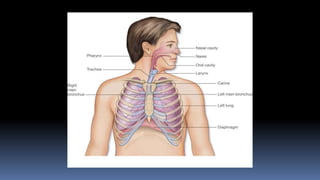
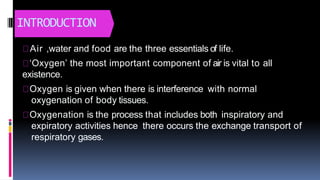
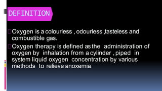
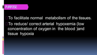
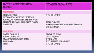
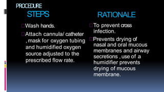
OXYGENATION
INTRODUCTION
Air ,water and food are the three essentials of life.
‘Oxygen’ the most important component of air is vital to all
existence.
Oxygen is given when there is interference with normal
oxygenation of body tissues.
Oxygenation is the process that includes both inspiratory and
expiratory activities hence there occurs the exchange transport of
respiratory gases.
DEFINITION
Oxygen is a colourless , odourless ,tasteless and
combustible gas.
Oxygen therapy is defined as the administration of
oxygen by inhalation from a cylinder , piped in
system liquid oxygen concentration by various
methods to relieve anoxemia.
PURPOSE
To facilitate normal metabolism of the tissues.
To reduce/ correct arterial hypoxemia (low
concentration of oxygen in the blood )and
tissue hypoxia
FACTORSINFLUENCINGOXYGENETION
There are mainly 4 factors causing alteration in
oxygenation ;
1) Physiological factors
2) Developmental factors
3) Life style
4) Environmental factors
Physiological factors
Decreased oxygen carrying capacity.
Decreased inspired oxygen
concentration(obstruction in airway tract
hypovolemia , increased metabolic rate).
Hyperventilation
Hypoventilation
Hypoxia
Condition affecting chest wall movement.
ALTERATION IN CARDIAC FUNCTIONING.
Illness and conditions affecting cardiac strength of
contraction ,blood flow through the heart or to the
heart muscles, and decreased peripheral circulation
cause alterations in cardiac functioning.
older adults experience alterations in cardiac
functions asa result of calcification of the conduction
pathways ,thicker and shifter heart valves caused by
lipid accumulation and fibrosis and decreases in the
number of pacemaker cells in the S.A node.
DISTURBANCESIN THECONDUCTION.
Electrical impulses that do not originate from the S.A
node cause conduction disturbances. these rhythm
disturbances are called dysrhythmias ,meaning a
deviation from the normal sinus heart rhythm.
Sinus tachycardia :Regular rhythm rate >100 –180 bts/min,
with normal p wave and normal QRScomplex.
Sinus bradycardia :Regular rhythm ratelessthan 60
bts/min, normal Pwave , normal PRinterval and QRS
complex.
Sinus dysrhythmia: sinus rhythm with cyclicvariation ,slows
during inspiration and increases with expiration rate of 60 -
100bts/min ;normal Pwave ,normal PRinterval ,normal QRS
complex.
Atrial fibrillation :no identifiable p wave,irregular cardiac
rate and rhythm.
Premature ventricularcontractions :IrregularRhythmWith
Ectopic Beats,RateOr Increased ,PWaveAbsent In Ectopic
Beat;PRIntervalAbsent.QRSComplexWidened And
Distorted ;TWaveIn Opposition ToRWave
ventricular tachycardia :Rhythm Slightly Irregular Rate100-200
bts/Min ,PWaveAbsent ,PRIntervalAbsent ,QRSWide And Bizarre
,>0.12 Seconds.
Ventricular fibrillation :uncoordinated electricalactivity .No
identifiable PQRSor Twave
Altered cardiacoutput :when the blood volume gets decreased
,the systematic and pulmonary circulation canresult in heart
failure.
congenital or acquired when sizeof the lumenof the valvesget
narrowed. It iscalled stenosis , when there iswidening of valves
that leadsto regurgitation.
 Angina pectoris :it istransient imbalance between
myocardial oxygen supply and demand. Thecondition
results in chest pain that isaching , sharp, tingling,
burning or the feeling of pressure.
 Myocardial infarction :It results from sudden
decrease in coronary blood flow or an increase in
myocardial oxygen demand with out adequate
coronary perfusion.
ALTERATION IN RESPIRATORY FUNCTIONING
 Hyperventilation : It is a state of excessive ventilation that
required to eliminate normal oxygen produced by cellular
metabolism. May be induced chemically by anxiety occurs as
the body tries to compensate for metabolic acidosis.
 Hypoventilation :It is a condition where there isless
ventilation ,not sufficient to eliminate carbon dioxide
produced by metabolic activities.
 Hypoxia :It is the inadequate oxygenation of the cells of
tissues that leads to the necrosis of the tissues and
ineffective functioning at that level.
Developmental factors :
The developmental stage of a patient and the
normal aging process affects tissue
oxygenation.
 Infant and toddlers
School age children's and adolescents.
Young and middle age adults.
Older adults
Life style factors:
Nutrition
Exercise
Smoking
Substance abuse
stress
Environmental factors:
 Residence location
 occupation
OXYGEN ADMINISTRATION
DEVICES
OXYGEN FLOW RATE
HIGH FLOW
VENTURI MASK
MECHANICAL AEROSOL SYSTEMS
HIGHFLOW HUMIDIFIER SYSTEM NON
REBREATHERMASK HIGHFLOW NASAL
CANNULA
4 TO 12(L/MIN)
10TO 15(L/MIN)
MAXIMUM 60 LPM VIA NASAL PRONGS
OR CANNULA
LOW FLOW
NASAL CANNULA ABOVE 5(L/MIN)
NASAL CATHETER UPTO 5(L/MIN)
TRANSTRACHEAL CATHETER 6 T0 12 LPM
FACE MASK 2 TO 6 LITRESPER MINUTE
TENT 8 TO 12( L/MIN)
METHODS OF OXYGEN ADMINISTRATION
NASAL CATHETER : It Involves
Inserting An Oxygen Catheter/Sample
Rubber Catheter Into The Nose Up to
The Nasopharynx. It Is Needs To Be
Changed At least Every Eight Hours
And Inserted Into The Other Nostril, It
IsAlso Painful And Can Cause Trauma.
Thus , It Is Less Desirable
NASAL CANNULA :
A Nasal Cannula Is a Simple Comfortable
Devices. The Cannula About 15cm (1/2
Inch) Long Protrude From The Centre Of
Disposable Tube And Are Inserted Into The
Nares.
TRANS-TRACHEALOXYGEN:
 TransTrachealOxygenation ,Oxygen Is Delivered Directly Into
TheTracheaViaA Catheter(SmallIntravenous Size)Into The
TracheaThrough ASurgicalOpening In TheLower Neck.
OXYGEN MASK :
 ItisaDeviceUsedToAdminister Humidified Oxygen.It Is
StrappedToFitSnuglyTo TheMouth And NoseAnd Is
SecuredIn PlaceWith A Strap.
OXYGENTENT:
WhenA Patient HasFacialInjuries Or For Any Other Reason
Cannot Tolerate An Oxygen Mask. Then This Method
Can BeUsed. The Tent Is First Flooded With Oxygen
And Then A Flow Of 4-5 Litres Per Minute Is Given. This
Will Maintain A Service Of 40% - 50% In The Tent.
OXYGEN CYLINDERS
GENERAL INSTRUCTIONS
oxygen should be treated as a drug ; the five rights
of medication administration also pertain to oxygen.
When using on oxygen cylinder or central supply
oxygen , use a regulator and humidifier.
Every part of the apparatus should be clean to
prevent infection.
Pay attention to conditions that can interfere with
the flow of oxygen from sources to the patient.this
may include tubing , loose connections and faulty
humidifying apparatus.
Airway keep a space oxygen cylinder in close vicinity.
Change nasal catheters at least every eight hours
or more often to prevent blockage of the nasal
catheter by a mucus.
Watch the patients receiving oxygen therapy
continuously to detect the early signs of oxygen
toxicity.
Since oxygen supports combustion ,fire protection
are to be taken when oxygen is on flow
PREPARATION OF THE PATIENT
PREPARATION OF
ARTICLES
PROCEDURE
STEPS
Wash hands.
Attach cannula/ catheter
and humidified oxygen
source adjusted to the
prescribed flow rate.
RATIONALE
To prevent cross
infection.
, mask for oxygen tubing Prevents drying of
nasal and oral mucous
membranes and airway
secretions , use of a
humidifier prevents
drying of mucous
membrane.
Place tips of cannula
into the patient nares, if
mask apply snuggly to
face.
Check cannula /
equipment every eight
hours.
Directs flow of oxygen
into the upper
respiratory tract,
prevents loss of oxygen.
Ensures patency of
cannula and oxygen
flow, also ensures safe
delivery of prescribed
oxygen.
Keep the humidification jar
filled at all times.
Observe the patients nares
and superior surface of
both ears and skin
breakdown.
Prevents inhalation of
dehumidified oxygen
prevents drying of
mucus membranes.
Oxygen therapy can dry
nasal mucosa, pressure
on ears from cannula
tubing's / elastic can
cause skin irritation
Check the oxygen flow
safe and the physicians
orders every eight hours.
Wash hands before
removing the oxygen
mask or tube.
Ensures delivery of the
prescribed oxygen
flow rate.
Reduces transmission
of micro-organisms.
Inspect the patient for
relief of symptom's
associated with hypoxia.
Record procedure in the
nurses notes.
Indicates the hypoxia is
reduced / treated.
Document correct use
of oxygen therapy and
the patients response.
AFTER CARE OF PATIENTS AND THE ARTICLES
 Stay with the patients till he / she is at care.
 Keep the patient warm and comfortable .
 Evaluate the patients progress by observing the
vital signs and symptom's.
 Watch the patients progress by observing the vital
signs and symptoms.
 Record procedure with date and time.
 Request for an arterial blood gas analysis at specified
intervals to make sure hypoxia is treated.
 Take all articles to the utility room.
 Clean nasal catheter with cold water then warm soapy
water and finally with clean water (if disposable ) boil
and store or send for sterilization.
Oxygenation

Oxygenation

  • 1.
  • 3.
    INTRODUCTION Air ,water andfood are the three essentials of life. ‘Oxygen’ the most important component of air is vital to all existence. Oxygen is given when there is interference with normal oxygenation of body tissues. Oxygenation is the process that includes both inspiratory and expiratory activities hence there occurs the exchange transport of respiratory gases.
  • 4.
    DEFINITION Oxygen is acolourless , odourless ,tasteless and combustible gas. Oxygen therapy is defined as the administration of oxygen by inhalation from a cylinder , piped in system liquid oxygen concentration by various methods to relieve anoxemia.
  • 5.
    PURPOSE To facilitate normalmetabolism of the tissues. To reduce/ correct arterial hypoxemia (low concentration of oxygen in the blood )and tissue hypoxia
  • 6.
    FACTORSINFLUENCINGOXYGENETION There are mainly4 factors causing alteration in oxygenation ; 1) Physiological factors 2) Developmental factors 3) Life style 4) Environmental factors
  • 7.
    Physiological factors Decreased oxygencarrying capacity. Decreased inspired oxygen concentration(obstruction in airway tract hypovolemia , increased metabolic rate). Hyperventilation Hypoventilation Hypoxia Condition affecting chest wall movement.
  • 8.
    ALTERATION IN CARDIACFUNCTIONING. Illness and conditions affecting cardiac strength of contraction ,blood flow through the heart or to the heart muscles, and decreased peripheral circulation cause alterations in cardiac functioning. older adults experience alterations in cardiac functions asa result of calcification of the conduction pathways ,thicker and shifter heart valves caused by lipid accumulation and fibrosis and decreases in the number of pacemaker cells in the S.A node.
  • 9.
    DISTURBANCESIN THECONDUCTION. Electrical impulsesthat do not originate from the S.A node cause conduction disturbances. these rhythm disturbances are called dysrhythmias ,meaning a deviation from the normal sinus heart rhythm. Sinus tachycardia :Regular rhythm rate >100 –180 bts/min, with normal p wave and normal QRScomplex. Sinus bradycardia :Regular rhythm ratelessthan 60 bts/min, normal Pwave , normal PRinterval and QRS complex.
  • 11.
    Sinus dysrhythmia: sinusrhythm with cyclicvariation ,slows during inspiration and increases with expiration rate of 60 - 100bts/min ;normal Pwave ,normal PRinterval ,normal QRS complex. Atrial fibrillation :no identifiable p wave,irregular cardiac rate and rhythm. Premature ventricularcontractions :IrregularRhythmWith Ectopic Beats,RateOr Increased ,PWaveAbsent In Ectopic Beat;PRIntervalAbsent.QRSComplexWidened And Distorted ;TWaveIn Opposition ToRWave
  • 12.
    ventricular tachycardia :RhythmSlightly Irregular Rate100-200 bts/Min ,PWaveAbsent ,PRIntervalAbsent ,QRSWide And Bizarre ,>0.12 Seconds. Ventricular fibrillation :uncoordinated electricalactivity .No identifiable PQRSor Twave Altered cardiacoutput :when the blood volume gets decreased ,the systematic and pulmonary circulation canresult in heart failure. congenital or acquired when sizeof the lumenof the valvesget narrowed. It iscalled stenosis , when there iswidening of valves that leadsto regurgitation.
  • 13.
     Angina pectoris:it istransient imbalance between myocardial oxygen supply and demand. Thecondition results in chest pain that isaching , sharp, tingling, burning or the feeling of pressure.  Myocardial infarction :It results from sudden decrease in coronary blood flow or an increase in myocardial oxygen demand with out adequate coronary perfusion.
  • 14.
    ALTERATION IN RESPIRATORYFUNCTIONING  Hyperventilation : It is a state of excessive ventilation that required to eliminate normal oxygen produced by cellular metabolism. May be induced chemically by anxiety occurs as the body tries to compensate for metabolic acidosis.  Hypoventilation :It is a condition where there isless ventilation ,not sufficient to eliminate carbon dioxide produced by metabolic activities.  Hypoxia :It is the inadequate oxygenation of the cells of tissues that leads to the necrosis of the tissues and ineffective functioning at that level.
  • 15.
    Developmental factors : Thedevelopmental stage of a patient and the normal aging process affects tissue oxygenation.  Infant and toddlers School age children's and adolescents. Young and middle age adults. Older adults
  • 16.
  • 17.
    Environmental factors:  Residencelocation  occupation
  • 18.
    OXYGEN ADMINISTRATION DEVICES OXYGEN FLOWRATE HIGH FLOW VENTURI MASK MECHANICAL AEROSOL SYSTEMS HIGHFLOW HUMIDIFIER SYSTEM NON REBREATHERMASK HIGHFLOW NASAL CANNULA 4 TO 12(L/MIN) 10TO 15(L/MIN) MAXIMUM 60 LPM VIA NASAL PRONGS OR CANNULA LOW FLOW NASAL CANNULA ABOVE 5(L/MIN) NASAL CATHETER UPTO 5(L/MIN) TRANSTRACHEAL CATHETER 6 T0 12 LPM FACE MASK 2 TO 6 LITRESPER MINUTE TENT 8 TO 12( L/MIN)
  • 19.
    METHODS OF OXYGENADMINISTRATION NASAL CATHETER : It Involves Inserting An Oxygen Catheter/Sample Rubber Catheter Into The Nose Up to The Nasopharynx. It Is Needs To Be Changed At least Every Eight Hours And Inserted Into The Other Nostril, It IsAlso Painful And Can Cause Trauma. Thus , It Is Less Desirable
  • 20.
    NASAL CANNULA : ANasal Cannula Is a Simple Comfortable Devices. The Cannula About 15cm (1/2 Inch) Long Protrude From The Centre Of Disposable Tube And Are Inserted Into The Nares.
  • 22.
    TRANS-TRACHEALOXYGEN:  TransTrachealOxygenation ,OxygenIs Delivered Directly Into TheTracheaViaA Catheter(SmallIntravenous Size)Into The TracheaThrough ASurgicalOpening In TheLower Neck.
  • 24.
    OXYGEN MASK : ItisaDeviceUsedToAdminister Humidified Oxygen.It Is StrappedToFitSnuglyTo TheMouth And NoseAnd Is SecuredIn PlaceWith A Strap.
  • 26.
    OXYGENTENT: WhenA Patient HasFacialInjuriesOr For Any Other Reason Cannot Tolerate An Oxygen Mask. Then This Method Can BeUsed. The Tent Is First Flooded With Oxygen And Then A Flow Of 4-5 Litres Per Minute Is Given. This Will Maintain A Service Of 40% - 50% In The Tent.
  • 28.
  • 30.
    GENERAL INSTRUCTIONS oxygen shouldbe treated as a drug ; the five rights of medication administration also pertain to oxygen. When using on oxygen cylinder or central supply oxygen , use a regulator and humidifier. Every part of the apparatus should be clean to prevent infection.
  • 31.
    Pay attention toconditions that can interfere with the flow of oxygen from sources to the patient.this may include tubing , loose connections and faulty humidifying apparatus. Airway keep a space oxygen cylinder in close vicinity.
  • 32.
    Change nasal cathetersat least every eight hours or more often to prevent blockage of the nasal catheter by a mucus. Watch the patients receiving oxygen therapy continuously to detect the early signs of oxygen toxicity. Since oxygen supports combustion ,fire protection are to be taken when oxygen is on flow
  • 33.
  • 34.
  • 35.
    PROCEDURE STEPS Wash hands. Attach cannula/catheter and humidified oxygen source adjusted to the prescribed flow rate. RATIONALE To prevent cross infection. , mask for oxygen tubing Prevents drying of nasal and oral mucous membranes and airway secretions , use of a humidifier prevents drying of mucous membrane.
  • 36.
    Place tips ofcannula into the patient nares, if mask apply snuggly to face. Check cannula / equipment every eight hours. Directs flow of oxygen into the upper respiratory tract, prevents loss of oxygen. Ensures patency of cannula and oxygen flow, also ensures safe delivery of prescribed oxygen.
  • 37.
    Keep the humidificationjar filled at all times. Observe the patients nares and superior surface of both ears and skin breakdown. Prevents inhalation of dehumidified oxygen prevents drying of mucus membranes. Oxygen therapy can dry nasal mucosa, pressure on ears from cannula tubing's / elastic can cause skin irritation
  • 38.
    Check the oxygenflow safe and the physicians orders every eight hours. Wash hands before removing the oxygen mask or tube. Ensures delivery of the prescribed oxygen flow rate. Reduces transmission of micro-organisms.
  • 39.
    Inspect the patientfor relief of symptom's associated with hypoxia. Record procedure in the nurses notes. Indicates the hypoxia is reduced / treated. Document correct use of oxygen therapy and the patients response.
  • 40.
    AFTER CARE OFPATIENTS AND THE ARTICLES  Stay with the patients till he / she is at care.  Keep the patient warm and comfortable .  Evaluate the patients progress by observing the vital signs and symptom's.  Watch the patients progress by observing the vital signs and symptoms.  Record procedure with date and time.
  • 41.
     Request foran arterial blood gas analysis at specified intervals to make sure hypoxia is treated.  Take all articles to the utility room.  Clean nasal catheter with cold water then warm soapy water and finally with clean water (if disposable ) boil and store or send for sterilization.