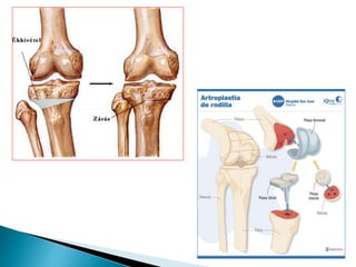
 Osteonecrosis is a disorder resulting from a
temporary or permanent loss of blood supply to
the bone. When the blood supply is disrupted,
the bone tissues (osteo) begin to break down
(necrosis). This can weaken the bone and
eventually result in its collapse. If this occurs
near a joint, it can lead to the collapse of the
joint surface, resulting in pain and inflammation
(arthritis). Osteonecrosis is also referred to as
avascular necrosis or “AVN,” “aseptic necrosis,”
and “ischemic bone necrosis.”
 La osteonecrosis puede producir dolor agudo y
discapacidad, al igual que la artritis.
 Más del 90% de los casos de osteonecrosis se
producen debido al uso de medicamentos
glucocorticoides (algunas veces denominados
corticosteroides) o por tomar demasiado alcohol.
 Si bien la osteonecrosis puede producirse en
prácticamente cualquier hueso del cuerpo, las
caderas, las rodillas y los hombros son los sitios
más comúnmente afectados.
 Es más fácil prevenir la osteonecrosis que
tratarla.
 Las causas más frecuentes de osteonecrosis
son:
 Trauma grave (lesión), el cual interrumpe el
suministro de sangre al hueso
 Medicamentos corticosteroides (como
prednisona, cortisona o metilprednisolona),
principalmente cuando se utilizan por mucho
tiempo o en una dosis muy alta
 Exceso en el consumo de alcohol
 Lupus
 Enfermedad por descompresión (también las
“flexiones” que se producen con el buceo)
 Trastornos sanguíneos como la anemia
falciforme
 Infección por VIH (el virus que produce el
SIDA)
 Terapia de radiación
 Bifosfonatos, vinculados a la osteonecrosis de
la mandíbula
 Corticosteroids (such as prednisone and
dexamethasone) are commonly used for
treatment of many cancers, such as leukemia and
lymphoma. Dexamethasone is also sometimes
used for treatment of nausea and vomiting
associated with chemotherapy and to control
brain swelling. There is no clear explanation as to
how corticosteroids cause osteonecrosis, but it is
believed that they may interfere with the body’s
ability to break down fatty substances. These
substances can clog the blood vessels, causing
them to narrow. This reduces the amount of
blood that gets into the bone.
 People in the early stages of osteonecrosis may not have
any symptoms. However, as the disorder progresses, most
people will experience some joint pain. At first, the person
may only experience pain when bearing weight on the
affected bone or joint. As the disorder progresses,
symptoms may be present even at rest. Pain may develop
gradually and its intensity can range from mild to severe.
 If osteonecrosis progresses and the bone and surrounding
joint surfaces collapse, the pain can increase considerably
and may become severe enough to limit movement in the
affected joint. The period of time between the first
symptoms of osteonecrosis and the loss of joint function
is different for each person and ranges from several
months to years.
 Los médicos sospechan de osteonecrosis cuando una persona
que presenta factores de riesgo en los huesos siente dolor
“localizado”
 Dolor en la cadera por osteonecrosis = dolor en la ingle.
 El dolor por osteonecrosis en cadera o rodilla generalmente es
peor con la carga de peso o al caminar.
 An x-ray is usually the first test to be done when osteonecrosis
is suspected.
 MRI is one of the most useful tools in diagnosing osteonecrosis
 Bone scans.
 CT scan
 Surgical procedures such as a bone biopsy can conclusively
diagnose osteonecrosis.
 Desafortunadamente no hay una prueba clara
que indique la mejor manera de tratar la
osteonecrosis, pero lo mejor es iniciar el
tratamiento ante las primeras manifestaciones,
antes del colapso del hueso.
 Treatment can be conservative or surgical. In
order to decide the best treatment for a patient,
the following factors are considered:
◦ • The person’s age
◦ • The stage of the disorder (early or late)
◦ • The location and the amount of bone affected (small or
large)
◦ • The status of cancer and cancer treatment
• Medication - to reduce pain
• Reduced weight bearing - to slow the damage and promote
natural healing.
• Crutches may be recommended to limit weight or pressure
on the affected joint.
• Range of motion exercises – to keep the joints flexible. This
is also important tomaintain movement and increase
circulation in the joints. This can promote healing and may
relieve pain. Physical therapists can teach the correct
exercises.
• Electrical stimulation - to induce bone growth
 Conservative treatments may be used alone or in
combination, but they may not provide lasting
improvement. Some people may require surgery to
permanently repair or replace the joint.
 • Core decompression – is a surgery that removes the inner layer of
bone.
This may reduce pressure within the bone and create an open area for
new blood vessels to grow. Sometimes a piece of healthy bone with good
blood vessels (bone graft) is put in this area to speed up the process. This
procedure works best in the early stages of osteonecrosis and should help
relieve pain and promote healing.
 • Osteotomy – is a surgery that involves taking out a piece of bone,
usually a wedge, to reposition the bone so that the tissue lacking blood
supply (avascular area) bears less weight than an adjacent healthy area.
 • Arthroplasty – is also referred to as joint replacement. The affected
bone is removed and replaced with an artificial joint. This treatment may
be needed in the late stages of osteonecrosis and when a joint is
destroyed.
 Evitar el exceso de alcohol y el consumo de
tabaco
 CORTICOIDES a dosis pequeñas y menor
tiempo posible (consultar con su médico)
 Higiene dental (infección dental)
 El diagnóstico y tratamiento tempranos
podrían mejorar el resultado.
 National Institute of Arthritis and
Musculoskeletal and Skin Diseases
www.niams.nih.gov/hi/topics/osteonecrosis/index.htm
 American Academy of Orthopaedic Surgeons
www.aaos.org
 American College of Rheumatology
www.rheumatology.org/REF

Osteonecrosis

  • 2.
     Osteonecrosis isa disorder resulting from a temporary or permanent loss of blood supply to the bone. When the blood supply is disrupted, the bone tissues (osteo) begin to break down (necrosis). This can weaken the bone and eventually result in its collapse. If this occurs near a joint, it can lead to the collapse of the joint surface, resulting in pain and inflammation (arthritis). Osteonecrosis is also referred to as avascular necrosis or “AVN,” “aseptic necrosis,” and “ischemic bone necrosis.”
  • 3.
     La osteonecrosispuede producir dolor agudo y discapacidad, al igual que la artritis.  Más del 90% de los casos de osteonecrosis se producen debido al uso de medicamentos glucocorticoides (algunas veces denominados corticosteroides) o por tomar demasiado alcohol.  Si bien la osteonecrosis puede producirse en prácticamente cualquier hueso del cuerpo, las caderas, las rodillas y los hombros son los sitios más comúnmente afectados.  Es más fácil prevenir la osteonecrosis que tratarla.
  • 4.
     Las causasmás frecuentes de osteonecrosis son:  Trauma grave (lesión), el cual interrumpe el suministro de sangre al hueso  Medicamentos corticosteroides (como prednisona, cortisona o metilprednisolona), principalmente cuando se utilizan por mucho tiempo o en una dosis muy alta  Exceso en el consumo de alcohol
  • 5.
     Lupus  Enfermedadpor descompresión (también las “flexiones” que se producen con el buceo)  Trastornos sanguíneos como la anemia falciforme  Infección por VIH (el virus que produce el SIDA)  Terapia de radiación  Bifosfonatos, vinculados a la osteonecrosis de la mandíbula
  • 6.
     Corticosteroids (suchas prednisone and dexamethasone) are commonly used for treatment of many cancers, such as leukemia and lymphoma. Dexamethasone is also sometimes used for treatment of nausea and vomiting associated with chemotherapy and to control brain swelling. There is no clear explanation as to how corticosteroids cause osteonecrosis, but it is believed that they may interfere with the body’s ability to break down fatty substances. These substances can clog the blood vessels, causing them to narrow. This reduces the amount of blood that gets into the bone.
  • 7.
     People inthe early stages of osteonecrosis may not have any symptoms. However, as the disorder progresses, most people will experience some joint pain. At first, the person may only experience pain when bearing weight on the affected bone or joint. As the disorder progresses, symptoms may be present even at rest. Pain may develop gradually and its intensity can range from mild to severe.  If osteonecrosis progresses and the bone and surrounding joint surfaces collapse, the pain can increase considerably and may become severe enough to limit movement in the affected joint. The period of time between the first symptoms of osteonecrosis and the loss of joint function is different for each person and ranges from several months to years.
  • 8.
     Los médicossospechan de osteonecrosis cuando una persona que presenta factores de riesgo en los huesos siente dolor “localizado”  Dolor en la cadera por osteonecrosis = dolor en la ingle.  El dolor por osteonecrosis en cadera o rodilla generalmente es peor con la carga de peso o al caminar.  An x-ray is usually the first test to be done when osteonecrosis is suspected.  MRI is one of the most useful tools in diagnosing osteonecrosis  Bone scans.  CT scan  Surgical procedures such as a bone biopsy can conclusively diagnose osteonecrosis.
  • 9.
     Desafortunadamente nohay una prueba clara que indique la mejor manera de tratar la osteonecrosis, pero lo mejor es iniciar el tratamiento ante las primeras manifestaciones, antes del colapso del hueso.  Treatment can be conservative or surgical. In order to decide the best treatment for a patient, the following factors are considered: ◦ • The person’s age ◦ • The stage of the disorder (early or late) ◦ • The location and the amount of bone affected (small or large) ◦ • The status of cancer and cancer treatment
  • 10.
    • Medication -to reduce pain • Reduced weight bearing - to slow the damage and promote natural healing. • Crutches may be recommended to limit weight or pressure on the affected joint. • Range of motion exercises – to keep the joints flexible. This is also important tomaintain movement and increase circulation in the joints. This can promote healing and may relieve pain. Physical therapists can teach the correct exercises. • Electrical stimulation - to induce bone growth  Conservative treatments may be used alone or in combination, but they may not provide lasting improvement. Some people may require surgery to permanently repair or replace the joint.
  • 11.
     • Coredecompression – is a surgery that removes the inner layer of bone. This may reduce pressure within the bone and create an open area for new blood vessels to grow. Sometimes a piece of healthy bone with good blood vessels (bone graft) is put in this area to speed up the process. This procedure works best in the early stages of osteonecrosis and should help relieve pain and promote healing.  • Osteotomy – is a surgery that involves taking out a piece of bone, usually a wedge, to reposition the bone so that the tissue lacking blood supply (avascular area) bears less weight than an adjacent healthy area.  • Arthroplasty – is also referred to as joint replacement. The affected bone is removed and replaced with an artificial joint. This treatment may be needed in the late stages of osteonecrosis and when a joint is destroyed.
  • 14.
     Evitar elexceso de alcohol y el consumo de tabaco  CORTICOIDES a dosis pequeñas y menor tiempo posible (consultar con su médico)  Higiene dental (infección dental)  El diagnóstico y tratamiento tempranos podrían mejorar el resultado.
  • 15.
     National Instituteof Arthritis and Musculoskeletal and Skin Diseases www.niams.nih.gov/hi/topics/osteonecrosis/index.htm  American Academy of Orthopaedic Surgeons www.aaos.org  American College of Rheumatology www.rheumatology.org/REF