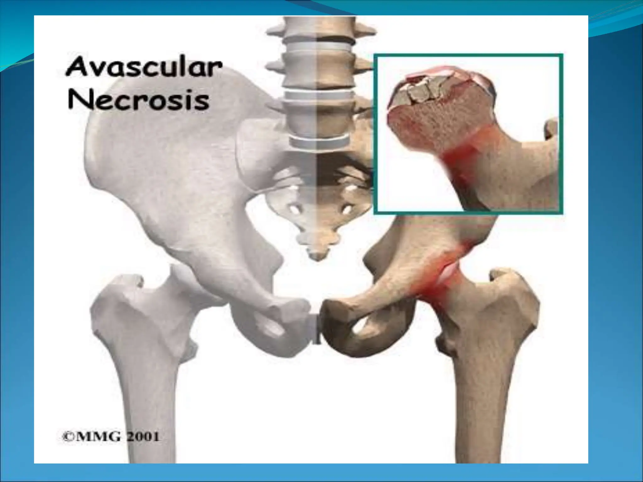
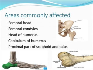
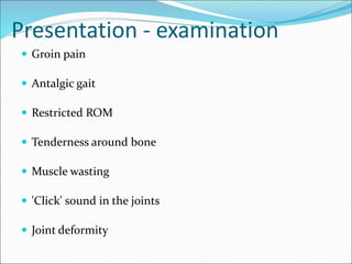
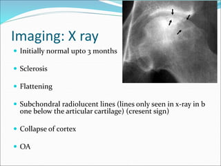
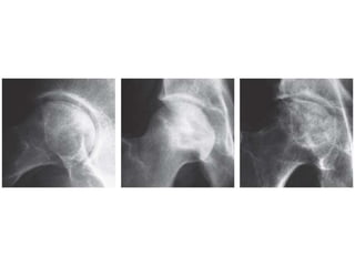
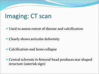
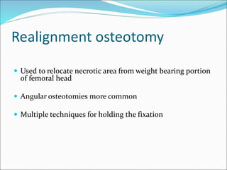
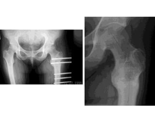
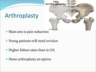
Sonal
BPT 3rd year
Outline
 Definition
 Pathophysiology
 Aetiology
 Presentation
 Imaging
 Staging
 Management
Definition
 Cellular death of bone components secondary to interr
uption of blood supply
 Consequent collapse of bone components
 Pain, loss of function of joints
Areas commonly affected
• Femoral head
• Femoral condyles
• Head of humerus
• Capitulum of humerus
• Proximal part of scaphoid and talus
Predisposing factors
• Distance from vascular territory of bone
• Enclosed by cartilage limiting vascularity.
• Vascular sinusoids (type of capillaries which has open
pores which increases their permeability, and seen in b
one marrow, liver etc).
Are compressed by increased pressure and volume of
marrow tissue.
Pathophysiology
 Interruption of blood flow to bone
 Affect bones with single terminal blood supply:
 Talus
 Carpals, tarsals
 Proximal humerus
 Head of femur
 Femoral condyles
 Results in necrosis of Bone marrow, medullary cavity (
place where bone marrow resides) and cortical bone( t
ype of lamellar bone, with densely packed lamellae)
 Final pathway from multiple causes
Pathways to necrosis
 Bone necrosis after 12 – 48 hrs of anoxia
 Reactive new bone formation around necrotic bone
 Granulation tissue over necrosed bone – sclerosis
 Structural failure in heavily stressed part of the necroti
c segment – linear fractures
 Crack may break through the articular cartilage and at su
rgery necrotic part lifts off like cracked shell of hard boile
d egg.
 Articular cartilage retains it's thickness but in final stages
deformity and destruction of joint surface is seen.
 The medullary cavity is the central cavity of bone shafts
where red bone marrow and/or yellow bone marrow
(adipose tissue) is stored. It has walls composed of
spongy bone (cancellous bone) and is lined with a thin,
vascular membrane (endosteum).
Traumatic causes
 In fractures and dislocations of hip the retinacular vessels
supplying the femoral head are torn. If in addition, there is
damage to or thrombosis of the ligamentum Teres, osteone
crosis is inevitable.
 In fractures of scaphoid, proximal part is more prone to n
ecrosis
• Fracture of talus, avascular necrosis is commonly seen. It is
because the anastomotic ring of blood vessels run postero-
laterally. Therefore blood supply is often cut off during neck
fractures.
Non-Traumatic causes
• Intravascular thrombosis
• High dose of corticosteroids and alcohol abuse leads to
hyperlipidemia and fatty degeneration of liver.
• Hypercoaguable states- thrombophilia
• Systemic lupus erythrematosis - deficiency of anti-
phospholipid.
• Hematologic disorder- sickle cell anemia ( enhanced
coagulability)
Extravascular marrow swelling
• High dosage of alcohol and corticosteroids leads to fat cell
swelling in the marrow.
• This leads to intra-osseous pressure increase and compression
of sinusoids as in femoral head.
Caisson disease and Dysbaric osteonecrosis
• Decompression disease seen in deep - sea divers and
compress air workers.
• Under increased pressure, the blood and other tissues (fat)
becomes super saturated with nitrogen.
• These gas bubbles leads to tissue damage and prolonged tissue
damage leads to swelling of marrow fat cells and decrease
blood supply due to toxicity.
Gaucher's disease
• Lack of enzyme glucocerebrocide in the macrophages of the
reticuloendothelial system.
• Affects spleen and bone marrow.
• Leads to sinusoidal compression in hip.
Radiation therapy
• Prolonged exposure to radiations leads to bone death.
• This is due to combined effects of damage to small blood
vessel, marrow cells and bone cells
Malignancy
• Corticosteroids
• Bisphosphonates (long term IV use leads to osteonecrosis of
jaw)
• Certain types of chemotherapy
• Radiotherapy
• Patients who are being treated for acute myeloid
leukaemia, acute lymphoblastic leukaemia, lymphomas,
testicular cancer, ovarian cancer, breast cancer, and
multiple myeloma have an increased risk of developing
AVN regardless of the treatment they receive
• Bone Marrow or Stem Cell Transplantation
Presentation - History
 Trauma
 Corticosteroid use
 Alcohol intake
 Medical conditions – malignancy, thrombophilia, SLE, SCD
Presentation - examination
 Groin pain
 Antalgic gait
 Restricted ROM
 Tenderness around bone
 Muscle wasting
 'Click' sound in the joints
 Joint deformity
Imaging: X ray
 Initially normal upto 3 months
 Sclerosis
 Flattening
 Subchondral radiolucent lines (lines only seen in x-ray in b
one below the articular cartilage) (cresent sign)
 Collapse of cortex
 OA
Imaging: CT scan
 Used to assess extent of disease and calcification
 Clearly shows articular deformity
 Calcification and bone collapse
 Central sclerosis in femoral head produces star shaped
structure (asterisk sign)
Imaging: MRI
 90% sensitive
 Changes detected early
Radionuclide scan
 Donut sign – central reduced uptake with surrounding
rim of increased uptake
 More sensitive than plain films in early AVN
 Less sensitive than MRI
 Necrotic zone surrounded by reactive new bone forma
tion
Histology
 Definitive diagnosis
 Usually a confirmatory during surgery for treatment
 Occasionally biopsy of sclerotic lesion
 Necrosis of cortical bone is followed by a regenerative
process in surrounding tissues.
Intramedullary pressures
 Cannula into metaphysis
 Measure at rest and after saline injection
 Femoral head:
 10 – 20 mmHg, increasing by 15 mmHg after saline
 Markedly increased values in AVN (3 to 4 fold)
 Less marked increase in OA
Ficat and the Steinberg Classification
Stage 1 • X-ray - no changes
• Diagnosis will be based on
intra-osseous pressure and
bone biopsy
Stage 2 Femoral head contour was
still normal but there were
early signs of Reactive
changes in the subchondral
bone
Stage 3 Clear evidence of
osteonecrosis and
structural damage and
distortion of bone
outline.
Stage 4 Collapse of articular
surface and signs of OA.
Management principles
 Early stages (I & II):
 Bisphosphonates prevent collapse
 Unloading osteotomies
 Medullary decompression + bone grafting
 Intermediate stage (III):
 Realignment osteototmies, Decompression, Curettage
 Arthrodesis
 Late stage (IV):
 Analgesia, activity modification
 Arthrodesis
 Total joint replacement
Management - conservative
 Offloading affected joints with use of crutches
 Immobilisation
 Analgesia
 Bisphosphonates delays bone break down in femur
 Statins in patients on high dose corticosteroids – reduc
ed lipid deposition
Core decompression
 8 – 10 mm anterolateral core of bone
 Filled with bone graft (vascularised/non vascularised)
 Decompresses medullary cavity, reduces pain
 Vascularised graft may reverse necrosis
Realignment osteotomy
 Used to relocate necrotic area from weight bearing portion
of femoral head
 Angular osteotomies more common
 Multiple techniques for holding the fixation
Arthroplasty
 Main aim is pain reduction
 Young patients will need revision
 Higher failure rates than in OA
 Hemi arthroplasty an option
AVASCULAR NECROSIS

AVASCULAR NECROSIS

  • 1.
  • 3.
    Outline  Definition  Pathophysiology Aetiology  Presentation  Imaging  Staging  Management
  • 4.
    Definition  Cellular deathof bone components secondary to interr uption of blood supply  Consequent collapse of bone components  Pain, loss of function of joints
  • 5.
    Areas commonly affected •Femoral head • Femoral condyles • Head of humerus • Capitulum of humerus • Proximal part of scaphoid and talus
  • 6.
    Predisposing factors • Distancefrom vascular territory of bone • Enclosed by cartilage limiting vascularity. • Vascular sinusoids (type of capillaries which has open pores which increases their permeability, and seen in b one marrow, liver etc). Are compressed by increased pressure and volume of marrow tissue.
  • 7.
    Pathophysiology  Interruption ofblood flow to bone  Affect bones with single terminal blood supply:  Talus  Carpals, tarsals  Proximal humerus  Head of femur  Femoral condyles  Results in necrosis of Bone marrow, medullary cavity ( place where bone marrow resides) and cortical bone( t ype of lamellar bone, with densely packed lamellae)  Final pathway from multiple causes
  • 8.
    Pathways to necrosis Bone necrosis after 12 – 48 hrs of anoxia  Reactive new bone formation around necrotic bone  Granulation tissue over necrosed bone – sclerosis  Structural failure in heavily stressed part of the necroti c segment – linear fractures
  • 9.
     Crack maybreak through the articular cartilage and at su rgery necrotic part lifts off like cracked shell of hard boile d egg.  Articular cartilage retains it's thickness but in final stages deformity and destruction of joint surface is seen.
  • 11.
     The medullarycavity is the central cavity of bone shafts where red bone marrow and/or yellow bone marrow (adipose tissue) is stored. It has walls composed of spongy bone (cancellous bone) and is lined with a thin, vascular membrane (endosteum).
  • 12.
    Traumatic causes  Infractures and dislocations of hip the retinacular vessels supplying the femoral head are torn. If in addition, there is damage to or thrombosis of the ligamentum Teres, osteone crosis is inevitable.  In fractures of scaphoid, proximal part is more prone to n ecrosis
  • 13.
    • Fracture oftalus, avascular necrosis is commonly seen. It is because the anastomotic ring of blood vessels run postero- laterally. Therefore blood supply is often cut off during neck fractures.
  • 15.
    Non-Traumatic causes • Intravascularthrombosis • High dose of corticosteroids and alcohol abuse leads to hyperlipidemia and fatty degeneration of liver. • Hypercoaguable states- thrombophilia • Systemic lupus erythrematosis - deficiency of anti- phospholipid. • Hematologic disorder- sickle cell anemia ( enhanced coagulability)
  • 17.
    Extravascular marrow swelling •High dosage of alcohol and corticosteroids leads to fat cell swelling in the marrow. • This leads to intra-osseous pressure increase and compression of sinusoids as in femoral head. Caisson disease and Dysbaric osteonecrosis • Decompression disease seen in deep - sea divers and compress air workers. • Under increased pressure, the blood and other tissues (fat) becomes super saturated with nitrogen.
  • 18.
    • These gasbubbles leads to tissue damage and prolonged tissue damage leads to swelling of marrow fat cells and decrease blood supply due to toxicity. Gaucher's disease • Lack of enzyme glucocerebrocide in the macrophages of the reticuloendothelial system. • Affects spleen and bone marrow. • Leads to sinusoidal compression in hip.
  • 19.
    Radiation therapy • Prolongedexposure to radiations leads to bone death. • This is due to combined effects of damage to small blood vessel, marrow cells and bone cells Malignancy • Corticosteroids • Bisphosphonates (long term IV use leads to osteonecrosis of jaw) • Certain types of chemotherapy • Radiotherapy
  • 20.
    • Patients whoare being treated for acute myeloid leukaemia, acute lymphoblastic leukaemia, lymphomas, testicular cancer, ovarian cancer, breast cancer, and multiple myeloma have an increased risk of developing AVN regardless of the treatment they receive • Bone Marrow or Stem Cell Transplantation
  • 21.
    Presentation - History Trauma  Corticosteroid use  Alcohol intake  Medical conditions – malignancy, thrombophilia, SLE, SCD
  • 22.
    Presentation - examination Groin pain  Antalgic gait  Restricted ROM  Tenderness around bone  Muscle wasting  'Click' sound in the joints  Joint deformity
  • 23.
    Imaging: X ray Initially normal upto 3 months  Sclerosis  Flattening  Subchondral radiolucent lines (lines only seen in x-ray in b one below the articular cartilage) (cresent sign)  Collapse of cortex  OA
  • 25.
    Imaging: CT scan Used to assess extent of disease and calcification  Clearly shows articular deformity  Calcification and bone collapse  Central sclerosis in femoral head produces star shaped structure (asterisk sign)
  • 27.
    Imaging: MRI  90%sensitive  Changes detected early
  • 29.
    Radionuclide scan  Donutsign – central reduced uptake with surrounding rim of increased uptake  More sensitive than plain films in early AVN  Less sensitive than MRI  Necrotic zone surrounded by reactive new bone forma tion
  • 31.
    Histology  Definitive diagnosis Usually a confirmatory during surgery for treatment  Occasionally biopsy of sclerotic lesion  Necrosis of cortical bone is followed by a regenerative process in surrounding tissues.
  • 32.
    Intramedullary pressures  Cannulainto metaphysis  Measure at rest and after saline injection  Femoral head:  10 – 20 mmHg, increasing by 15 mmHg after saline  Markedly increased values in AVN (3 to 4 fold)  Less marked increase in OA
  • 33.
    Ficat and theSteinberg Classification Stage 1 • X-ray - no changes • Diagnosis will be based on intra-osseous pressure and bone biopsy Stage 2 Femoral head contour was still normal but there were early signs of Reactive changes in the subchondral bone
  • 34.
    Stage 3 Clearevidence of osteonecrosis and structural damage and distortion of bone outline. Stage 4 Collapse of articular surface and signs of OA.
  • 36.
    Management principles  Earlystages (I & II):  Bisphosphonates prevent collapse  Unloading osteotomies  Medullary decompression + bone grafting  Intermediate stage (III):  Realignment osteototmies, Decompression, Curettage  Arthrodesis  Late stage (IV):  Analgesia, activity modification  Arthrodesis  Total joint replacement
  • 37.
    Management - conservative Offloading affected joints with use of crutches  Immobilisation  Analgesia  Bisphosphonates delays bone break down in femur  Statins in patients on high dose corticosteroids – reduc ed lipid deposition
  • 38.
    Core decompression  8– 10 mm anterolateral core of bone  Filled with bone graft (vascularised/non vascularised)  Decompresses medullary cavity, reduces pain  Vascularised graft may reverse necrosis
  • 40.
    Realignment osteotomy  Usedto relocate necrotic area from weight bearing portion of femoral head  Angular osteotomies more common  Multiple techniques for holding the fixation
  • 42.
    Arthroplasty  Main aimis pain reduction  Young patients will need revision  Higher failure rates than in OA  Hemi arthroplasty an option