This document provides a practice question sample set for the medical licensing exam preparation platform Osmosis. It includes two sample questions, one testing basic science knowledge of myocardial infarction histology, and the other testing diagnosis of Dressler Syndrome, a post-myocardial infarction condition. The basic science question asks which histological elements would be found on microscopic examination of a patient's heart tissue given autopsy findings and timeline. The diagnosis question presents a patient presenting 8 weeks after a myocardial infarction with fever, sore throat, shortness of breath and back pain to test recognition of Dressler Syndrome.


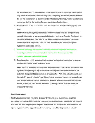
![● Beginning of granulation tissue
formation at margins
10 – 14 days ● Redgray and
depressed
borders
● Mature granulation tissue with
type I collagen[1]
2 – 8 weeks ● Graywhite
granulation
tissue
● Increased collagen deposition
● Decreased cellularity
More than 2
months
Completed scarring Dense collagenous scar formed
https://en.wikipedia.org/wiki/Timeline_of_myocardial_infarction_pathology
References:
● Bishop JE, Greenbaum R, Gibson DG, Yacoub M, Laurent GJ. Enhanced deposition of
predominantly type I collagen in myocardial disease. J Mol Cell Cardiol.
1990;22:1157–1165
● Rangogiannis NG: The immune system and cardiac repair. Pharmacol Res. 2008, 58:
88111
● Campobasso CP, Dell’Erba AS, Addante A, Zotti F, Marzullo A, Colonna MF: Sudden
cardiac death and myocardial ischemia indicators: a comparative study of four
immunohistochemical markers. Am J Forensic Med Pathol. 2008, 29: 154161.
#2. Diagnosis Question
Theme: Cardiovascular
Disease Diagnosis Tested: Myocardial Infarction and diagnosis of the postmyocardial
infarction syndrome, also known as Dressler Syndrome](https://image.slidesharecdn.com/aa6ba683-9e1d-4935-8ec1-219651a94a46-160619203300/85/OsmosisSampleQuestions_Pamer-6-320.jpg)