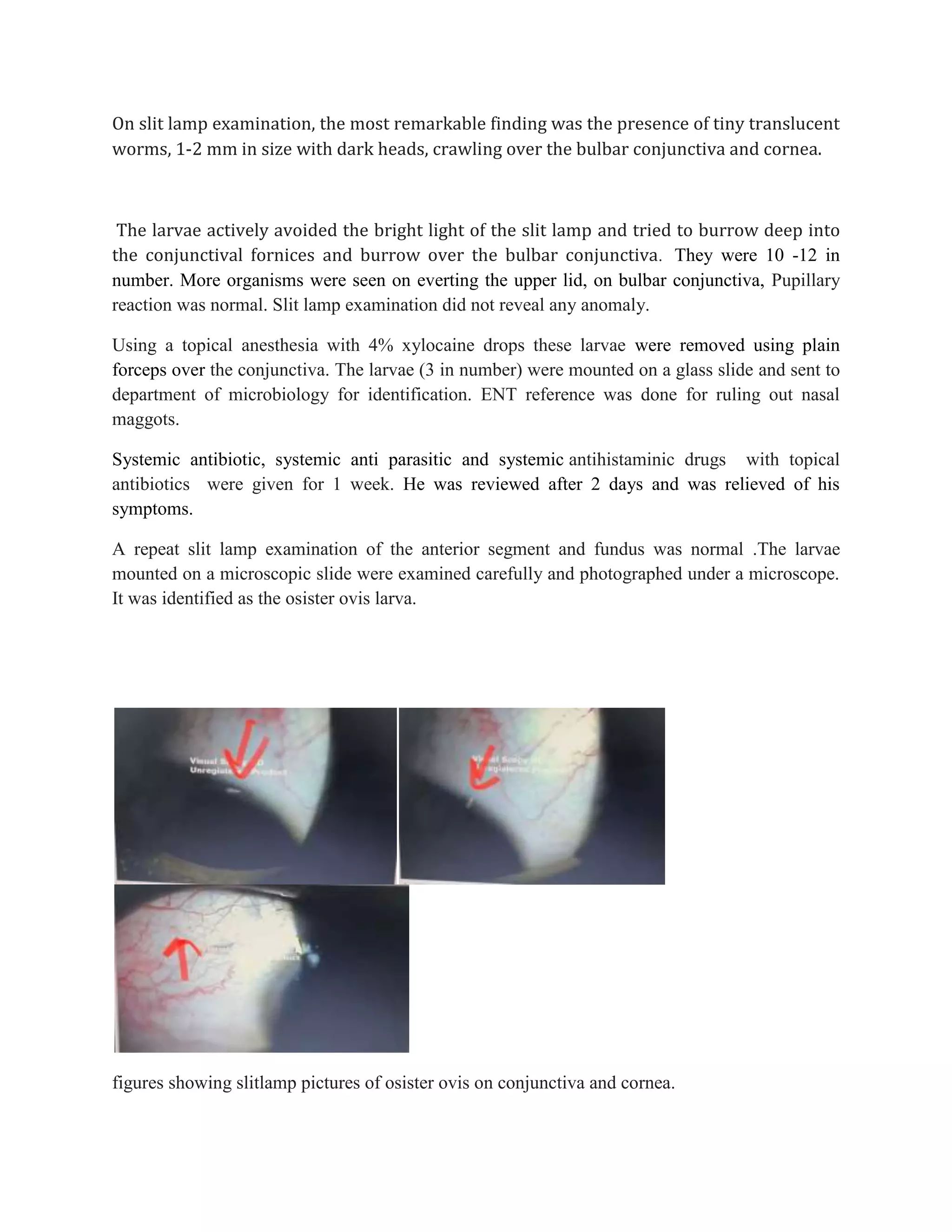
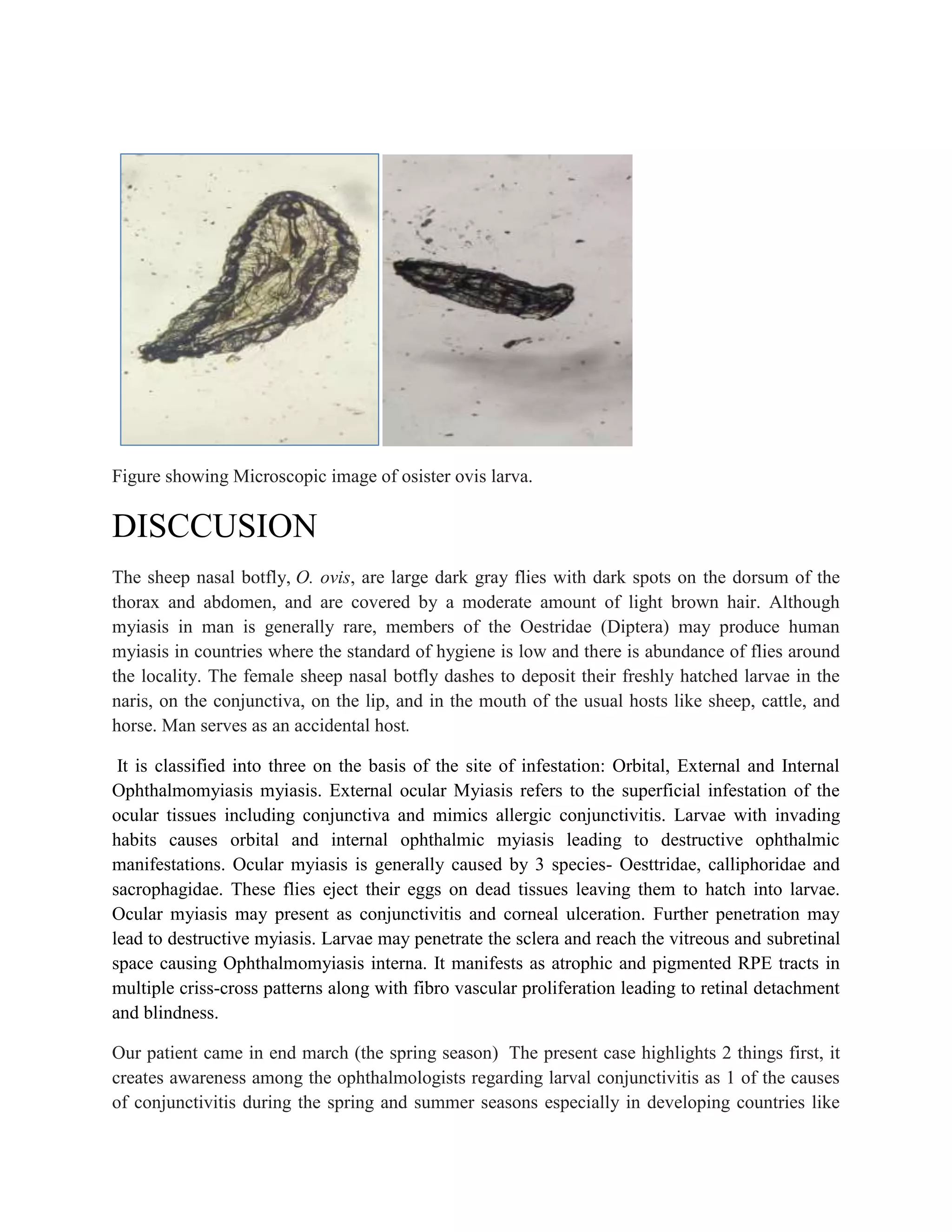
Ophthalmomyiasis: A Case Report
Abstract: Myiasis of different organs has been reported off and on from various regions in the world.
We report a human case of external ophthalmomyiasis caused by the larvae of a sheep nasal botfly, Oestrus
ovis . A 19 -year-old male presented with severe symptoms of conjunctivitis and foreign body sensation . The
larvae, 3 in number, were observed in the bulbar conjunctiva, and following removal the symptoms of eye
inflammation improved within a few hours.
Keywords: Oestrus ovis, myiasis, ophthalmomyiasis.
Introduction
Myiasis is the infestation of humans and animals with maggots (larvae) of certain flies . Skin is the most
common organ of infestation, but maggots have been removed from eyes, ears, nose, intestine and
urogenital tract. Ophthalmomyiasis refers to the infestation of the eye by the larvae. If the infestation is
confined to the ocular surface or periorbital tissues, it is termed as ophthalmomyiasis externa; whereas,
intraocular penetration of larva is referred to as ophthalmomyiasis interna. Ophthalmomyiasis externa
caused by Oestrus ovis (the sheep nasal botfly).
CASE REPORT
A 19 year male came to our outpatient department in 29th march 2023 with a 2-day history
of foreign body sensation, burning, and excessive watering from his left eye. He gave a
history of something falling in his eye while he was passing by on the road. He was
absolutely fine before that, and there was no significant history of ocular or medical
problems preceding this.
On examination it was found that his visual acuity was 6/9 in both eyes with pinhole 6/6.
Eyelids of the affected left eye were absolutely normal. The conjunctiva was mildly
congested with profuse lacrimation. Extraocular movements were full.
On slit lamp examination, the most remarkable finding was the presence of tiny translucent
worms, 1-2 mm in size with dark heads, crawling over the bulbar conjunctiva and cornea.
The larvae actively avoided the bright light of the slit lamp and tried to burrow deep into
the conjunctival fornices and burrow over the bulbar conjunctiva. They were 10 -12 in
number. More organisms were seen on everting the upper lid, on bulbar conjunctiva, Pupillary
reaction was normal. Slit lamp examination did not reveal any anomaly.
Using a topical anesthesia with 4% xylocaine drops these larvae were removed using plain
forceps over the conjunctiva. The larvae (3 in number) were mounted on a glass slide and sent to
department of microbiology for identification. ENT reference was done for ruling out nasal
maggots.
Systemic antibiotic, systemic anti parasitic and systemic antihistaminic drugs with topical
antibiotics were given for 1 week. He was reviewed after 2 days and was relieved of his
symptoms.
A repeat slit lamp examination of the anterior segment and fundus was normal .The larvae
mounted on a microscopic slide were examined carefully and photographed under a microscope.
It was identified as the osister ovis larva.
figures showing slitlamp pictures of osister ovis on conjunctiva and cornea.
Figure showing Microscopic image of osister ovis larva.
DISCCUSION
The sheep nasal botfly, O. ovis, are large dark gray flies with dark spots on the dorsum of the
thorax and abdomen, and are covered by a moderate amount of light brown hair. Although
myiasis in man is generally rare, members of the Oestridae (Diptera) may produce human
myiasis in countries where the standard of hygiene is low and there is abundance of flies around
the locality. The female sheep nasal botfly dashes to deposit their freshly hatched larvae in the
naris, on the conjunctiva, on the lip, and in the mouth of the usual hosts like sheep, cattle, and
horse. Man serves as an accidental host.
It is classified into three on the basis of the site of infestation: Orbital, External and Internal
Ophthalmomyiasis myiasis. External ocular Myiasis refers to the superficial infestation of the
ocular tissues including conjunctiva and mimics allergic conjunctivitis. Larvae with invading
habits causes orbital and internal ophthalmic myiasis leading to destructive ophthalmic
manifestations. Ocular myiasis is generally caused by 3 species- Oesttridae, calliphoridae and
sacrophagidae. These flies eject their eggs on dead tissues leaving them to hatch into larvae.
Ocular myiasis may present as conjunctivitis and corneal ulceration. Further penetration may
lead to destructive myiasis. Larvae may penetrate the sclera and reach the vitreous and subretinal
space causing Ophthalmomyiasis interna. It manifests as atrophic and pigmented RPE tracts in
multiple criss-cross patterns along with fibro vascular proliferation leading to retinal detachment
and blindness.
Our patient came in end march (the spring season) The present case highlights 2 things first, it
creates awareness among the ophthalmologists regarding larval conjunctivitis as 1 of the causes
of conjunctivitis during the spring and summer seasons especially in developing countries like
India, where the general standard of hygiene is low and there are a large number of flies around.
Second, irrigation of the conjunctiva with normal saline is unsuccessful in washing out the larvae
because the larvae grab the conjunctiva firmly with the help of a pair of oral hooks and numerous
hooks on each segment. Therefore, after anesthetizing the conjunctiva, a thorough examination
of the eye under magnification and prompt removal of the larvae manually with forceps should
be done to avoid disastrous complications of internal ophthalmomyiasis.
A number of procedures have been described for the removal of maggots like use of turpentine oil, liquid
paraffin, betadine, mechanical removal and surgical debridement of the tissue.Systemic treatment with
oral antibiotics is indicated to prevent secondary bacterial infection. Anti parasitic drugs like ivermectin
may be used in the dosage of 200µg/kg.
CONCLUSION
Though Ophthalmomyiasis is a relatively rare condition, it can lead to blindness if not diagnosed
and treated well in time. High index of suspicion is needed for its diagnosis. Meticulous removal
of all visible larvae is important to prevent penetration of the globe and thus to prevent blinding
complications.
Reference
1. Sharma M, Sareen A, Negi SSOphthalmomyiasis: A Case Report.DJO 2020;30:52-53
2. Pandey A, Madan M, Asthana AK, Das A, Kumar S, Jain K. External ophthalmomyiasis
caused by Oestrus ovis: a rare case report from India. Korean J Parasitol. 2009
Mar;47(1):57-9. doi: 10.3347/kjp.2009.47.1.57. Epub 2009 Mar 12. PMID: 19290093;
PMCID: PMC2655336.
3. Smillie I, Gubbi PK, Cocks. Nasal ophthalmomyiasis: a case report. J Laryngol Otol,
2010; 124(8):934-5.
4. Cameron JA, Shoukrey NM, Al Garni AA. Conjunctival ophthalmomyiasis caused by
sheep nasal botfly (Oestrus ovis). Am J Ophthalmol, 1991; 112(3):331-4.
5. Pandey A, Madan M, Asthana AK, Das A, et al. External ophthalmomyiasis caused by
Oestrus ovis: a rare case report from India. Korean J Ophthalmol, 2009; 47(1):57-9.
6. Sigauke E, Beebe WE, Gander RM, Cavouti D, et al. Case Report: Ophthalomyiasis
externa in Dallas County, Texas. Am J Trop Med Hyg, 2003; 68(1):46-7.
7. Khurana S, Biswas M, Bhatti H.Orbiatl myiasis : Three cases from North India. Indian J
Med Microbiol, 2010; 28(3):57-61.
8. Ashenhurst M, Pictuche S. Management of Ophthalmomyiasis externa; a case report. Can
J Ophthalmol, 2004; 39(3):285-7.
Ophthalmomyiasis case report.docx

Ophthalmomyiasis case report.docx

  • 1.
    Ophthalmomyiasis: A CaseReport Abstract: Myiasis of different organs has been reported off and on from various regions in the world. We report a human case of external ophthalmomyiasis caused by the larvae of a sheep nasal botfly, Oestrus ovis . A 19 -year-old male presented with severe symptoms of conjunctivitis and foreign body sensation . The larvae, 3 in number, were observed in the bulbar conjunctiva, and following removal the symptoms of eye inflammation improved within a few hours. Keywords: Oestrus ovis, myiasis, ophthalmomyiasis. Introduction Myiasis is the infestation of humans and animals with maggots (larvae) of certain flies . Skin is the most common organ of infestation, but maggots have been removed from eyes, ears, nose, intestine and urogenital tract. Ophthalmomyiasis refers to the infestation of the eye by the larvae. If the infestation is confined to the ocular surface or periorbital tissues, it is termed as ophthalmomyiasis externa; whereas, intraocular penetration of larva is referred to as ophthalmomyiasis interna. Ophthalmomyiasis externa caused by Oestrus ovis (the sheep nasal botfly). CASE REPORT A 19 year male came to our outpatient department in 29th march 2023 with a 2-day history of foreign body sensation, burning, and excessive watering from his left eye. He gave a history of something falling in his eye while he was passing by on the road. He was absolutely fine before that, and there was no significant history of ocular or medical problems preceding this. On examination it was found that his visual acuity was 6/9 in both eyes with pinhole 6/6. Eyelids of the affected left eye were absolutely normal. The conjunctiva was mildly congested with profuse lacrimation. Extraocular movements were full.
  • 2.
    On slit lampexamination, the most remarkable finding was the presence of tiny translucent worms, 1-2 mm in size with dark heads, crawling over the bulbar conjunctiva and cornea. The larvae actively avoided the bright light of the slit lamp and tried to burrow deep into the conjunctival fornices and burrow over the bulbar conjunctiva. They were 10 -12 in number. More organisms were seen on everting the upper lid, on bulbar conjunctiva, Pupillary reaction was normal. Slit lamp examination did not reveal any anomaly. Using a topical anesthesia with 4% xylocaine drops these larvae were removed using plain forceps over the conjunctiva. The larvae (3 in number) were mounted on a glass slide and sent to department of microbiology for identification. ENT reference was done for ruling out nasal maggots. Systemic antibiotic, systemic anti parasitic and systemic antihistaminic drugs with topical antibiotics were given for 1 week. He was reviewed after 2 days and was relieved of his symptoms. A repeat slit lamp examination of the anterior segment and fundus was normal .The larvae mounted on a microscopic slide were examined carefully and photographed under a microscope. It was identified as the osister ovis larva. figures showing slitlamp pictures of osister ovis on conjunctiva and cornea.
  • 3.
    Figure showing Microscopicimage of osister ovis larva. DISCCUSION The sheep nasal botfly, O. ovis, are large dark gray flies with dark spots on the dorsum of the thorax and abdomen, and are covered by a moderate amount of light brown hair. Although myiasis in man is generally rare, members of the Oestridae (Diptera) may produce human myiasis in countries where the standard of hygiene is low and there is abundance of flies around the locality. The female sheep nasal botfly dashes to deposit their freshly hatched larvae in the naris, on the conjunctiva, on the lip, and in the mouth of the usual hosts like sheep, cattle, and horse. Man serves as an accidental host. It is classified into three on the basis of the site of infestation: Orbital, External and Internal Ophthalmomyiasis myiasis. External ocular Myiasis refers to the superficial infestation of the ocular tissues including conjunctiva and mimics allergic conjunctivitis. Larvae with invading habits causes orbital and internal ophthalmic myiasis leading to destructive ophthalmic manifestations. Ocular myiasis is generally caused by 3 species- Oesttridae, calliphoridae and sacrophagidae. These flies eject their eggs on dead tissues leaving them to hatch into larvae. Ocular myiasis may present as conjunctivitis and corneal ulceration. Further penetration may lead to destructive myiasis. Larvae may penetrate the sclera and reach the vitreous and subretinal space causing Ophthalmomyiasis interna. It manifests as atrophic and pigmented RPE tracts in multiple criss-cross patterns along with fibro vascular proliferation leading to retinal detachment and blindness. Our patient came in end march (the spring season) The present case highlights 2 things first, it creates awareness among the ophthalmologists regarding larval conjunctivitis as 1 of the causes of conjunctivitis during the spring and summer seasons especially in developing countries like
  • 4.
    India, where thegeneral standard of hygiene is low and there are a large number of flies around. Second, irrigation of the conjunctiva with normal saline is unsuccessful in washing out the larvae because the larvae grab the conjunctiva firmly with the help of a pair of oral hooks and numerous hooks on each segment. Therefore, after anesthetizing the conjunctiva, a thorough examination of the eye under magnification and prompt removal of the larvae manually with forceps should be done to avoid disastrous complications of internal ophthalmomyiasis. A number of procedures have been described for the removal of maggots like use of turpentine oil, liquid paraffin, betadine, mechanical removal and surgical debridement of the tissue.Systemic treatment with oral antibiotics is indicated to prevent secondary bacterial infection. Anti parasitic drugs like ivermectin may be used in the dosage of 200µg/kg. CONCLUSION Though Ophthalmomyiasis is a relatively rare condition, it can lead to blindness if not diagnosed and treated well in time. High index of suspicion is needed for its diagnosis. Meticulous removal of all visible larvae is important to prevent penetration of the globe and thus to prevent blinding complications. Reference 1. Sharma M, Sareen A, Negi SSOphthalmomyiasis: A Case Report.DJO 2020;30:52-53 2. Pandey A, Madan M, Asthana AK, Das A, Kumar S, Jain K. External ophthalmomyiasis caused by Oestrus ovis: a rare case report from India. Korean J Parasitol. 2009 Mar;47(1):57-9. doi: 10.3347/kjp.2009.47.1.57. Epub 2009 Mar 12. PMID: 19290093; PMCID: PMC2655336. 3. Smillie I, Gubbi PK, Cocks. Nasal ophthalmomyiasis: a case report. J Laryngol Otol, 2010; 124(8):934-5. 4. Cameron JA, Shoukrey NM, Al Garni AA. Conjunctival ophthalmomyiasis caused by sheep nasal botfly (Oestrus ovis). Am J Ophthalmol, 1991; 112(3):331-4. 5. Pandey A, Madan M, Asthana AK, Das A, et al. External ophthalmomyiasis caused by Oestrus ovis: a rare case report from India. Korean J Ophthalmol, 2009; 47(1):57-9. 6. Sigauke E, Beebe WE, Gander RM, Cavouti D, et al. Case Report: Ophthalomyiasis externa in Dallas County, Texas. Am J Trop Med Hyg, 2003; 68(1):46-7. 7. Khurana S, Biswas M, Bhatti H.Orbiatl myiasis : Three cases from North India. Indian J Med Microbiol, 2010; 28(3):57-61. 8. Ashenhurst M, Pictuche S. Management of Ophthalmomyiasis externa; a case report. Can J Ophthalmol, 2004; 39(3):285-7.