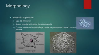
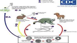
Created by:
Assem Karam “950”
Abderrahman Sami “952”
Abderrahman Sharara “953”
Abdullah Eraf “955”
Omar Elgazzar “957”
Omar Ayman “958”
THIS PRESENTATION COVERS
Acanthamoeba spp Baylisascaris procyonis Cysticercosis
Filariasis Onchocerca volvulus Sparganosis
Thelazia californiensis Toxocariasis Toxoplasmosis
Acanthamoeba spp
AMOEBIC KERATITIS AND CORNEAL ULCER
Morphology
 Amoeboid trophozoite:
 Size: 22-30 micron
 Shape: irregular with spine like pseudopodia
 Content: single nucleus with large central karyosome and central contractile
vacuole.
Morphology
 Cyst:
 Size: about 18 micron in diameter
 Shape: Rounded
 Structure: With double waal with slightly wrinkled outer cyst
wall an polyhedral inner cyst wall.
 Cysts are dispersed by wind to contaminate environment.
Life cycle
 The life cycle consists of two stages:
 An actively feeding and dividing cyst
 A dormant cyst
 Cysts are formed under unfavorable conditions such as food deprivation,
desiccation and changes in temperature and pH.
 Acanthamoeba spp. have been found in soil; fresh, brackish, and sea water;
sewage; swimming pools; contact lens equipment; medicinal pools; dental
treatment units; dialysis machines; heating, ventilating, and air
conditioning systems; mammalian cell cultures; vegetables; human nostrils
and throats; and human and animal brain, skin, and lung tissues.
Life cycle
 The trophozoites replicate by mitosis. The trophozoites
are the infective forms, although both cysts and
trophozoites gain entry into the body through various
means. Entry can occur through the eye , the nasal
passages to the lower respiratory tract , or ulcerated or
broken skin .
 When Acanthamoeba spp. enters the eye it can cause
severe keratitis in otherwise healthy individuals,
particularly contact lens users . When it enters the
respiratory system or through the skin, it can invade the
central nervous system by hematogenous dissemination
causing granulomatous amebic encephalitis (GAE) or
disseminated disease , or skin lesions in individuals with
compromised immune systems. Acanthamoeba spp. cysts
and trophozoites are found in tissue.
Disease Production
AMOEBIC KERATITIS & CORNEAL ULCER
Mode of infection
1. Inhalation of trophozoites or cysts (infective stages) through nasal
passages and lung.
2. Entrance through skin lesions and genitourinary tract.
3. Introduction through corneal abrasions especially with use of contact
lens.
Pathogenesis
 It is an opportunistic pathogen occurs in immunosuppressed chronically ill
patients with AIDS , TB, Radiotherapy and chemotherapy or to immune
privileged sites e.g. Cornea.
 Tissue invasion is slow leading to subacute or chronic illness and marked
Granulomatous reaction.
Pathogenesis
Opportunistic
diseases
Granulomatous
amoebic encephalitis
(GAE)
Disseminated
granulomatous
amoebic disease
Non-opportunistic
diseases
Amoebic keratitis
and corneal ulcer.
Amoebic Keratitis and corneal ulcers
 It occurs in healthy individuals.
 Especially in patients using contact lenses.
 The condition starts with trauma to cornea
with use of lenses and subsequent exposure
to water contaminated with cyst or
trophozoites.
 Clinically:
1. It is chronic progressive ulcerative diseases of the
eye.
2. Painful corneal ulcer, complete corneal opacity and
blindness
3. Congested conjunctiva
Diagnosis
Clinical diagnosis
 History and clinical picture.
 CNS manifestations in IC patient or chronic corneal
ulcers unresponsive to antibiotic in contact lenses
wearer.
Diagnosis
Laboratory diagnosis
I. CSF examination:
 Free of bacteria
 Increased protein content and decreased or normal glucose content
 Microscopic examination: Examination of wet mount stained smear to detect
trophozoites or cysts.
 Culture.
II. Examination of corneal scraping or skin biopsy.
Treatment & control
TTT
 Sulfadiazine is the drug of choice.
Control
 Proper cleaning of contact lenses
 No wearing of lenses during swimming
 CSF examination:
 Examination of corneal scraping or skin biopsy.
Baylisascariasis
Baylisascaris procyonis (Raccoon round worm)
Epidemiology
▪ DH: Raccoons and rarely dogs.
▪ IH: Highly infectious; it’s the most common cause ofVLM in animals.
▪ Man acts as an “accidental host” to this parasite resulting in OLM andVLM.
Life cycle
▪ An adult worm lives and reproduces in the intestine of its DH, the raccoon.
▪ The female worm can produce between 115,000-179,000 eggs per day.
▪ Eggs are excreted along with feces, and become infective in the soil after 2–4 weeks.
▪ If ingested by another raccoon, the life cycle repeats.
Life cycle
▪ However, if these eggs are ingested by an IH (small mammals, birds) the larvae of B.
procyonis will penetrate the gut wall of the host and migrate into tissues.
▪ Larvae tend to migrate to the brain, cause damage, and affect the behavior of the IH, making
it an easier prey for raccoons.
▪ Reproduction does not occur in these paratenic hosts; however, if a raccoon preys on an
infected paratenic host, the encysted larvae can become adults in the raccoon and the cycle
resumes.
Disease Production
Baylisascariasis
Infection in humans
▪ Human infection with B. procyonis has been relatively rare, with about 13 cases reported
since 1980.
▪ However, disease caused by this parasite can be extremely dangerous, causing death or
severe symptoms.
▪ Out of the 13 cases, 5 were fatal and the remaining victims were left with severe neurological
damage.
Migration of Baylisascaris procyonis into the vitreous
▪ Baylisascaris procyonis is believed to cause DUSN (Diffuse unilateral subacute neuroretinitis).
▪ It’s characterized early by visual loss, vitritis, papillitis, and recurrent crops of grey‐white
retinal lesions and later by progressive visual loss, optic atrophy, retinal vessel narrowing, and
diffuse retinal pigment epithelial degeneration.
clinically
• Nausea
• Tiredness
• Liver enlargement
• Lack of attention to people and
surroundings
• Loss of muscle control
• Loss of coordination
• Blindness
• Coma
laboratory
• identification of larvae in tissue
examination.
• Serological tests
Diagnosis
Differential diagnosis
▪ T. canis
▪ T. cati
▪ A. lumbricoides
▪ Gnathostoma
▪ Angiostrongylus
▪ Ancylostoma
Prognosis and treatment
▪ Even with treatment, prognosis is poor.
▪ Albendazole is able to treat adult worms living in the intestines, but are less potent against
migrating larvae.
▪ treatment is more effective before the larvae have reached the brain; however, migration to
the brain was shown to occur only 3 days after ingestion.
HUMAN SPARGANOSIS
Spirometra mansoni
1 Habitat Small intestine of dogs
and cats
Never
found n
human
intestine
2 DH Dogs and Cats
3 I. Host 1st :. Cyclops
2nd :. Frogs – snakes –
birds
4 Larva name Sparganum mansoni
Morphology
-scolex : almond shape with 2
slits like suckers .
-mature segment :
Broader than longer
Median CGP
Bilobed ovary posteriorly .
Testes at dorso-lateral fields
HUMAN SPARGANOSIS
Disease Production
1 Definition Infection f human tissue by larval stage of
Spirometra mansoni which s named
Sparganum mansoni .
* Man acts as blind intermediate host .
2 Habitat Subcutaneous tissue – eyes – liver – lung
– brain
3 Infective stage 1- Procercoid larva
2- Sparganum mansoni
Life cycle
Mode of infection
-Drinking : water containing cyclops infected with
procercoid larva .
-Eating :. Improperly cooked frog , snake containing
Sparganum mansoni larva .
-Application of poultices or foments .
Pathogenesis
Sparganum larva induces chronic inflammation
ending in swelling formation .
Clinical picture
Skin :. Inflammatory painful, tender –
Abscess formation
Eye :. Periorbital edema – conjunctivitis
Diagnosis
Sparganosis is typically diagnosed following surgical removal of the worms, although
the infection may also be diagnosed by identification of eosinophilia or identification of
the parasite in a tissue specimen. If such biopsy and excision procedures are not
feasible, the antisparganum ELISA test may be used. In theory, a pre-operative
diagnosis could be made by identification of exposure history and a painful, migratory,
subcutaneous nodule. Sparganosis usually presents as a single nodule, while other
cestode infections such as cysticercosis typically present as multiple nodules.
Preoperative diagnosis, however, is rare.
Treatment
Prevention and control
Surgical removal of Sparganum
1- properly cooking of frog or bird meat
2- avod use frog as poultices .
TOXOCARIASIS &
OCCULAR LARVAE
MIGRANS (OLM)
Toxocara Cati & Toxocara canis
Overview
 It is a disease caused by non human Ascaris called Toxocara species. Either Toxocara canis
or Toxocara cati.
 Definitive host:
 T. canis: Dog
 T. Cati: Cat
 Man acts as a accidental host (blind intermediate host or dead end host).
 Habitat: Small intestine of Dogs and cats.
Morphology
 Adults of both species have complete digestive
systems and three lips surrounding mouth
anteriorly.
 T. canis are found only within dogs and foxes and
the males are 4–6 cm in length, with a curved
posterior end. The males each have spicules and
one “tubular testis.” Females can be as long as
15 cm, with the vulva stretching one third of their
body length. The females do not curve at the
posterior end.
 T. cati adult females are approximately 10 cm
long, while males are typically 6 cm or less. The
T. cati adults only occur within cats, and male T.
cati are curved at the posterior end.
Life cycle
Inside dogs and cats
 Cats, dogs and foxes can become infected with Toxocara through the ingestion of eggs or by
transmission of the larvae from a mother to her offspring either transplacentally or through
lactation.
 Eggs hatch as second stage larvae in the intestines of the cat, dog or fox host.
 Larvae enter the bloodstream and migrate to the lungs, where they are coughed up and
swallowed.
 The larvae mature into adults within the small intestine of a cat, dog or fox, where mating and
egg laying occurs.
 Eggs are passed in the feces and only become infective after several weeks outside of a host.
During this incubation period, molting from first to second stage larva takes place within the
egg.
Life cycle
Inside Humans
 Eggs containing second stage larvae
will also hatch in the small intestine of
an accidental host, such as a human,
after accidental ingestion of infective
eggs. The larvae will then migrate
through the organs and tissues of the
accidental host, most commonly the
lungs, liver, eyes, and brain. Since
second stage larvae cannot mature in
accidental hosts, after this period of
migration, Toxocara larvae will encyst
as second stage larvae stimulating
tissue reaction and granuloma
formation.
DISEASE PRODUCTION
Toxocariasis
Epidemiology
 Commonly seen in young children because of close
contact with puppies and kittens and because of habit
of pica.
Mode of transmission
 Accidental ingestion of embryonated egg of T. canis
and T. cati containing second stage rhabditiform
larvae (infective stage):
 Contaminated vegetables
 Food contaminated by house flies
 Contaminated water
 Pica
 Contaminated hands from polluted soil.
INFECTIVE STAGES
Size: 60 x 45 micron
Shape: Oval
Shell: Thick and with coarse mamillations
Colour: Brown
Content: Second stage rhabditiform larvae
Clinical picture
 It depends on site and severity of infection.
 Most infections are asymptomatic.
 While A light Toxocara burden in young adults is thought to
induce a low immune response, allowing a larva to enter the host’s
eye leading to OLM. OLM often occurs in just one eye and from a
single larva migrating into and encysting within the orbit.
 Loss of vision occurs over days or weeks (due to chronic
inflammation of posterior chamber of eye leading to retinal
inflammation , retinal fibrosis, retinal detachment).
 Squint caused by haemorrhage at or near macula 
 Other signs and symptoms
 Red Eyes
 White pupil
 Retinal granulomas.
Diagnosis
Clinical diagnosis
 History of contact with dogs and cats and from signs and symptoms.
Laboratory diagnosis
i. Immunodiagnosis: Serum and vitreous Toxocara antibody
determination are available done by ELISA.
ii. Direct diagnosis: It depends on histological examination of tissue
sections from accessible granuloma which may show nematode
larvae at center of eosinophilic granulomatous reaction.
iii. Blood picture: Persistent eosinophilia.
Differential diagnosis
 Retinal tumour (Retinoblastoma).
 Other parasitic diseases causing choroiditis(toxoplasmosis).
Treatment
 Diethylcarbamazine (DEC): The dose must be increased gradually to avoid allergic reaction
caused by dying larvae.
 Corticosteroids: To reduce inflammatory reaction.
 Laser photocoagulation: Destruction of larvae in the eye.
N.B. Loss of vision may be arrested but lost vision can’t be restored.
 Health education (Proper washing of raw vegetables and hands before meals).
 Prevent children from eating dirt or playing with puppies and kittens.
 Proper disposal of animals’ feces.
 Control of stray dogs and cats.
 House hold pets should be dewormed periodically.
TOXOPLASMOSIS
Toxoplasma gondii
By: Omar Elgazzar “957”
Toxoplasma gondii is an obligate intracellular, parasitic protozoan that causes the
disease toxoplasmosis. Found worldwide, T. gondii is capable of infecting virtually
all warm-blooded animals, but felids such as domestic cats are the only
known definitive hosts in which the parasite can undergo sexual reproduction.
During different periods of its life cycle, individual parasites convert into various
cellular stages, with each stage characterized by a distinct
cellular morphology, biochemistry, and behavior, these stages include:
Tachyzoites Merozoites Bradyzoites Sporozoites
TACHYZOITES
Motile, and quickly multiplying, tachyzoites are responsible
for expanding the population of the parasite in the
host. When a host consumes a tissue cyst (containing
bradyzoites) or an oocyst (containing sporozoites), the
bradyzoites or sporozoites stage-convert into tachyzoites
upon infecting the intestinal epithelium of the host. During
the initial, acute period of infection, tachyzoites spread
throughout the body via the blood stream. During the later,
latent (chronic) stages of infection, tachyzoites stage-
convert to bradyzoites to form tissue cysts.
MEROZOITS
Like tachyzoites, merozoites divide quickly, and
are responsible for expanding the population of
the parasite inside the cat intestine prior to sexual
reproduction. When a feline definitive host
consumes a tissue cyst (containing bradyzoites),
bradyzoites convert into merozoites inside
intestinal epithelial cells. Following a brief period
of rapid population growth in the intestinal
epithelium, merozoites convert into the
noninfectious sexual stages of the parasite to
undergo sexual reproduction, eventually resulting
in the formation of zygote-containing oocysts.
BRADYZOITES
Bradyzoites are the slowly dividing
stage of the parasite that make up
tissue cysts. When an uninfected host
consumes a tissue cyst, bradyzoites
released from the cyst infect intestinal
epithelial cells before converting to the
proliferative tachyzoite stage. Following
the initial period of proliferation
throughout the host body, tachyzoites
then convert back to bradyzoites, which
reproduce inside host cells to form
tissue cysts in the new host.
SPOROZOITES
Sporozoites are the stage of the parasite residing within
oocysts. When a human or other warm-blooded host
consumes an oocyst, sporozoites are released from it,
infecting epithelial cells before converting to the
proliferative tachyzoite stage.
The lifecycle of T. gondii can be broadly summarized into two components:
1) a sexual component that occurs only within cats (felids, wild or domestic)
2) an asexual component that can occur within virtually all warm-blooded animals,
including humans, cats, and birds.
Because T. gondii can sexually reproduce only within cats, they are defined as the
definitive host of T. gondii. All other hosts – hosts in which only asexual reproduction
can occur – are defined as intermediate hosts.
SEXUAL REPRODUCTION IN THE
FELINE DEFINITIVE HOST
When a member of the cat family is
infected with T. gondii (e.g. by
consuming an infected mouse laden with
the parasite's tissue cysts), the parasite
survives passage through the stomach,
eventually infecting epithelial cells of the
cat's small intestine. Inside these
intestinal cells, the parasites undergo
sexual development and reproduction,
producing millions of thick-
walled, zygote-containing cysts known
as oocysts.
FELINE SHEDDING OF OOCYSTS
Infected epithelial cells eventually rupture and release oocysts into the intestinal
lumen, whereupon they are shed in the cat's feces. Oocysts can then spread to soil,
water, food, or anything potentially contaminated with the feces. Highly resilient,
oocysts can survive and remain infective for many months in cold and dry climates.
Ingestion of oocysts by humans or other warm-blooded animals is one of the
common routes of infection. Humans can be exposed to oocysts by, for example,
consuming unwashed vegetables or contaminated water, or by handling the feces
(litter) of an infected cat. Although cats can also be infected by ingesting oocysts, they
are much less sensitive to oocyst infection than are intermediate hosts.
INITIAL INFECTION OF THE
INTERME-DIATE HOST
• When an oocyst or tissue cyst is ingested by a human or other warm-blooded
animal, the resilient cyst wall is dissolved by proteolytic enzymes in the stomach and
small intestine, freeing sporozoites from within the oocyst.
• The parasites first invade cells in and surrounding the intestinal epithelium, and
inside these cells, the parasites differentiate into tachyzoites, the motile and quickly
multiplying cellular stage of T. gondii.
• Tissue cysts in tissues such as brain and muscle tissue, form approximately 7–10
days after initial infection.
ASEXUAL REPRODUCTION IN THE
INTERMEDIATE HOST
Inside host cells, the tachyzoites replicate inside
specialized vacuoles (called the parasitophorous
vacuoles) created during parasitic entry into the
cell. Tachyzoites multiply inside this vacuole until
the host cell dies and ruptures, releasing and
spreading the tachyzoites via the bloodstream to
all organs and tissues of the body, including
the brain.
DISEASE PRODUCTION
Toxoplasmosis
Acute
Chronic
Congenital
Up to half of the world's population are infected by toxoplasmosis but have no
symptoms. In the United States about 23% are affected and in some areas of the
world this is up to 95%. About 200,000 cases of congenital toxoplasmosis occur a
year.
• Toxoplasmosis can be acquired by:
• ingestion of sporozoites-containing oocysts or bradyzoites
• transplacental from infected mother to fetus (only during acute phase).
• Humans can be exposed to oocysts by, for example:
• consuming unwashed vegetables or contaminated water
• by handling the feces (litter) of an infected cat.
• Although cats can also be infected by ingesting oocysts, they are much less
sensitive to oocyst infection than are intermediate hosts.
INFECTIVE STAGES
Toxoplasma gondii oocyst in
faecal flotation
Toxoplasma gondii tissue cyst in
mouse brain, Giemsa stain
SPOROZOITE-CONTAINING OOCYST BRADYZOITE-CONTAINING TISSUE CYST
• After releasing of tachyzoites into bloodstream they cause
acute manifestations (only in immune suppressed patients).
• Then they are transformed into bradyzoites under the
pressure performed by the host immune response.
• Inside host cells, clusters of these bradyzoites are known as
tissue cysts & the cyst wall is formed by the parasitophorous
vacuole membrane.
• Although bradyzoite-containing tissue cysts can form in
virtually any organ, tissue cysts predominantly form and
persist in the brain, the eyes, and striated muscle (including
the heart).
• However, specific tissue tropisms can vary between species.
ACUTE TOXOPLASMOSIS
• The toxoplasmic trophozoites causing acute toxoplasmosis are referred to
as tachyzoites, and are typically found in bodily fluids.
ACUTE TOXOPLASMOSIS
Signs & Symptoms:
• Acute toxoplasmosis is often asymptomatic in healthy adults.
• However, symptoms may manifest and are often influenza-like:
swollen lymph nodes, headaches, fever, and fatigue, or muscle aches and pains that last
for a month or more.
• People with weakened immune systems(e.g. Young children
and immunocompromised people are likely to experience:
headache, confusion, poor coordination, seizures, lung problems that may resemble
tuberculosis or Pneumocystis jiroveci pneumonia (a common opportunistic infection that
occurs in people with AIDS), or blurred vision caused by severe inflammation of the retina.
LATENT TOXOPLASMOSIS
• In most immunocompetent people, the infection enters a latent phase, during which
only bradyzoites(tissue cysts) are present.
• Most infants who are infected while in the womb have no symptoms at birth, but
may develop symptoms later in life.
LATENT TOXOPLASMOSIS
Complications:
• tissue cysts and even lesions can occur in the retinas causing unilateral decrease in
visual acuity.
• Alveolar lining of the lungs (where an acute infection may mimic a Pneumocystis
jirovecii infection).
• Heart & skeletal muscle affection .
• CNS, including the brain; Cysts form in the CNS (brain tissue) upon infection with T.
gondii and persist for the lifetime of the host.
• While rare, skin lesions may occur in the acquired form of the disease, includ-
ing roseola and eruptions, prurigo-like nodules, urticaria, and maculopap-ular
lesions. Newborns may have punctate macules, ecchymosis, or “blue-berry muffin”
lesions.
LATENT TOXOPLASMOSIS
Toxoplasmic retinochoroiditis:
• Traditionally, it was thought that most active ocular toxoplasmosis represented
reactivation of congenital toxoplasmosis acquired transplacentally from the mother,
but recently, however, it has been shown that acquired infections occur more
frequently than previously suspected.
• Toxoplasmic retinochoroiditis is a recurrent disease in two-thirds of patients.
• The clinical presentation of ocular toxoplasmosis depends on patient age, and the
location, size and severity of retinochoroiditis.
• Ocular manifestations include floaters and blurred vision. Decreased visual acuity
may occur as a result of macular involvement or severe vitreous inflammation. In
immunocompromised patients, the clinical presentation may be rather atypical.
LATENT TOXOPLASMOSIS
Color fundus (left), infrared (center) and autofluorescence (right) photographs of a 27-year-old
man demonstrating an acute diffuse neuroretinitis (white arrows). Color fundus photographs
show a pre-retinal hemorrhage (arrow head) and an old toxoplasma retinochoroiditis scar
superior to the macula (black arrow). The corresponding autofluorescence image shows the
extent of hyper-autofluorescence (white arrows), which clearly demonstrates the lesion border,
which may not be detectable clinically.
CONGENITAL TOXOPLASMOSIS
Congenital disease occurs due to the acquisition of the organism by a pregnant
woman exposed to tissue cysts or oocytes in uncooked meat or substances
contaminated with cat feces.
CONGENITAL TOXOPLASMOSIS
• Spontaneous abortion may result if the disease is acquired during the first trimester.
• Congenital toxoplasmosis may lead to hydrocephalus, seizures, lymphadenopathy,
hepatosplenomegaly, rash, and fever. However, retinochoroiditis is the most
common manifestation, occurring in 3/4 of cases.
• In congenital toxoplasmosis, the disease is bilateral in 65%-85% of cases and
involves the macula in 58%.
• Chronic or recurrent maternal infection during pregnancy is not thought to confer a
risk of congenital toxoplasmosis because maternal immunity protects against fetal
transmission.
• Diagnosis of toxoplasmosis in humans is made by biological, serological,
histological, or molecular methods, or by some combination of the above.
• Toxoplasmosis can be difficult to distinguish from primary central nervous system
lymphoma. It mimics several other infectious diseases so clinical signs are non-
specific and are not sufficiently characteristic for a definite diagnosis.
• As a result, the diagnosis is made by a trial of therapy
(pyrimethamine, sulfadiazine, and folinic acid (USAN: leucovorin)), if the drugs
produce no effect clinically and no improvement on repeat imaging.
DIAGNOSIS OF ACUTE & CHRONIC
TOXOPLASMOSIS
• T. gondii in may also be detected in blood, amniotic fluid or cerebrospinal fluid by
using polymerase chain reaction. T. gondii may exist in a host as an inactive cyst that
would likely evade detection.
• Serological testing can detect T. gondii antibodies in the blood serum, using
methods including the Sabin–Feldman dye test (DT), the indirect hemagglu-tination
assay, (IFA), the direct agglutination test, (LAT), (ELISA) and (IAAT).
• IgG : appear within the first 2 weeks after infection, typically remain detectable for life,
albeit at low levels and may cross the placenta; and is measured either by or by
titration as the rising titre indicates a recent infection.
• IgM : rise 1-2 weeks after acquiring a primary infection, typically remain detectable for
less than 1 year, and do not cross the placenta.
• Both IgG & IgM are not convenient to be used to determine chronic infection.
avidity test
Dx OF ACUTE & CHRONIC
TOXOPLASMOSIS
Avidity test
In the first response to infection, toxoplasma-specific IgG has a low affinity for the toxoplasma
antigen; in the following weeks and month, IgG affinity for the antigen increases. Based on the
IgG avidity test, if the IgG in the infected individual has a high affinity, it means that the
infection began three to five months before testing. This is particularly useful in congenital
infection, where pregnancy status and gestational age at time of infection determines
treatment.
Dx OF OCULAR TOXOPLASMOSIS
• An active toxoplasmic retinochoroiditis is whitish and moderately exudative with ill-defined
borders and involves the macula in a majority of patients.
• Vitreous inflammation is virtually always present.
• Mild to moderate anterior segment inflammation may or may not be a pres-enting feature.
• Retinal vasculitis in the vicinity of an active lesion or in the distant retina may also be seen.
• Retinochoroiditis in patients with HIV/AIDS may show atypical features, such as large
confluent areas of retinochoroidal necrosis, active bilateral lesions, optic neuritis, punctate
outer retinitis, neuroretinitis, papillitis and/or pseudoretinitis.
• Retinochoroidal shunt and even choroidal neovascularization, gliosis and vitreous tractional
bands may develop.
Dx OF OCULAR TOXOPLASMOSIS
Differential Diagnosis:
Recurrent toxoplasmic retinochoroiditis adjacent to a scar area may be confused with
serpiginous choroiditis. Necrotizing retinitis due to CMV, herpes simplex virus, herpes
zoster virus, fungal retinitis (candidiasis, blastomycosis), septic retinitis, ocular
toxocariasis, sarcoidosis, syphilis and tuberculosis are other diagnoses to exclude
when considering toxoplasmosis. Punctate outer retinal toxoplasmosis is an atypical
form of ocular toxoplasmosis that may be confused with other white dot syndromes.
Dx OF CONGENITAL TOXOPLAS-
MOSIS
• During pregnancy, serological testing is recommended at three week intervals.
• Prenatal diagnosis based on testing of amniotic fluid and ultrasound examinations.
• neonatal diagnosis based on molecular testing of placenta and cord blood and
comparative mother-child serologic tests.
• early childhood diagnosis based on neurologic and ophthalmologic examinations
and a serologic survey during the first year of life.
• Many PCR-based techniques have been developed to diagnose toxoplasmosis using
clinical specimens that include amniotic fluid, blood, cerebrospinal fluid
and tissue biopsy.
Dx OF CONGENITAL TOXOPLAS-
MOSIS
Limitations of serological tests:
it may fail to detect the active phase of T. gondii infection because the specific anti-
Toxoplasma IgG or IgM may not be produced until after several weeks of infection.
• Treatment is often only recommended for people with serious health proble-ms,
such as people with HIV whose CD4 counts are under 200 cells/mm3, be-cause
the disease is most serious when one's immune system is weak.
• Ttimethoprim/sulfamethoxazole is the drug of choice to prevent toxoplasm-osis,
but not for treating active disease.
• A new study (May 2012) shows a promising new way to treat the active and latent
form of this disease using two endochin-like quinolones.
TTT OF ACUTE STAGE
• The medications prescribed for acute toxoplasmosis are the following:
• Pyrimethamine — an antimalarial medication.
• Sulfadiazine — an antibiotic used in combination with pyrimethamine to treat
toxoplasmosis
• combination therapy is usually given with folic acid supplements to reduce incidence of
thrombocytopaenia
• combination therapy is most useful in the setting of HIV.
• Clindamycin.
• Spiramycin — an antibiotic used most often for pregnant women to prevent the
infection of their children.
• other antibiotics, such as minocycline, have seen some use as a salvage therapy.
TTT OF LATENT STAGE
• In people with latent toxoplasmosis, the cysts are immune to these treatments, as
the antibiotics do not reach the bradyzoites in sufficient concentration.
• The medications prescribed for latent toxoplasmosis are:
• Atovaquone — an antibiotic that has been used to kill Toxoplasma cysts
inside AIDS patients.
• Clindamycin — an antibiotic that, in combination with atovaquone, seemed to optimally
kill cysts in mice.
TTT OF CONGENITAL TOXOPLA-
SMOSIS
• When a pregnant woman is diagnosed with acute toxoplasmosis, amnioce-ntesis
can be used to determine whether the fetus has been infected or not. When a
pregnant woman develops acute toxoplasmosis, the tachyzoites ha-ve
approximately a 30% chance of entering the placental tissue, and from there
entering and infecting the fetus. As gestational age at the time of infe-ction
increases, the chance of fetal infection also increases.
• If the parasite has not yet reached the fetus, spiramycin can help to prevent
placental transmission.
• If the fetus has been infected, the pregnant woman can be treated
with pyrimethamine and sulfadiazine, with folinic acid, after the first trimester.
• cooking meat to a safe temperature (i.e. one sufficient to kill Toxoplasma)
• peeling or thoroughly washing fruits and vegetables before eating.
• cleaning cooking surfaces and utensils after they have contacted raw meat,
poultry, seafood, or unwashed fruits or vegetables.
• pregnant women avoiding changing cat litter or, if no one else is available to
change the cat litter, using gloves, then washing hands thoroughly.
• not feeding raw or undercooked meat to cats to prevent acquisition of
Toxoplasma.
THANK YOU

Ophthalmic Parasitosis

  • 1.
    Created by: Assem Karam“950” Abderrahman Sami “952” Abderrahman Sharara “953” Abdullah Eraf “955” Omar Elgazzar “957” Omar Ayman “958”
  • 2.
    THIS PRESENTATION COVERS Acanthamoebaspp Baylisascaris procyonis Cysticercosis Filariasis Onchocerca volvulus Sparganosis Thelazia californiensis Toxocariasis Toxoplasmosis
  • 3.
  • 4.
    Morphology  Amoeboid trophozoite: Size: 22-30 micron  Shape: irregular with spine like pseudopodia  Content: single nucleus with large central karyosome and central contractile vacuole.
  • 5.
    Morphology  Cyst:  Size:about 18 micron in diameter  Shape: Rounded  Structure: With double waal with slightly wrinkled outer cyst wall an polyhedral inner cyst wall.  Cysts are dispersed by wind to contaminate environment.
  • 6.
    Life cycle  Thelife cycle consists of two stages:  An actively feeding and dividing cyst  A dormant cyst  Cysts are formed under unfavorable conditions such as food deprivation, desiccation and changes in temperature and pH.  Acanthamoeba spp. have been found in soil; fresh, brackish, and sea water; sewage; swimming pools; contact lens equipment; medicinal pools; dental treatment units; dialysis machines; heating, ventilating, and air conditioning systems; mammalian cell cultures; vegetables; human nostrils and throats; and human and animal brain, skin, and lung tissues.
  • 7.
    Life cycle  Thetrophozoites replicate by mitosis. The trophozoites are the infective forms, although both cysts and trophozoites gain entry into the body through various means. Entry can occur through the eye , the nasal passages to the lower respiratory tract , or ulcerated or broken skin .  When Acanthamoeba spp. enters the eye it can cause severe keratitis in otherwise healthy individuals, particularly contact lens users . When it enters the respiratory system or through the skin, it can invade the central nervous system by hematogenous dissemination causing granulomatous amebic encephalitis (GAE) or disseminated disease , or skin lesions in individuals with compromised immune systems. Acanthamoeba spp. cysts and trophozoites are found in tissue.
  • 9.
  • 10.
    Mode of infection 1.Inhalation of trophozoites or cysts (infective stages) through nasal passages and lung. 2. Entrance through skin lesions and genitourinary tract. 3. Introduction through corneal abrasions especially with use of contact lens.
  • 11.
    Pathogenesis  It isan opportunistic pathogen occurs in immunosuppressed chronically ill patients with AIDS , TB, Radiotherapy and chemotherapy or to immune privileged sites e.g. Cornea.  Tissue invasion is slow leading to subacute or chronic illness and marked Granulomatous reaction.
  • 12.
  • 13.
    Amoebic Keratitis andcorneal ulcers  It occurs in healthy individuals.  Especially in patients using contact lenses.  The condition starts with trauma to cornea with use of lenses and subsequent exposure to water contaminated with cyst or trophozoites.  Clinically: 1. It is chronic progressive ulcerative diseases of the eye. 2. Painful corneal ulcer, complete corneal opacity and blindness 3. Congested conjunctiva
  • 14.
    Diagnosis Clinical diagnosis  Historyand clinical picture.  CNS manifestations in IC patient or chronic corneal ulcers unresponsive to antibiotic in contact lenses wearer.
  • 15.
    Diagnosis Laboratory diagnosis I. CSFexamination:  Free of bacteria  Increased protein content and decreased or normal glucose content  Microscopic examination: Examination of wet mount stained smear to detect trophozoites or cysts.  Culture. II. Examination of corneal scraping or skin biopsy.
  • 16.
    Treatment & control TTT Sulfadiazine is the drug of choice. Control  Proper cleaning of contact lenses  No wearing of lenses during swimming  CSF examination:  Examination of corneal scraping or skin biopsy.
  • 17.
  • 18.
    Epidemiology ▪ DH: Raccoonsand rarely dogs. ▪ IH: Highly infectious; it’s the most common cause ofVLM in animals. ▪ Man acts as an “accidental host” to this parasite resulting in OLM andVLM.
  • 19.
    Life cycle ▪ Anadult worm lives and reproduces in the intestine of its DH, the raccoon. ▪ The female worm can produce between 115,000-179,000 eggs per day. ▪ Eggs are excreted along with feces, and become infective in the soil after 2–4 weeks. ▪ If ingested by another raccoon, the life cycle repeats.
  • 20.
    Life cycle ▪ However,if these eggs are ingested by an IH (small mammals, birds) the larvae of B. procyonis will penetrate the gut wall of the host and migrate into tissues. ▪ Larvae tend to migrate to the brain, cause damage, and affect the behavior of the IH, making it an easier prey for raccoons. ▪ Reproduction does not occur in these paratenic hosts; however, if a raccoon preys on an infected paratenic host, the encysted larvae can become adults in the raccoon and the cycle resumes.
  • 22.
  • 23.
    Infection in humans ▪Human infection with B. procyonis has been relatively rare, with about 13 cases reported since 1980. ▪ However, disease caused by this parasite can be extremely dangerous, causing death or severe symptoms. ▪ Out of the 13 cases, 5 were fatal and the remaining victims were left with severe neurological damage.
  • 24.
    Migration of Baylisascarisprocyonis into the vitreous ▪ Baylisascaris procyonis is believed to cause DUSN (Diffuse unilateral subacute neuroretinitis). ▪ It’s characterized early by visual loss, vitritis, papillitis, and recurrent crops of grey‐white retinal lesions and later by progressive visual loss, optic atrophy, retinal vessel narrowing, and diffuse retinal pigment epithelial degeneration.
  • 25.
    clinically • Nausea • Tiredness •Liver enlargement • Lack of attention to people and surroundings • Loss of muscle control • Loss of coordination • Blindness • Coma laboratory • identification of larvae in tissue examination. • Serological tests Diagnosis
  • 26.
    Differential diagnosis ▪ T.canis ▪ T. cati ▪ A. lumbricoides ▪ Gnathostoma ▪ Angiostrongylus ▪ Ancylostoma
  • 27.
    Prognosis and treatment ▪Even with treatment, prognosis is poor. ▪ Albendazole is able to treat adult worms living in the intestines, but are less potent against migrating larvae. ▪ treatment is more effective before the larvae have reached the brain; however, migration to the brain was shown to occur only 3 days after ingestion.
  • 28.
  • 29.
    1 Habitat Smallintestine of dogs and cats Never found n human intestine 2 DH Dogs and Cats 3 I. Host 1st :. Cyclops 2nd :. Frogs – snakes – birds 4 Larva name Sparganum mansoni
  • 30.
    Morphology -scolex : almondshape with 2 slits like suckers . -mature segment : Broader than longer Median CGP Bilobed ovary posteriorly . Testes at dorso-lateral fields
  • 31.
  • 32.
    1 Definition Infectionf human tissue by larval stage of Spirometra mansoni which s named Sparganum mansoni . * Man acts as blind intermediate host . 2 Habitat Subcutaneous tissue – eyes – liver – lung – brain 3 Infective stage 1- Procercoid larva 2- Sparganum mansoni
  • 33.
  • 35.
    Mode of infection -Drinking: water containing cyclops infected with procercoid larva . -Eating :. Improperly cooked frog , snake containing Sparganum mansoni larva . -Application of poultices or foments .
  • 36.
    Pathogenesis Sparganum larva induceschronic inflammation ending in swelling formation . Clinical picture Skin :. Inflammatory painful, tender – Abscess formation Eye :. Periorbital edema – conjunctivitis
  • 37.
    Diagnosis Sparganosis is typicallydiagnosed following surgical removal of the worms, although the infection may also be diagnosed by identification of eosinophilia or identification of the parasite in a tissue specimen. If such biopsy and excision procedures are not feasible, the antisparganum ELISA test may be used. In theory, a pre-operative diagnosis could be made by identification of exposure history and a painful, migratory, subcutaneous nodule. Sparganosis usually presents as a single nodule, while other cestode infections such as cysticercosis typically present as multiple nodules. Preoperative diagnosis, however, is rare. Treatment Prevention and control Surgical removal of Sparganum 1- properly cooking of frog or bird meat 2- avod use frog as poultices .
  • 38.
    TOXOCARIASIS & OCCULAR LARVAE MIGRANS(OLM) Toxocara Cati & Toxocara canis
  • 39.
    Overview  It isa disease caused by non human Ascaris called Toxocara species. Either Toxocara canis or Toxocara cati.  Definitive host:  T. canis: Dog  T. Cati: Cat  Man acts as a accidental host (blind intermediate host or dead end host).  Habitat: Small intestine of Dogs and cats.
  • 40.
    Morphology  Adults ofboth species have complete digestive systems and three lips surrounding mouth anteriorly.  T. canis are found only within dogs and foxes and the males are 4–6 cm in length, with a curved posterior end. The males each have spicules and one “tubular testis.” Females can be as long as 15 cm, with the vulva stretching one third of their body length. The females do not curve at the posterior end.  T. cati adult females are approximately 10 cm long, while males are typically 6 cm or less. The T. cati adults only occur within cats, and male T. cati are curved at the posterior end.
  • 41.
    Life cycle Inside dogsand cats  Cats, dogs and foxes can become infected with Toxocara through the ingestion of eggs or by transmission of the larvae from a mother to her offspring either transplacentally or through lactation.  Eggs hatch as second stage larvae in the intestines of the cat, dog or fox host.  Larvae enter the bloodstream and migrate to the lungs, where they are coughed up and swallowed.  The larvae mature into adults within the small intestine of a cat, dog or fox, where mating and egg laying occurs.  Eggs are passed in the feces and only become infective after several weeks outside of a host. During this incubation period, molting from first to second stage larva takes place within the egg.
  • 42.
    Life cycle Inside Humans Eggs containing second stage larvae will also hatch in the small intestine of an accidental host, such as a human, after accidental ingestion of infective eggs. The larvae will then migrate through the organs and tissues of the accidental host, most commonly the lungs, liver, eyes, and brain. Since second stage larvae cannot mature in accidental hosts, after this period of migration, Toxocara larvae will encyst as second stage larvae stimulating tissue reaction and granuloma formation.
  • 44.
  • 45.
    Epidemiology  Commonly seenin young children because of close contact with puppies and kittens and because of habit of pica. Mode of transmission  Accidental ingestion of embryonated egg of T. canis and T. cati containing second stage rhabditiform larvae (infective stage):  Contaminated vegetables  Food contaminated by house flies  Contaminated water  Pica  Contaminated hands from polluted soil.
  • 46.
    INFECTIVE STAGES Size: 60x 45 micron Shape: Oval Shell: Thick and with coarse mamillations Colour: Brown Content: Second stage rhabditiform larvae
  • 47.
    Clinical picture  Itdepends on site and severity of infection.  Most infections are asymptomatic.  While A light Toxocara burden in young adults is thought to induce a low immune response, allowing a larva to enter the host’s eye leading to OLM. OLM often occurs in just one eye and from a single larva migrating into and encysting within the orbit.  Loss of vision occurs over days or weeks (due to chronic inflammation of posterior chamber of eye leading to retinal inflammation , retinal fibrosis, retinal detachment).  Squint caused by haemorrhage at or near macula   Other signs and symptoms  Red Eyes  White pupil  Retinal granulomas.
  • 48.
    Diagnosis Clinical diagnosis  Historyof contact with dogs and cats and from signs and symptoms. Laboratory diagnosis i. Immunodiagnosis: Serum and vitreous Toxocara antibody determination are available done by ELISA. ii. Direct diagnosis: It depends on histological examination of tissue sections from accessible granuloma which may show nematode larvae at center of eosinophilic granulomatous reaction. iii. Blood picture: Persistent eosinophilia. Differential diagnosis  Retinal tumour (Retinoblastoma).  Other parasitic diseases causing choroiditis(toxoplasmosis).
  • 49.
    Treatment  Diethylcarbamazine (DEC):The dose must be increased gradually to avoid allergic reaction caused by dying larvae.  Corticosteroids: To reduce inflammatory reaction.  Laser photocoagulation: Destruction of larvae in the eye. N.B. Loss of vision may be arrested but lost vision can’t be restored.
  • 50.
     Health education(Proper washing of raw vegetables and hands before meals).  Prevent children from eating dirt or playing with puppies and kittens.  Proper disposal of animals’ feces.  Control of stray dogs and cats.  House hold pets should be dewormed periodically.
  • 51.
  • 52.
    Toxoplasma gondii isan obligate intracellular, parasitic protozoan that causes the disease toxoplasmosis. Found worldwide, T. gondii is capable of infecting virtually all warm-blooded animals, but felids such as domestic cats are the only known definitive hosts in which the parasite can undergo sexual reproduction.
  • 53.
    During different periodsof its life cycle, individual parasites convert into various cellular stages, with each stage characterized by a distinct cellular morphology, biochemistry, and behavior, these stages include: Tachyzoites Merozoites Bradyzoites Sporozoites
  • 54.
    TACHYZOITES Motile, and quicklymultiplying, tachyzoites are responsible for expanding the population of the parasite in the host. When a host consumes a tissue cyst (containing bradyzoites) or an oocyst (containing sporozoites), the bradyzoites or sporozoites stage-convert into tachyzoites upon infecting the intestinal epithelium of the host. During the initial, acute period of infection, tachyzoites spread throughout the body via the blood stream. During the later, latent (chronic) stages of infection, tachyzoites stage- convert to bradyzoites to form tissue cysts.
  • 55.
    MEROZOITS Like tachyzoites, merozoitesdivide quickly, and are responsible for expanding the population of the parasite inside the cat intestine prior to sexual reproduction. When a feline definitive host consumes a tissue cyst (containing bradyzoites), bradyzoites convert into merozoites inside intestinal epithelial cells. Following a brief period of rapid population growth in the intestinal epithelium, merozoites convert into the noninfectious sexual stages of the parasite to undergo sexual reproduction, eventually resulting in the formation of zygote-containing oocysts.
  • 56.
    BRADYZOITES Bradyzoites are theslowly dividing stage of the parasite that make up tissue cysts. When an uninfected host consumes a tissue cyst, bradyzoites released from the cyst infect intestinal epithelial cells before converting to the proliferative tachyzoite stage. Following the initial period of proliferation throughout the host body, tachyzoites then convert back to bradyzoites, which reproduce inside host cells to form tissue cysts in the new host.
  • 57.
    SPOROZOITES Sporozoites are thestage of the parasite residing within oocysts. When a human or other warm-blooded host consumes an oocyst, sporozoites are released from it, infecting epithelial cells before converting to the proliferative tachyzoite stage.
  • 58.
    The lifecycle ofT. gondii can be broadly summarized into two components: 1) a sexual component that occurs only within cats (felids, wild or domestic) 2) an asexual component that can occur within virtually all warm-blooded animals, including humans, cats, and birds. Because T. gondii can sexually reproduce only within cats, they are defined as the definitive host of T. gondii. All other hosts – hosts in which only asexual reproduction can occur – are defined as intermediate hosts.
  • 60.
    SEXUAL REPRODUCTION INTHE FELINE DEFINITIVE HOST When a member of the cat family is infected with T. gondii (e.g. by consuming an infected mouse laden with the parasite's tissue cysts), the parasite survives passage through the stomach, eventually infecting epithelial cells of the cat's small intestine. Inside these intestinal cells, the parasites undergo sexual development and reproduction, producing millions of thick- walled, zygote-containing cysts known as oocysts.
  • 61.
    FELINE SHEDDING OFOOCYSTS Infected epithelial cells eventually rupture and release oocysts into the intestinal lumen, whereupon they are shed in the cat's feces. Oocysts can then spread to soil, water, food, or anything potentially contaminated with the feces. Highly resilient, oocysts can survive and remain infective for many months in cold and dry climates. Ingestion of oocysts by humans or other warm-blooded animals is one of the common routes of infection. Humans can be exposed to oocysts by, for example, consuming unwashed vegetables or contaminated water, or by handling the feces (litter) of an infected cat. Although cats can also be infected by ingesting oocysts, they are much less sensitive to oocyst infection than are intermediate hosts.
  • 62.
    INITIAL INFECTION OFTHE INTERME-DIATE HOST • When an oocyst or tissue cyst is ingested by a human or other warm-blooded animal, the resilient cyst wall is dissolved by proteolytic enzymes in the stomach and small intestine, freeing sporozoites from within the oocyst. • The parasites first invade cells in and surrounding the intestinal epithelium, and inside these cells, the parasites differentiate into tachyzoites, the motile and quickly multiplying cellular stage of T. gondii. • Tissue cysts in tissues such as brain and muscle tissue, form approximately 7–10 days after initial infection.
  • 63.
    ASEXUAL REPRODUCTION INTHE INTERMEDIATE HOST Inside host cells, the tachyzoites replicate inside specialized vacuoles (called the parasitophorous vacuoles) created during parasitic entry into the cell. Tachyzoites multiply inside this vacuole until the host cell dies and ruptures, releasing and spreading the tachyzoites via the bloodstream to all organs and tissues of the body, including the brain.
  • 64.
  • 65.
    Up to halfof the world's population are infected by toxoplasmosis but have no symptoms. In the United States about 23% are affected and in some areas of the world this is up to 95%. About 200,000 cases of congenital toxoplasmosis occur a year.
  • 66.
    • Toxoplasmosis canbe acquired by: • ingestion of sporozoites-containing oocysts or bradyzoites • transplacental from infected mother to fetus (only during acute phase). • Humans can be exposed to oocysts by, for example: • consuming unwashed vegetables or contaminated water • by handling the feces (litter) of an infected cat. • Although cats can also be infected by ingesting oocysts, they are much less sensitive to oocyst infection than are intermediate hosts.
  • 67.
    INFECTIVE STAGES Toxoplasma gondiioocyst in faecal flotation Toxoplasma gondii tissue cyst in mouse brain, Giemsa stain SPOROZOITE-CONTAINING OOCYST BRADYZOITE-CONTAINING TISSUE CYST
  • 68.
    • After releasingof tachyzoites into bloodstream they cause acute manifestations (only in immune suppressed patients). • Then they are transformed into bradyzoites under the pressure performed by the host immune response. • Inside host cells, clusters of these bradyzoites are known as tissue cysts & the cyst wall is formed by the parasitophorous vacuole membrane. • Although bradyzoite-containing tissue cysts can form in virtually any organ, tissue cysts predominantly form and persist in the brain, the eyes, and striated muscle (including the heart). • However, specific tissue tropisms can vary between species.
  • 69.
    ACUTE TOXOPLASMOSIS • Thetoxoplasmic trophozoites causing acute toxoplasmosis are referred to as tachyzoites, and are typically found in bodily fluids.
  • 70.
    ACUTE TOXOPLASMOSIS Signs &Symptoms: • Acute toxoplasmosis is often asymptomatic in healthy adults. • However, symptoms may manifest and are often influenza-like: swollen lymph nodes, headaches, fever, and fatigue, or muscle aches and pains that last for a month or more. • People with weakened immune systems(e.g. Young children and immunocompromised people are likely to experience: headache, confusion, poor coordination, seizures, lung problems that may resemble tuberculosis or Pneumocystis jiroveci pneumonia (a common opportunistic infection that occurs in people with AIDS), or blurred vision caused by severe inflammation of the retina.
  • 71.
    LATENT TOXOPLASMOSIS • Inmost immunocompetent people, the infection enters a latent phase, during which only bradyzoites(tissue cysts) are present. • Most infants who are infected while in the womb have no symptoms at birth, but may develop symptoms later in life.
  • 72.
    LATENT TOXOPLASMOSIS Complications: • tissuecysts and even lesions can occur in the retinas causing unilateral decrease in visual acuity. • Alveolar lining of the lungs (where an acute infection may mimic a Pneumocystis jirovecii infection). • Heart & skeletal muscle affection . • CNS, including the brain; Cysts form in the CNS (brain tissue) upon infection with T. gondii and persist for the lifetime of the host. • While rare, skin lesions may occur in the acquired form of the disease, includ- ing roseola and eruptions, prurigo-like nodules, urticaria, and maculopap-ular lesions. Newborns may have punctate macules, ecchymosis, or “blue-berry muffin” lesions.
  • 73.
    LATENT TOXOPLASMOSIS Toxoplasmic retinochoroiditis: •Traditionally, it was thought that most active ocular toxoplasmosis represented reactivation of congenital toxoplasmosis acquired transplacentally from the mother, but recently, however, it has been shown that acquired infections occur more frequently than previously suspected. • Toxoplasmic retinochoroiditis is a recurrent disease in two-thirds of patients. • The clinical presentation of ocular toxoplasmosis depends on patient age, and the location, size and severity of retinochoroiditis. • Ocular manifestations include floaters and blurred vision. Decreased visual acuity may occur as a result of macular involvement or severe vitreous inflammation. In immunocompromised patients, the clinical presentation may be rather atypical.
  • 74.
    LATENT TOXOPLASMOSIS Color fundus(left), infrared (center) and autofluorescence (right) photographs of a 27-year-old man demonstrating an acute diffuse neuroretinitis (white arrows). Color fundus photographs show a pre-retinal hemorrhage (arrow head) and an old toxoplasma retinochoroiditis scar superior to the macula (black arrow). The corresponding autofluorescence image shows the extent of hyper-autofluorescence (white arrows), which clearly demonstrates the lesion border, which may not be detectable clinically.
  • 75.
    CONGENITAL TOXOPLASMOSIS Congenital diseaseoccurs due to the acquisition of the organism by a pregnant woman exposed to tissue cysts or oocytes in uncooked meat or substances contaminated with cat feces.
  • 76.
    CONGENITAL TOXOPLASMOSIS • Spontaneousabortion may result if the disease is acquired during the first trimester. • Congenital toxoplasmosis may lead to hydrocephalus, seizures, lymphadenopathy, hepatosplenomegaly, rash, and fever. However, retinochoroiditis is the most common manifestation, occurring in 3/4 of cases. • In congenital toxoplasmosis, the disease is bilateral in 65%-85% of cases and involves the macula in 58%. • Chronic or recurrent maternal infection during pregnancy is not thought to confer a risk of congenital toxoplasmosis because maternal immunity protects against fetal transmission.
  • 77.
    • Diagnosis oftoxoplasmosis in humans is made by biological, serological, histological, or molecular methods, or by some combination of the above. • Toxoplasmosis can be difficult to distinguish from primary central nervous system lymphoma. It mimics several other infectious diseases so clinical signs are non- specific and are not sufficiently characteristic for a definite diagnosis. • As a result, the diagnosis is made by a trial of therapy (pyrimethamine, sulfadiazine, and folinic acid (USAN: leucovorin)), if the drugs produce no effect clinically and no improvement on repeat imaging.
  • 78.
    DIAGNOSIS OF ACUTE& CHRONIC TOXOPLASMOSIS • T. gondii in may also be detected in blood, amniotic fluid or cerebrospinal fluid by using polymerase chain reaction. T. gondii may exist in a host as an inactive cyst that would likely evade detection. • Serological testing can detect T. gondii antibodies in the blood serum, using methods including the Sabin–Feldman dye test (DT), the indirect hemagglu-tination assay, (IFA), the direct agglutination test, (LAT), (ELISA) and (IAAT). • IgG : appear within the first 2 weeks after infection, typically remain detectable for life, albeit at low levels and may cross the placenta; and is measured either by or by titration as the rising titre indicates a recent infection. • IgM : rise 1-2 weeks after acquiring a primary infection, typically remain detectable for less than 1 year, and do not cross the placenta. • Both IgG & IgM are not convenient to be used to determine chronic infection. avidity test
  • 79.
    Dx OF ACUTE& CHRONIC TOXOPLASMOSIS Avidity test In the first response to infection, toxoplasma-specific IgG has a low affinity for the toxoplasma antigen; in the following weeks and month, IgG affinity for the antigen increases. Based on the IgG avidity test, if the IgG in the infected individual has a high affinity, it means that the infection began three to five months before testing. This is particularly useful in congenital infection, where pregnancy status and gestational age at time of infection determines treatment.
  • 80.
    Dx OF OCULARTOXOPLASMOSIS • An active toxoplasmic retinochoroiditis is whitish and moderately exudative with ill-defined borders and involves the macula in a majority of patients. • Vitreous inflammation is virtually always present. • Mild to moderate anterior segment inflammation may or may not be a pres-enting feature. • Retinal vasculitis in the vicinity of an active lesion or in the distant retina may also be seen. • Retinochoroiditis in patients with HIV/AIDS may show atypical features, such as large confluent areas of retinochoroidal necrosis, active bilateral lesions, optic neuritis, punctate outer retinitis, neuroretinitis, papillitis and/or pseudoretinitis. • Retinochoroidal shunt and even choroidal neovascularization, gliosis and vitreous tractional bands may develop.
  • 81.
    Dx OF OCULARTOXOPLASMOSIS Differential Diagnosis: Recurrent toxoplasmic retinochoroiditis adjacent to a scar area may be confused with serpiginous choroiditis. Necrotizing retinitis due to CMV, herpes simplex virus, herpes zoster virus, fungal retinitis (candidiasis, blastomycosis), septic retinitis, ocular toxocariasis, sarcoidosis, syphilis and tuberculosis are other diagnoses to exclude when considering toxoplasmosis. Punctate outer retinal toxoplasmosis is an atypical form of ocular toxoplasmosis that may be confused with other white dot syndromes.
  • 82.
    Dx OF CONGENITALTOXOPLAS- MOSIS • During pregnancy, serological testing is recommended at three week intervals. • Prenatal diagnosis based on testing of amniotic fluid and ultrasound examinations. • neonatal diagnosis based on molecular testing of placenta and cord blood and comparative mother-child serologic tests. • early childhood diagnosis based on neurologic and ophthalmologic examinations and a serologic survey during the first year of life. • Many PCR-based techniques have been developed to diagnose toxoplasmosis using clinical specimens that include amniotic fluid, blood, cerebrospinal fluid and tissue biopsy.
  • 83.
    Dx OF CONGENITALTOXOPLAS- MOSIS Limitations of serological tests: it may fail to detect the active phase of T. gondii infection because the specific anti- Toxoplasma IgG or IgM may not be produced until after several weeks of infection.
  • 84.
    • Treatment isoften only recommended for people with serious health proble-ms, such as people with HIV whose CD4 counts are under 200 cells/mm3, be-cause the disease is most serious when one's immune system is weak. • Ttimethoprim/sulfamethoxazole is the drug of choice to prevent toxoplasm-osis, but not for treating active disease. • A new study (May 2012) shows a promising new way to treat the active and latent form of this disease using two endochin-like quinolones.
  • 85.
    TTT OF ACUTESTAGE • The medications prescribed for acute toxoplasmosis are the following: • Pyrimethamine — an antimalarial medication. • Sulfadiazine — an antibiotic used in combination with pyrimethamine to treat toxoplasmosis • combination therapy is usually given with folic acid supplements to reduce incidence of thrombocytopaenia • combination therapy is most useful in the setting of HIV. • Clindamycin. • Spiramycin — an antibiotic used most often for pregnant women to prevent the infection of their children. • other antibiotics, such as minocycline, have seen some use as a salvage therapy.
  • 86.
    TTT OF LATENTSTAGE • In people with latent toxoplasmosis, the cysts are immune to these treatments, as the antibiotics do not reach the bradyzoites in sufficient concentration. • The medications prescribed for latent toxoplasmosis are: • Atovaquone — an antibiotic that has been used to kill Toxoplasma cysts inside AIDS patients. • Clindamycin — an antibiotic that, in combination with atovaquone, seemed to optimally kill cysts in mice.
  • 87.
    TTT OF CONGENITALTOXOPLA- SMOSIS • When a pregnant woman is diagnosed with acute toxoplasmosis, amnioce-ntesis can be used to determine whether the fetus has been infected or not. When a pregnant woman develops acute toxoplasmosis, the tachyzoites ha-ve approximately a 30% chance of entering the placental tissue, and from there entering and infecting the fetus. As gestational age at the time of infe-ction increases, the chance of fetal infection also increases. • If the parasite has not yet reached the fetus, spiramycin can help to prevent placental transmission. • If the fetus has been infected, the pregnant woman can be treated with pyrimethamine and sulfadiazine, with folinic acid, after the first trimester.
  • 88.
    • cooking meatto a safe temperature (i.e. one sufficient to kill Toxoplasma) • peeling or thoroughly washing fruits and vegetables before eating. • cleaning cooking surfaces and utensils after they have contacted raw meat, poultry, seafood, or unwashed fruits or vegetables. • pregnant women avoiding changing cat litter or, if no one else is available to change the cat litter, using gloves, then washing hands thoroughly. • not feeding raw or undercooked meat to cats to prevent acquisition of Toxoplasma.
  • 89.

Editor's Notes

  • #39 NOTE: Want a different image on this slide? Select the picture and delete it. Now click the Pictures icon in the placeholder to insert your own image.