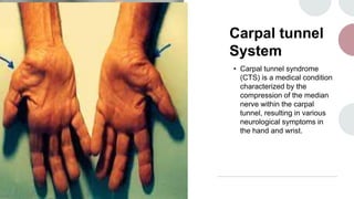
1. Carpal tunnel
System
• Carpal tunnel syndrome
(CTS) is a medical condition
characterized by the
compression of the median
nerve within the carpal
tunnel, resulting in various
neurological symptoms in
the hand and wrist.
2. • Signs and Symptoms:
• Pain, tingling, and numbness in the
thumb, index, middle, and ring
fingers.Weakness in the hand,
leading to difficulty gripping
objects.Symptoms often worsen at
night or during repetitive hand
movements.
3. Causes
Repetitive hand and wrist
motions, common in certain
occupations.Pregnancy,
hormonal changes, and fluid
retention.Wrist injuries or
fractures.Medical conditions
like diabetes, hypothyroidism,
and rheumatoid arthritis.
4. Pathogenesis
• :The median nerve, responsible
for sensation and movement in
the hand, becomes compressed
due to swelling or
irritation.Pressure on the nerve
within the carpal tunnel leads to
the characteristic
symptoms.Over time, the nerve
compression can result in nerve
damage if left untreated.
5. Management
• Conservative measures include
wrist splints, which help keep the
wrist in a neutral position and
alleviate pressure.Nonsteroidal
anti-inflammatory drugs (NSAIDs)
can help reduce pain and
inflammation.Corticosteroid
injections can provide short-term
relief by reducing inflammation.If
symptoms persist, surgical
intervention (carpal tunnel
release) may be considered to
relieve pressure on the nerve.
6. Rehabilitation
• :Post-surgery,
• patients might need physical therapy to regain
strength and range of motion.
• Gradual exercises to improve grip strength and
wrist flexibility.
• .Remember, treatment plans should be tailored
to each patient's individual condition and needs.
As a doctor, your role involves accurate
diagnosis, considering various factors, and
guiding patients through appropriate treatments
and rehabilitation strategies.
7.
8. Compartment Syndrome:
Compartment syndrome is a medical condition
characterized by increased pressure within a
confined muscle compartment of the body. This
heightened pressure can impede proper blood
circulation, leading to tissue damage and potentially
permanent injury if not promptly addressed.
9. Signs and Symptoms:
• Common signs and symptoms of compartment
syndrome include intense pain that is out of
proportion to the injury, swelling, tightness, a
sensation of fullness, numbness or tingling, and
weakness in the affected area. The pain may
worsen with movement or stretching of the
muscles within the compartment.
10. Causes and Pathogenesis:
• Compartment syndrome can be caused by various
factors, such as trauma from fractures, crush
injuries, or intense physical activity. The condition
arises when pressure within a muscle
compartment increases to a level that restricts
blood flow. This pressure buildup can lead to
ischemia (lack of blood supply) and tissue damage.
11. Investigations
• : Doctors diagnose compartment syndrome based
on clinical evaluation, including assessing
symptoms and physical examination findings.
Compartment pressure measurements can be
taken using specialized devices to determine if
pressure levels are dangerously high. Imaging
techniques like MRI or ultrasound may help
visualize the affected area and identify the extent
of damage.
12. Treatment
• : Immediate treatment is crucial to prevent
further tissue damage. The primary
intervention is a surgical procedure called
fasciotomy. During a fasciotomy, the
surgeon makes incisions in the fascia, the
tough connective tissue surrounding the
muscle compartment, to relieve pressure
and restore blood flow. This surgical release
allows the damaged tissues to recover and
minimizes the risk of permanent damage.
13. Rehabilitation
• : After surgical treatment, rehabilitation
plays a vital role in restoring functionality
and preventing complications. Physical
therapy is often prescribed to help regain
muscle strength, range of motion, and
coordination in the affected area. The
rehabilitation process may involve controlled
exercises and activities tailored to the
patient's specific needs and recovery
progress.
14.
15. Neuropathy
• Neuropathy is a condition characterized by
damage to peripheral nerves, resulting in a variety
of symptoms. These symptoms can range from
pain and tingling to numbness and weakness. The
condition is often associated with various
underlying causes and requires careful diagnosis
and management.
16. Causes
• Neuropathy can arise from different causes,
including diabetes, infections like Lyme disease or
HIV, exposure to toxins such as alcohol or
chemotherapy drugs, autoimmune disorders, and
genetic factors. Identifying the specific cause is
crucial for effective treatment.
17. Sign and symptoms
• Signs of neuropathy manifest
as sensory disturbances,
such as sharp or burning
pain, tingling sensations
(paresthesia), numbness, and
muscle weakness. These
symptoms often occur in the
extremities, starting in the
toes or fingers and spreading
upward.
18. pathogenesis
• Neuropathy's pathogenesis involves damage to
nerve fibers. Inflammation and compromised blood
flow to the nerves can contribute to their
degeneration. This damage disrupts the nerve's
ability to transmit signals properly, leading to the
characteristic symptoms.
19. investigations
• Diagnosing neuropathy requires
comprehensive investigations. Nerve
conduction studies and
electromyography assess nerve and
muscle function. Blood tests can
identify potential underlying causes
like diabetes, vitamin deficiencies, or
infections. These tests help pinpoint
the source of the neuropathy.
20. Treatment
• Treatment strategies vary based on the
underlying cause. Managing the root
cause, such as controlling blood sugar
levels in diabetes, is essential.
Medications like pain relievers, anti-
seizure drugs, or antidepressants can
help manage neuropathic pain.
Physical therapy aims to improve
muscle strength, mobility, and overall
quality of life.
21. Rehabilitation
• plays a crucial role in neuropathy management.
Physiotherapy exercises can help restore nerve
function and strengthen muscles. Pain
management techniques, such as medication
adjustments or topical treatments, alleviate
discomfort. Lifestyle changes like maintaining a
balanced diet and avoiding harmful substances aid
in overall nerve health.
22.
23. Muscular dystrophy
• General Consideration:
• Muscular dystrophy is a group of
genetic disorders characterized by
progressive muscle weakness and
wasting. It's caused by mutations in
genes responsible for producing vital
muscle proteins. These mutations
lead to the breakdown of muscle
fibers and a subsequent decline in
muscle function.
24.
25. Clinical Features
• : The clinical presentation of muscular
dystrophy can vary depending on the
specific type, but common features
include:
• Muscle Weakness: Gradual and
progressive muscle weakness, often
starting in the legs and pelvis and
extending to other muscle groups.
• Motor Difficulties: Difficulty with mobility,
walking, and performing everyday tasks
due to muscle weakness.
26. • Contractures:
• Muscles becoming shorter and tighter, limiting joint
movement and flexibility.
• : Feeling tired and fatigued due to the effort
required for basic movements.
• Muscle Atrophy: Wasting or shrinking of muscles
due to the loss of muscle fibers.
• Respiratory Issues: Some types of muscular
dystrophy can affect the muscles involved in
breathing, leading to respiratory problems.
• Cardiac Involvement: Certain types can impact the
heart muscles, leading to cardiomyopathy and
heart-related issues.
27. Causes and Pathogenesis:
• Muscular dystrophy is primarily caused by mutations in
specific genes that encode for proteins essential for
muscle structure and function. The most common types
of muscular dystrophy are caused by mutations in the
dystrophin gene. Without these crucial proteins, muscle
fibers weaken, become more susceptible to damage,
and struggle to regenerate properly. Over time, this
cycle of muscle breakdown and repair deficiency leads
to progressive muscle wasting and weakness.
28. How to investigate
Diagnosing muscular dystrophy involves a combination of clinical assessments and
tests, including:
Physical Examination: Assessing muscle weakness, range of motion, and other
clinical features.
Medical History: Gathering information about family history and the progression of
symptoms.
Genetic Testing: Identifying mutations in specific genes associated with different
types of muscular dystrophy.
Muscle Biopsy: Removing a small sample of muscle tissue for examination under a
microscope to assess muscle fiber structure and protein levels
Imaging: Techniques like MRI can reveal muscle changes and atrophy
Electromyography (EMG): Measures electrical activity in muscles, helping to assess
muscle and nerve function.
29. Management
• : While there is no cure for muscular dystrophy, management strategies
focus on improving quality of life and slowing disease progression:
• Physical Therapy: Helps maintain muscle strength and flexibility,
preventing contractures.Occupational Therapy: Assists in adapting to
daily tasks and using assistive devices.
• Medications: Some drugs may help manage specific symptoms and
complications.
• Respiratory Support: Breathing aids and devices might be necessary for
those with respiratory involvement.
• Surgery: Orthopedic interventions can address contractures and
scoliosis.
• gene Therapy: Experimental approaches aim to replace or correct faulty
genes in some types of muscular dystrophy.
30. Rehabilitation
• : Rehabilitation is a crucial aspect of managing
muscular dystrophy:
• Physical Therapy: Tailored exercise programs to
maintain muscle strength and mobility
• .Occupational Therapy: Techniques to enhance
independence in daily activities.
• Speech Therapy: For those with muscular
dystrophy affecting speech and swallowing.