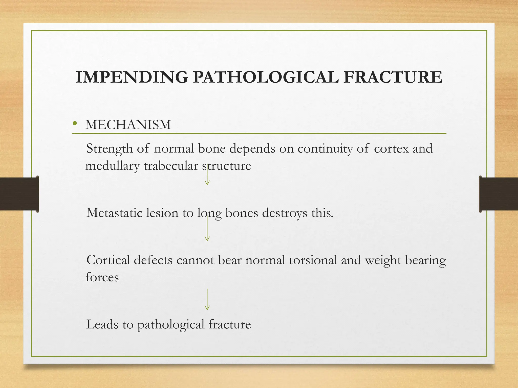
ONCOLOGICAL
EMERGENCIES
PRESENTER : DR MAYA POOJARI
MODERATOR : DR PRATHIMA
FLOW OF PRESENTATION
 DEFINITION
 CLASSIFICATION
 ONCOLOGICAL EMERGENCIES REQUIRING RT
1. SUPERIOR VENA CAVA SYNDROME
2. SPINAL CORD COMPRESSION
3. RAISED INTRACRANIAL PRESSURE
4. IMPENDING PATHOLOGICAL FRACTURE
5. HEMORRHAGIC SOLID TUMORS
 METABOLIC EMERGENCIES
1. TUMOR LYSIS SYNDROME
2. HYPERCALCEMIA
 INFECTIONS - FEBRILE NEUTROPENIA
DEFINITION
“A clinical condition resulting from metabolic ,
neurologic , cardiovascular , hematologic , and /or
infectious change caused by cancer or its treatment
that requires immediate intervention to prevent loss
of life or quality of life.”
ONCOLOGICAL EMERGENCIES
CLASSIFICATION
• METABOLIC
• NEUROLOGIC
• CARDIOVASCULAR
• HEMATOLOGICAL
• INFECTIONS
ONCOLOGICAL EMERGENCIES
CLASSIFICATION
• METABOLIC
 TUMOUR LYSIS SYNDROME
 HYPERCALCEMIA OF MALIGNANCY
 HYPONATREMIA
 LACTIC ACIDOSIS
 CANCER RELATED HEMOLYTIC UREMIC SYNDROME
• NEUROLOGIC
 SPINAL CORD COMPRESSION
 RAISED ICT
 IMPENDING PATHOLOGICAL FRACTURE
 CARDIOVASCULAR
 MALIGNANT PERICARDIAL AND PLEURAL
EFFUSION
 SUPERIOR VENA CAVAL OBSTRUCTION.
• HEMATOLOGICAL
 HYPERVISCOSITY SYNDROME
 BLEEDING.
 HYPERLEUKOCYTOSIS
• INFECTIONS
 FEBRILE NEUTROPENIA
Oncologic emergencies requiring
RT
• Superior vena cava obstruction
• Spinal cord compression
• Raised intracranial pressure
• Impending pathological fracture
• Bleeding solid tumours
Superior vena cava syndrome
• It results from partial or complete obstruction
of blood flow through superior vena cava to
right atrium causing reduction of venous return
from head, neck and upper extremities.
• CAUSES OF SVCO- Either by direct
compression/ invasion or thrombosis
GRADE CATEGORY INCIDEN
CE %
DEFINITION
0 ASYMPTOMATIC 10 RADIOGRAPHIC SVCO IN ABSENCE
OF SYMPTOMS
1 MILD 25 VASCULAR DISTENSION, CYANOSIS,
PLETHORA
2 MODERATE 50 EDEMA IN HEAD OR NECK WITH
FUNCTIONAL IMPAIREMENT
3 SEVERE 10 MILD OR MODERATE CEREBRAL
EDEMA, MILD/MODERATE
LARYNEAL EDEMA OR DIMINISHED
CARDIAC RESERVE
4 LIFE
THREATENING
5 SIGNIFICANT CEREBRAL EDEMA OR
SIGNIFICANT LARYNGEAL EDEMA
OR SIGNIFICANT HEMODYNAMIC
COMPROMISE
5 FATAL <1 DEATH
CLINICAL GRADING SYSTEM
SVCO GRADING( RADIOLOGICAL)
• GRADE 0- SVC narrowing without clinical evidence of SVC syndrome
• GRADE 1-
o Ia- mild SVC narrowing without collaterals
o Ib- moderate SVC narrowing without collaterals.
• GRADE 2- severe SVC narrowing above azygos with azygos vein
serving as partial collateral.
• GRADE 3- severe SVC obstruction below azygos arch.
• GRADE 4- SVC obstruction at the azygos arch.
GRADE 2 GRADE 3
GRADE 4
ETIOLOGY
• MALIGNANCY is the most common cause
1. LUNG CANCER- small cell and squamous histology
2. LYMPHOMA- Non hodgkins lymphoma
3. GERM CELL TUMOUR
4. METASTASIS- breast cancer
5. THYMOMA
6. CATHETER INDUCED
7. OTHERS
• RESTROSTERNAL GOITRE
• HISTOPLASMOSIS
• MEDIASTINAL FIBROSIS
• AORTIC ANEURYSM
• SYPHILIS
SIGNS AND SYMPTOMS
1. DYSPNOEA
2. FACIAL SWELLING
3. HEAD FULLNESS
4. COUGH
5. ARM SWELLING
6. CHEST PAIN
1. VENOUS DISTENSION OF
NECK
2. VENOUS DISTENSION OF
CHEST WALL
3. FACIAL EDEMA
4. CYANOSIS
5. PLETHORA OF FACE
6. EDEMA OF ARMS
ACUTE- Develops within days
SUBACUTE- 6 Weeks
CHRONIC- > 6 Weeks
CLINICAL EVALUATION:
HISTORY AND PHYSICAL
EVALUTION
CHEST X-RAY AND CT SCAN
MALIGNANT SVC
SYNDROME
GRADE 4
SYMPTOMS
TREATMENT
WITHOUT
BIOPSY
CONFIRMATI
ON
GRADE 1,2,3 SYMTOMS
TUMOUR BIOPSY, STAGING, TUMOUR AND STAGE SPECIFIC
TREATMENT PLAN
SURGICALLY
MANAGED
TUMOUR
Eg: thymoma,
retrosternal goitre
CHEMO-
RADIOSENSITIVE
Eg: SCLC,
lymphoma, germ cell
GRADE 1&2
DEFINITIVE
TREATMENT SAME
AS WITHOUT SVCO
GRADE 3
EARLY RT
POOR TREATMENT
OPTIONS
Eg: malignant pleural
mesothelioma
Poor performace status
GRADE 1&2
RT
SUPPORTIV
E CARE
GRADE 3
STENT
MANAGEMENT OF SVCO
• Current management- tissue diagnosis prior to initiation of therapy except in life
threatening situation.
• MEDICAL DECOMPREESION-
• Oxygenation
• Steroids- little evidence to support benefit.
Helpful to reduce symptoms in steroid responsive tumours.
( initiation prior to pathological confirmation can affect accurate diagnosis).
• CHEMOTHERAPY - small cell lung , Lymphoma , germ cell tumour.
 Local consolidation with RT after few cycles of chemotherapy.
 RELIEF OF SVCS- occurs within 1-2 weeks of initiation of therapy
 % of partial symptom relief- 60-77%
MANAGEMENT OF SVCO
• RADIOTHERAPY-
 Mechanism-
• Decreasing pressure on SVC by decreasing surrounding or invading mass
• Development of collaterals after pressure in mediastinum decreased
 Technique - 2D or 3DCRT via AP/PA portal
 Treatment volume
• Definitive ( in absence of distant metastatic disease)- all gross disease
with clinical and planned target volume
• Palliative- Gross disease responsible for SVCO+ 2cm margin.
 Dose- 20Gy/5fr OR 30 Gy / 10Fr
 RELIEF OF SVCS- occurs within 3-30days of initiation of therapy
 % of partial symptom relief- 60-90%
• ENDOVASCULAR STENTING AND
ANGIOPLASTY WITH THROMBOLYSIS.
RELIEF OF SVCS- occurs within 0-72hrs of initiation
of therapy
• % of partial symptom relief-80-95%
• SURGERY- limited role-
sternotomy or thoracotomy with extensive resection of
tumour and reconstruction of SVC.
SPINAL CORD COMPRESSION
• Occurs in three ways
1. Extradural – within spine but outside the dura that covers spinal cord, nerve roots
and spinal fluid .
Examples-
a) Malignant: Metastatis to bone, Ewings sarcoma, Osteosarcoma.
b) Benign: Hemangiomas, Giant cell tumour, Osteoblastoma
2. Intradural extramedullary- within dural sac but outside spinal cord
Examples- Meningomas, Nerve sheath tumours ( Schwannomas and
Neurofibromas)
3. Intradural intramedullary – within spinal cord
Examples- astrocytomas, ependymomas, hemangioblastomas
ETIOLOGY
• Most common malignancy causing
cord compression:
o Multiple Myeloma
o Lung cancer
o Breast cancer
o Prostate cancer
o Non hodgkins Lymphomas
o Neuroblastoma
o Primary vertebral osteosarcoma
o Ewings sarcoma
o Germ cell tumour
o Others- Epidural hematoma, abscess
• Commonest site-
1. Thoracic spine- 70%
common with lung
and breast cancer
2. Lumbar spine- 20%
common with
colorectal and
prostate cancer
3. Cervical and sacral
spine < 10%
MECHANISM OF CORD COMPRESSION
MECHANISM OF CORD
COMPRESSION
1. Direct extension of metastasis of vertebral body to
epidural space.
2. Pathological fracture or spine instability due to metastasis
3. Disease extension within spinal canal
4. Extension of paravertebral lesion
• Direct extension to spinal canal eg:pancoast tumour
• Through vertebral foramen.eg- lymphoma and
neuroblastoma
PATHOGENESIS:
• Impairement of venous drainage
Intramedullary vasogenic edema
Rise in interstitial pressure
Cord perfusion compromised
Necrosis
• In case of Pathological fracture or posterior displacement of bony
fragment mainly due to- Mechanical cord compression
Clinical presentation
• Pain
1. Infiltration of periosteum
2. Infiltration of nerve root
3. Irritation of long tracts of spinal cord
4. Paravertebral muscle spasm
• Neurological symptoms- evovles within weeks to months of
onset of back pain.
• Motor dysfunction- earliest sign, occurs before sensory
disturbance.
Bilsky grading system
• Score 0- bone only disease .
• Score 1 – epidural extension and impingement on the thecal
sac without spinal cord compression.
• Score 2 is epidural extension without spinal cord
compression or obliteration of cerebrospinal fluid space .
• Score 3 is spinal cord compression, deformation of the spinal
cord, and obliteration of cerebrospinal fluid space.
• Grades 2 and 3 represent high-grade MSCC and should be
considered an oncologic emergency.
• Management depends on:
1. Histological type
2. Duration of onset of symptoms
3. Neurological deficits.
4. Age
5. Performance status of patient.
MANAGEMENT
STEROID-
• Facilitate pain relief
• Reduce vasogenic edema
• Prevent additional cord damage
 Dosage- there is no additional benefit by starting 100mg 0r 96mg loading dose
compared to 16 mg OD or 4mg QID( Greenberg et al, Vecht et al, Heimdal et al).
Ambulation rate after treatment was similar- 57.9% vs 57.1%,
Toxicity- 28.6% vs 7.6%.
SURGERY-
• Radioresistant tumours - Posterior laminectomy with stabilisation of vertebral
bodies
RT DOSE- 8Gy/1fr or 20Gy/5fr or 30Gy/1Ofr
• Treatment volume- includes one
vertebrae above and one vertebrae
below the compression.
• REASON-
Vertebra develops from caudal part of
one sclerotoma and cranial portion of
next sclerotome and mesenchymal cells
in between forms intervertebral disc .
RADIOTHERAPY-Mainstay of treatment
Prevents progression
Provides stabilisation
Pain relief.
RAISED INTRACRANIAL PRESSURE
• Mechanism :
1. Direct mass effect- by primary or secondary brain
tumour, peritumoral edema, abscess or hemorrhage
2. Vasogenic edema- increased leakage of plasma
filtrate through leaky capillaries within tumour
3. Cytotoxic edema- breakdown of ATP dependent
membrane ion transport- leads to intracellular
water entrapment .
• MECHANISM :
1. Increase in cerebral parenchymal pressure- cause direct
mass effect
a) Lung cancer
b) Breast cancer
c) Melanoma
d) Renal cell cancer
e) Choriocarcinoma
f) Papillary thyroid cancer
Others
• Herpes simplex encephalitis
• Cerebral toxoplasmosis
• Brain abscess
• Subdural hematoma
2. Cerebral spinal fluid obstruction-
a) Subependymal astrocytoma
b) Thalamic tumours
c) Medulloblastoma
d) Pontine glioma
e) Meningioma
f) Central neurocytoma
g) Ependymoma
h) Meningioma .
3. Venous outflow obstruction-
h) Dural sinus thrombosis
i) Metastasis to base of skull
j) Dural mass lesion occluding sinus
4. Disorders affecting CSF reabsorption-
Protein degradation products or cells released into CSF
might block arachiniod granulation- obstructing reabsorption
• Opportunistic meningeal infection-
Eg: Cryptococcus Neoformans meningitis
• Ependymoma
• Others- schwannoma, meningioma, neurofibroma.
5. Disorders affecting CSF production – Rare
Eg: choroid plexus papilloma
Clinical presentation
1. Headache
2. Early morning heaviness in head
3. Nausea
4. Projectile vomiting
5. Seizures
6. Drowsiness
7. Cognitive dysfunction.
MANAGEMENT
• General measures
1. Assessment and management of airway, breathing and circulation
2. Early intubation if GCS<8, Evidence of herniation, Apnea, inability to
maintain airway
3. Head elevation 15-30 degree
• Steriods
 Dosage – 8mg vs 16mg OD had similar degree of
improvement after 1 week of treatment.
• Hyperosmolar agents-When blood pressure is normal
 Inhibit water reabsorption in proximal convoluted
tubules and thin loop of henle
 Extracts water from intracellular compartment
increasing extracellular volume.
• Hypertonic saline-In case of hypotension, hypovolemia,
serum osmolality >320mOsm/kg, renal failure
• SURGERY- SOLITARY brain metastasis
• Anti-edema measures should be started before
initiating RT.
• RADIOTHERAPY- Mainstay of treatment:
1. Method- whole brain RT with 2 lateral opposed
fields
2. Dose- 20Gy/5fr or 30Gy/10fr.
IMPENDING PATHOLOGICAL FRACTURE
• MECHANISM
Strength of normal bone depends on continuity of cortex and
medullary trabecular structure
Metastatic lesion to long bones destroys this.
Cortical defects cannot bear normal torsional and weight bearing
forces
Leads to pathological fracture
MIREL’S SCORING SYSTEM
MIREL’S SCORE CLINICAL
RECOMMENDATION
≤7 Radiotherapy & observation
8 Use clinical judgement
≥ 9 Prophylactic fixation
MIREL’S CLINICAL RECOMMENDATION
• In imminent fracture
1. Immobilisation of affected bone
2. Followed by radiotherapy
• In frank pathological fracture-
1. Skeletal instability should be corrected with open or close reduction.
2. Radiotherapy to inhibit further tumour growth and avoid bone
destruction.
• RT DOSE – 8 Gy / 1 fr or 20 Gy / 5 Fr.
HEMORRAGIC SOLID
TUMOURS
• Most common in carcinoma of cervix , rectum ,
bladder , bronchus .
• Mechanism-
1. Mechanical trauma to fragile neovasculature within
tumour.
2. Extension of tumour to surrounding organ,
muscles or overlying skin.
Management
1. Replace loss in fluid compartment with intravenous fluids
and blood transfusion.
2. Immediate mechanical pressure to assessible bleeding
tumour sites.
3. Radiotherapy- acts by
• Compression of capillaries
• Shrinkage of tumour
• DOSE- 8Gy in single fraction or 20Gy/5fr or 30Gy/10fr.
TUMOR LYSIS SYNDROME
• Tumor lysis syndrome ( TLS) is the constellation of
metabolic abnormalities that occurs due to tumor cell
death with the release of intracellular contents .
ETIOLOGY
• Rapidly growing
• Chemosensitive
• Hematologic malignancies :
AML -3-4% , ALL – 5.2 % and NHL – 6.1%
Mortality rate of 0.9 %.
• High tumor burden ( bulky adenopathy, organomegaly , bulky
metastatic disease )
• Ionizing radiation including TBI in the transplant setting ,
Embolization , RFA , monoclonal antibody therapy , glucocorticoids .
DYNAMICS OF TUMOR LYSIS SYNDROME
CLASSIFICATION – CAIRO AND BISHOP
Classified into two groups :
1. Laboratory
2. Clinical
TREATMENT OF HYPERURICEMIA
HYPERCALCEMIA
• Occurs in 20% to 30% of all cancer patients at some
point in their disease course.
• Confers a poor prognosis – 80% of patients die
within 30 days .
• Most commonly seen in breast , lung , multiple
myeloma , lymphoma , prostate cancer.
• Hypercalcemia can be divided into the following four
types:
1. Local osteolytic hypercalcemia.
2. Humoral hypercalcemia of malignancy (secretion of
parathyroid hormone–related peptide).
3. 1,25-Dihydroxyvitamin D–secreting lymphomas .
4. Ectopic hyperparathyroidism .
PATHOGENESIS
SYMPTOMS OF HYPERCALCEMIA
• The well-known mnemonic “stones, bones, moans,
abdominal groans, and psychiatric overtones.”
• STONES – nephrolithiasis , renal insufficiency , polyuria .
• BONES – bone pain , weakness , myopathy .
• ABDOMINAL GROANS – constipation , anorexia ,
nausea , vomiting
• MOANS – CNS - hypertonia , confusion , ataxia , coma .
• PSYCHIATRIC OVERTONES – psychosis , depression .
INVESTIGATIONS
 CBP
 SR ELECTROLYTES – Ca, PO4 , Mg
 Always count for corrected calcium levels
 Corrected calcium = S. calcium + (4 – S.albumin ) x 0.8
 Ionised calcium is more important than total calcium .
 LFT & Sr proteins
 ECG
 PLASMA PTH & PTHrP.
TREATMENT OF
HYPERCALCEMIA
Hydration with normal saline (3 – 6L).
Bisphosphonates
RANK L inhibitors
Loop diuretics
Calcitonin ( 6- 8 U /kg SC or IM )
Dialysis if necessary .
BISPHOSPHONATES IN
HYPERCALCEMIA
 Zolendronate – 4 mg iv over 30 min
 Normalization of serum calcium in 4 to 10 days that lasts for 4 to 6 weeks in
90% patients
 Not much effective in PTHrP mediated hypercalcemia – calcitonin can be used
in refractory hypercalcemia .
FEBRILE NEUTROPENIA
• DEFINITION :
• FEVER : single oral temperature of 101F (38.3 C)
OR
- oral temp of 100.4 F (38 C ) lasting > 1 hr
• NEUTROPENIA :
- ANC <500/mm3
OR
- count of <1000 cells /mm3 with a predicted decrease to <500 cells
/ mm3 .
FACTORS INFLUENCING
RISK OF INFECTION
• Breach of skin and mucosal barriers :
1. IV Access devices
2. Mucositis
3. Surgery
4. Tumor growth
INITIAL EVALUATION
• HISTORY : time since last chemotherapy administration , major co
morbid illness , travel , H/O prior documented infections , etc .
• PHYSICAL EXAMINATION : to find any focus of infection
- IV access site , oropharynx , nasal cavity , skin including perivaginal
& perineal regions
• INVESTIGATIONS :
- CXR
- CBC , ELECTROLYTES , RFT , LFT
- BLOOD CULTURE – 2 SETS
- THROAT / WOUND SWAB AS INDICATED
- URINE / STOOL CULTURE ACC TO SYMPTOMS
IDSA RISK STRATIFICATION
HIGH RISK
• Inpatients
• Associated comorbidities
( hypotension , dehydration ,
hypoxia )
• Uncontrolled / progressive
cancer
• Sr. creat > 2mg/dl
• LFT >3 times normal
• HSCT /BMT Recipient
• Prolonged severe neutropenia
anticipated
LOW RISK
• Outpatients
• No associated comorbidities
• Good ps ( ECOG 0-1)
• Sr. creat <2 mg/dl
• LFT <3 times normal
• Non transplant , solid
tumor or lymphoma patient
.
• Anticipated duration of
neutropenia < 7 days.
MASCC SCORE
• Illness ( no or mild symptoms = 5 points ; severe = 3 )
• Absence of hypotension (5 points)
• COPD (4 points )
• Solid tumor or no previous fungal infection (4 points)
• Absence of dehydration (3 points)
• Outpatient status (3 points)
• Age < 60years (2 points)
• MASCC score >- 21 points ( of 26) are low risk & can be
considered for oral therapy .
HYPONATREMIA
MANAGEMENT
CRITERIA FOR SIADH
THANK YOU

ONCOLOGICAL_EMERGENCIES - it's management.pptx

  • 1.
    ONCOLOGICAL EMERGENCIES PRESENTER : DRMAYA POOJARI MODERATOR : DR PRATHIMA
  • 2.
    FLOW OF PRESENTATION DEFINITION  CLASSIFICATION  ONCOLOGICAL EMERGENCIES REQUIRING RT 1. SUPERIOR VENA CAVA SYNDROME 2. SPINAL CORD COMPRESSION 3. RAISED INTRACRANIAL PRESSURE 4. IMPENDING PATHOLOGICAL FRACTURE 5. HEMORRHAGIC SOLID TUMORS  METABOLIC EMERGENCIES 1. TUMOR LYSIS SYNDROME 2. HYPERCALCEMIA  INFECTIONS - FEBRILE NEUTROPENIA
  • 3.
    DEFINITION “A clinical conditionresulting from metabolic , neurologic , cardiovascular , hematologic , and /or infectious change caused by cancer or its treatment that requires immediate intervention to prevent loss of life or quality of life.”
  • 4.
    ONCOLOGICAL EMERGENCIES CLASSIFICATION • METABOLIC •NEUROLOGIC • CARDIOVASCULAR • HEMATOLOGICAL • INFECTIONS
  • 5.
    ONCOLOGICAL EMERGENCIES CLASSIFICATION • METABOLIC TUMOUR LYSIS SYNDROME  HYPERCALCEMIA OF MALIGNANCY  HYPONATREMIA  LACTIC ACIDOSIS  CANCER RELATED HEMOLYTIC UREMIC SYNDROME • NEUROLOGIC  SPINAL CORD COMPRESSION  RAISED ICT  IMPENDING PATHOLOGICAL FRACTURE
  • 6.
     CARDIOVASCULAR  MALIGNANTPERICARDIAL AND PLEURAL EFFUSION  SUPERIOR VENA CAVAL OBSTRUCTION. • HEMATOLOGICAL  HYPERVISCOSITY SYNDROME  BLEEDING.  HYPERLEUKOCYTOSIS • INFECTIONS  FEBRILE NEUTROPENIA
  • 7.
    Oncologic emergencies requiring RT •Superior vena cava obstruction • Spinal cord compression • Raised intracranial pressure • Impending pathological fracture • Bleeding solid tumours
  • 8.
    Superior vena cavasyndrome • It results from partial or complete obstruction of blood flow through superior vena cava to right atrium causing reduction of venous return from head, neck and upper extremities. • CAUSES OF SVCO- Either by direct compression/ invasion or thrombosis
  • 10.
    GRADE CATEGORY INCIDEN CE% DEFINITION 0 ASYMPTOMATIC 10 RADIOGRAPHIC SVCO IN ABSENCE OF SYMPTOMS 1 MILD 25 VASCULAR DISTENSION, CYANOSIS, PLETHORA 2 MODERATE 50 EDEMA IN HEAD OR NECK WITH FUNCTIONAL IMPAIREMENT 3 SEVERE 10 MILD OR MODERATE CEREBRAL EDEMA, MILD/MODERATE LARYNEAL EDEMA OR DIMINISHED CARDIAC RESERVE 4 LIFE THREATENING 5 SIGNIFICANT CEREBRAL EDEMA OR SIGNIFICANT LARYNGEAL EDEMA OR SIGNIFICANT HEMODYNAMIC COMPROMISE 5 FATAL <1 DEATH CLINICAL GRADING SYSTEM
  • 11.
    SVCO GRADING( RADIOLOGICAL) •GRADE 0- SVC narrowing without clinical evidence of SVC syndrome • GRADE 1- o Ia- mild SVC narrowing without collaterals o Ib- moderate SVC narrowing without collaterals. • GRADE 2- severe SVC narrowing above azygos with azygos vein serving as partial collateral. • GRADE 3- severe SVC obstruction below azygos arch. • GRADE 4- SVC obstruction at the azygos arch.
  • 12.
    GRADE 2 GRADE3 GRADE 4
  • 13.
    ETIOLOGY • MALIGNANCY isthe most common cause 1. LUNG CANCER- small cell and squamous histology 2. LYMPHOMA- Non hodgkins lymphoma 3. GERM CELL TUMOUR 4. METASTASIS- breast cancer 5. THYMOMA 6. CATHETER INDUCED 7. OTHERS • RESTROSTERNAL GOITRE • HISTOPLASMOSIS • MEDIASTINAL FIBROSIS • AORTIC ANEURYSM • SYPHILIS
  • 14.
    SIGNS AND SYMPTOMS 1.DYSPNOEA 2. FACIAL SWELLING 3. HEAD FULLNESS 4. COUGH 5. ARM SWELLING 6. CHEST PAIN 1. VENOUS DISTENSION OF NECK 2. VENOUS DISTENSION OF CHEST WALL 3. FACIAL EDEMA 4. CYANOSIS 5. PLETHORA OF FACE 6. EDEMA OF ARMS ACUTE- Develops within days SUBACUTE- 6 Weeks CHRONIC- > 6 Weeks
  • 17.
    CLINICAL EVALUATION: HISTORY ANDPHYSICAL EVALUTION CHEST X-RAY AND CT SCAN MALIGNANT SVC SYNDROME GRADE 4 SYMPTOMS TREATMENT WITHOUT BIOPSY CONFIRMATI ON GRADE 1,2,3 SYMTOMS TUMOUR BIOPSY, STAGING, TUMOUR AND STAGE SPECIFIC TREATMENT PLAN SURGICALLY MANAGED TUMOUR Eg: thymoma, retrosternal goitre CHEMO- RADIOSENSITIVE Eg: SCLC, lymphoma, germ cell GRADE 1&2 DEFINITIVE TREATMENT SAME AS WITHOUT SVCO GRADE 3 EARLY RT POOR TREATMENT OPTIONS Eg: malignant pleural mesothelioma Poor performace status GRADE 1&2 RT SUPPORTIV E CARE GRADE 3 STENT
  • 18.
    MANAGEMENT OF SVCO •Current management- tissue diagnosis prior to initiation of therapy except in life threatening situation. • MEDICAL DECOMPREESION- • Oxygenation • Steroids- little evidence to support benefit. Helpful to reduce symptoms in steroid responsive tumours. ( initiation prior to pathological confirmation can affect accurate diagnosis). • CHEMOTHERAPY - small cell lung , Lymphoma , germ cell tumour.  Local consolidation with RT after few cycles of chemotherapy.  RELIEF OF SVCS- occurs within 1-2 weeks of initiation of therapy  % of partial symptom relief- 60-77%
  • 19.
    MANAGEMENT OF SVCO •RADIOTHERAPY-  Mechanism- • Decreasing pressure on SVC by decreasing surrounding or invading mass • Development of collaterals after pressure in mediastinum decreased  Technique - 2D or 3DCRT via AP/PA portal  Treatment volume • Definitive ( in absence of distant metastatic disease)- all gross disease with clinical and planned target volume • Palliative- Gross disease responsible for SVCO+ 2cm margin.  Dose- 20Gy/5fr OR 30 Gy / 10Fr  RELIEF OF SVCS- occurs within 3-30days of initiation of therapy  % of partial symptom relief- 60-90%
  • 20.
    • ENDOVASCULAR STENTINGAND ANGIOPLASTY WITH THROMBOLYSIS. RELIEF OF SVCS- occurs within 0-72hrs of initiation of therapy • % of partial symptom relief-80-95% • SURGERY- limited role- sternotomy or thoracotomy with extensive resection of tumour and reconstruction of SVC.
  • 22.
    SPINAL CORD COMPRESSION •Occurs in three ways 1. Extradural – within spine but outside the dura that covers spinal cord, nerve roots and spinal fluid . Examples- a) Malignant: Metastatis to bone, Ewings sarcoma, Osteosarcoma. b) Benign: Hemangiomas, Giant cell tumour, Osteoblastoma 2. Intradural extramedullary- within dural sac but outside spinal cord Examples- Meningomas, Nerve sheath tumours ( Schwannomas and Neurofibromas) 3. Intradural intramedullary – within spinal cord Examples- astrocytomas, ependymomas, hemangioblastomas
  • 23.
    ETIOLOGY • Most commonmalignancy causing cord compression: o Multiple Myeloma o Lung cancer o Breast cancer o Prostate cancer o Non hodgkins Lymphomas o Neuroblastoma o Primary vertebral osteosarcoma o Ewings sarcoma o Germ cell tumour o Others- Epidural hematoma, abscess • Commonest site- 1. Thoracic spine- 70% common with lung and breast cancer 2. Lumbar spine- 20% common with colorectal and prostate cancer 3. Cervical and sacral spine < 10%
  • 24.
    MECHANISM OF CORDCOMPRESSION
  • 25.
    MECHANISM OF CORD COMPRESSION 1.Direct extension of metastasis of vertebral body to epidural space. 2. Pathological fracture or spine instability due to metastasis 3. Disease extension within spinal canal 4. Extension of paravertebral lesion • Direct extension to spinal canal eg:pancoast tumour • Through vertebral foramen.eg- lymphoma and neuroblastoma
  • 26.
    PATHOGENESIS: • Impairement ofvenous drainage Intramedullary vasogenic edema Rise in interstitial pressure Cord perfusion compromised Necrosis • In case of Pathological fracture or posterior displacement of bony fragment mainly due to- Mechanical cord compression
  • 27.
    Clinical presentation • Pain 1.Infiltration of periosteum 2. Infiltration of nerve root 3. Irritation of long tracts of spinal cord 4. Paravertebral muscle spasm • Neurological symptoms- evovles within weeks to months of onset of back pain. • Motor dysfunction- earliest sign, occurs before sensory disturbance.
  • 28.
    Bilsky grading system •Score 0- bone only disease . • Score 1 – epidural extension and impingement on the thecal sac without spinal cord compression. • Score 2 is epidural extension without spinal cord compression or obliteration of cerebrospinal fluid space . • Score 3 is spinal cord compression, deformation of the spinal cord, and obliteration of cerebrospinal fluid space. • Grades 2 and 3 represent high-grade MSCC and should be considered an oncologic emergency.
  • 30.
    • Management dependson: 1. Histological type 2. Duration of onset of symptoms 3. Neurological deficits. 4. Age 5. Performance status of patient.
  • 31.
    MANAGEMENT STEROID- • Facilitate painrelief • Reduce vasogenic edema • Prevent additional cord damage  Dosage- there is no additional benefit by starting 100mg 0r 96mg loading dose compared to 16 mg OD or 4mg QID( Greenberg et al, Vecht et al, Heimdal et al). Ambulation rate after treatment was similar- 57.9% vs 57.1%, Toxicity- 28.6% vs 7.6%. SURGERY- • Radioresistant tumours - Posterior laminectomy with stabilisation of vertebral bodies
  • 32.
    RT DOSE- 8Gy/1fror 20Gy/5fr or 30Gy/1Ofr • Treatment volume- includes one vertebrae above and one vertebrae below the compression. • REASON- Vertebra develops from caudal part of one sclerotoma and cranial portion of next sclerotome and mesenchymal cells in between forms intervertebral disc . RADIOTHERAPY-Mainstay of treatment Prevents progression Provides stabilisation Pain relief.
  • 34.
    RAISED INTRACRANIAL PRESSURE •Mechanism : 1. Direct mass effect- by primary or secondary brain tumour, peritumoral edema, abscess or hemorrhage 2. Vasogenic edema- increased leakage of plasma filtrate through leaky capillaries within tumour 3. Cytotoxic edema- breakdown of ATP dependent membrane ion transport- leads to intracellular water entrapment .
  • 35.
    • MECHANISM : 1.Increase in cerebral parenchymal pressure- cause direct mass effect a) Lung cancer b) Breast cancer c) Melanoma d) Renal cell cancer e) Choriocarcinoma f) Papillary thyroid cancer Others • Herpes simplex encephalitis • Cerebral toxoplasmosis • Brain abscess • Subdural hematoma
  • 36.
    2. Cerebral spinalfluid obstruction- a) Subependymal astrocytoma b) Thalamic tumours c) Medulloblastoma d) Pontine glioma e) Meningioma f) Central neurocytoma g) Ependymoma h) Meningioma . 3. Venous outflow obstruction- h) Dural sinus thrombosis i) Metastasis to base of skull j) Dural mass lesion occluding sinus
  • 37.
    4. Disorders affectingCSF reabsorption- Protein degradation products or cells released into CSF might block arachiniod granulation- obstructing reabsorption • Opportunistic meningeal infection- Eg: Cryptococcus Neoformans meningitis • Ependymoma • Others- schwannoma, meningioma, neurofibroma. 5. Disorders affecting CSF production – Rare Eg: choroid plexus papilloma
  • 38.
    Clinical presentation 1. Headache 2.Early morning heaviness in head 3. Nausea 4. Projectile vomiting 5. Seizures 6. Drowsiness 7. Cognitive dysfunction.
  • 39.
    MANAGEMENT • General measures 1.Assessment and management of airway, breathing and circulation 2. Early intubation if GCS<8, Evidence of herniation, Apnea, inability to maintain airway 3. Head elevation 15-30 degree
  • 40.
    • Steriods  Dosage– 8mg vs 16mg OD had similar degree of improvement after 1 week of treatment. • Hyperosmolar agents-When blood pressure is normal  Inhibit water reabsorption in proximal convoluted tubules and thin loop of henle  Extracts water from intracellular compartment increasing extracellular volume. • Hypertonic saline-In case of hypotension, hypovolemia, serum osmolality >320mOsm/kg, renal failure • SURGERY- SOLITARY brain metastasis
  • 41.
    • Anti-edema measuresshould be started before initiating RT. • RADIOTHERAPY- Mainstay of treatment: 1. Method- whole brain RT with 2 lateral opposed fields 2. Dose- 20Gy/5fr or 30Gy/10fr.
  • 42.
    IMPENDING PATHOLOGICAL FRACTURE •MECHANISM Strength of normal bone depends on continuity of cortex and medullary trabecular structure Metastatic lesion to long bones destroys this. Cortical defects cannot bear normal torsional and weight bearing forces Leads to pathological fracture
  • 43.
  • 44.
    MIREL’S SCORE CLINICAL RECOMMENDATION ≤7Radiotherapy & observation 8 Use clinical judgement ≥ 9 Prophylactic fixation MIREL’S CLINICAL RECOMMENDATION
  • 45.
    • In imminentfracture 1. Immobilisation of affected bone 2. Followed by radiotherapy • In frank pathological fracture- 1. Skeletal instability should be corrected with open or close reduction. 2. Radiotherapy to inhibit further tumour growth and avoid bone destruction. • RT DOSE – 8 Gy / 1 fr or 20 Gy / 5 Fr.
  • 46.
    HEMORRAGIC SOLID TUMOURS • Mostcommon in carcinoma of cervix , rectum , bladder , bronchus . • Mechanism- 1. Mechanical trauma to fragile neovasculature within tumour. 2. Extension of tumour to surrounding organ, muscles or overlying skin.
  • 47.
    Management 1. Replace lossin fluid compartment with intravenous fluids and blood transfusion. 2. Immediate mechanical pressure to assessible bleeding tumour sites. 3. Radiotherapy- acts by • Compression of capillaries • Shrinkage of tumour • DOSE- 8Gy in single fraction or 20Gy/5fr or 30Gy/10fr.
  • 48.
    TUMOR LYSIS SYNDROME •Tumor lysis syndrome ( TLS) is the constellation of metabolic abnormalities that occurs due to tumor cell death with the release of intracellular contents .
  • 49.
    ETIOLOGY • Rapidly growing •Chemosensitive • Hematologic malignancies : AML -3-4% , ALL – 5.2 % and NHL – 6.1% Mortality rate of 0.9 %. • High tumor burden ( bulky adenopathy, organomegaly , bulky metastatic disease ) • Ionizing radiation including TBI in the transplant setting , Embolization , RFA , monoclonal antibody therapy , glucocorticoids .
  • 50.
    DYNAMICS OF TUMORLYSIS SYNDROME
  • 51.
    CLASSIFICATION – CAIROAND BISHOP Classified into two groups : 1. Laboratory 2. Clinical
  • 53.
  • 55.
    HYPERCALCEMIA • Occurs in20% to 30% of all cancer patients at some point in their disease course. • Confers a poor prognosis – 80% of patients die within 30 days . • Most commonly seen in breast , lung , multiple myeloma , lymphoma , prostate cancer.
  • 56.
    • Hypercalcemia canbe divided into the following four types: 1. Local osteolytic hypercalcemia. 2. Humoral hypercalcemia of malignancy (secretion of parathyroid hormone–related peptide). 3. 1,25-Dihydroxyvitamin D–secreting lymphomas . 4. Ectopic hyperparathyroidism .
  • 57.
  • 58.
    SYMPTOMS OF HYPERCALCEMIA •The well-known mnemonic “stones, bones, moans, abdominal groans, and psychiatric overtones.” • STONES – nephrolithiasis , renal insufficiency , polyuria . • BONES – bone pain , weakness , myopathy . • ABDOMINAL GROANS – constipation , anorexia , nausea , vomiting • MOANS – CNS - hypertonia , confusion , ataxia , coma . • PSYCHIATRIC OVERTONES – psychosis , depression .
  • 59.
    INVESTIGATIONS  CBP  SRELECTROLYTES – Ca, PO4 , Mg  Always count for corrected calcium levels  Corrected calcium = S. calcium + (4 – S.albumin ) x 0.8  Ionised calcium is more important than total calcium .  LFT & Sr proteins  ECG  PLASMA PTH & PTHrP.
  • 60.
    TREATMENT OF HYPERCALCEMIA Hydration withnormal saline (3 – 6L). Bisphosphonates RANK L inhibitors Loop diuretics Calcitonin ( 6- 8 U /kg SC or IM ) Dialysis if necessary .
  • 61.
    BISPHOSPHONATES IN HYPERCALCEMIA  Zolendronate– 4 mg iv over 30 min  Normalization of serum calcium in 4 to 10 days that lasts for 4 to 6 weeks in 90% patients  Not much effective in PTHrP mediated hypercalcemia – calcitonin can be used in refractory hypercalcemia .
  • 62.
    FEBRILE NEUTROPENIA • DEFINITION: • FEVER : single oral temperature of 101F (38.3 C) OR - oral temp of 100.4 F (38 C ) lasting > 1 hr • NEUTROPENIA : - ANC <500/mm3 OR - count of <1000 cells /mm3 with a predicted decrease to <500 cells / mm3 .
  • 63.
    FACTORS INFLUENCING RISK OFINFECTION • Breach of skin and mucosal barriers : 1. IV Access devices 2. Mucositis 3. Surgery 4. Tumor growth
  • 64.
    INITIAL EVALUATION • HISTORY: time since last chemotherapy administration , major co morbid illness , travel , H/O prior documented infections , etc . • PHYSICAL EXAMINATION : to find any focus of infection - IV access site , oropharynx , nasal cavity , skin including perivaginal & perineal regions • INVESTIGATIONS : - CXR - CBC , ELECTROLYTES , RFT , LFT - BLOOD CULTURE – 2 SETS - THROAT / WOUND SWAB AS INDICATED - URINE / STOOL CULTURE ACC TO SYMPTOMS
  • 65.
    IDSA RISK STRATIFICATION HIGHRISK • Inpatients • Associated comorbidities ( hypotension , dehydration , hypoxia ) • Uncontrolled / progressive cancer • Sr. creat > 2mg/dl • LFT >3 times normal • HSCT /BMT Recipient • Prolonged severe neutropenia anticipated LOW RISK • Outpatients • No associated comorbidities • Good ps ( ECOG 0-1) • Sr. creat <2 mg/dl • LFT <3 times normal • Non transplant , solid tumor or lymphoma patient . • Anticipated duration of neutropenia < 7 days.
  • 66.
    MASCC SCORE • Illness( no or mild symptoms = 5 points ; severe = 3 ) • Absence of hypotension (5 points) • COPD (4 points ) • Solid tumor or no previous fungal infection (4 points) • Absence of dehydration (3 points) • Outpatient status (3 points) • Age < 60years (2 points) • MASCC score >- 21 points ( of 26) are low risk & can be considered for oral therapy .
  • 68.
  • 70.
  • 71.
  • 75.