Ocular Drug Delivery System.
•Download as PPT, PDF•
45 likes•4,535 views

The ocular drug delivery system.

Recommended
More Related Content
What's hot
What's hot (20)
Viewers also liked
Viewers also liked (20)
Similar to Ocular Drug Delivery System.
Similar to Ocular Drug Delivery System. (20)
Recently uploaded
Recently uploaded (20)
Ocular Drug Delivery System.
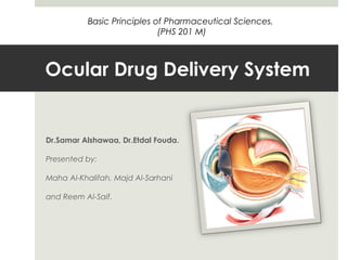
- 1. Ocular Drug Delivery System Dr.Samar Alshawaa, Dr.Etdal Fouda. Presented by: Maha Al-Khalifah, Majd Al-Sarhani and Reem Al-Saif. Basic Principles of Pharmaceutical Sciences. (PHS 201 M)
- 2. Table of Contents Introduction Structure of the eye. Eye layers. Lacrimal apparatus. Composition of the the eye. Mechanism of Drug Absorption. Barriers avoiding drug delivery. Bioavailability. Pharmacokinetics. Pharmacodynamics.
- 3. Introduction The eye is one of the most complex organs in the human body. The eye-ball is an organ protected from exogenous substances and external stress by various barriers. • Therefore, therapeutic drugs must be transported across several protective barriers regardless of which administration route is utilized, such as eye-drops, and subconjunctival, sub-tenon’s and intravitreal injection and/or implant. • More than 75% of applied ophthalmic solution is lost via nasolachrymal drainage and absorbed systemically via conjunctiva, hence ocular drug availability is very low .
- 4. Introduction To increase ocular bioavailability and prolong the retention time on the ocular surface, numerous ophthalmic vehicles such as viscous solutions, suspensions, emulsions, ointments, aqueous gels, and polymeric inserts, have been investigated for topical application to the eye. • Topical applied drugs do not reach the posterior segment of the eye (retina, vitreous, choroid), therefore, systemic administration, periocular or intraocular injections of drugs are normally applied in clinical therapeutics. Currently there is also rapidly growing interest in drug delivery systems (DDSs) to the posterior segment of the eye. This trend is toward a polymeric depot system implanted or injected directly into the vitreous, to obtain long-term, sustained release of drugs.
- 5. Structure of the eye
- 6. Structure of the eye Eyelids: The eyelids contain skeletal muscle that enable the eyelids to close and cover the front of the eye ball. Eyelashes along the border of each eyelid to help keep the dust away. The eyelids are lined with a thin membrane called conjunctiva, which is folded over the white of the eye and merges with the corneal epithelium. Eyeball : most of the eyeball is within and protected by an orbit formed by the lacrimal, maxilla, zygomatic, frontal, sphenoid and ethmoid bones. Six extrinsic muscles attached to this bony socket and to the surface of the eyeball.
- 7. Eye layers it has three layers: the outer sclera, middle choroid layer, and inner retina. The sclera is the thickest layer that is visible as white of the eye. The most interior portion is the cornea, which differs from the rest of the sclera in that it is transparent. the outermost layer of the eye and is made up of five layers of tissue itself. The cornea is clear, which allows light to enter through the pupil to shine on the retina. The cornea also helps protect the eye fro things like dirt and bacteria. The uvual tract is the middle layer of the eye and contains the iris, choroid, and ciliary body.
- 8. Eye layers The iris is the colored part of the eye and is made of muscles. These muscles contract and release to allow the proper amount of light through the pupil. The choroid contains blood vessels and is the main supply of blood to the eye. The ciliary body is where the clear liquid that coats the eye is formed. The retina is the layer at the back of the eye. This is where the photorecepters (rods and cones) are located. Light is reflected onto the retina through the pupil. The optic nerve is attached to the back of the retina, and this is how our brain gets the information from our eyes.
- 9. Eye Cavities There are two cavities within the eye: posterior and anterior. The posterior is found between the lens and the retina and contains vitreous humor. The anterior is found between the back of the cornea and the front of the lens, and contains aqueous humor.
- 10. Lacrimal apparatus Tears are produced by lacrimal glands, located at the upper, outer corner of the eyeball, within the orbit. Secretion of tears occur constantly, but increased by the presence of irritating chemical. Such as, onion vapor and dust or in certain emotional situations (sad or happy). Small ducts take tears to the anterior of the eyeball, and blinking spreads the tears and washes the surface of the eye.
- 11. Lacrimal apparatus Tears are mostly water with 1% sodium chloride. Similar to other body fluid. Tears contain lysozyme, an enzyme that inhibits the growth of most bacteria on the wet warm surface of the eye. At the medial corner of the eyelids are two small openings into the superior and inferior lacrimal canals. Theses ducts takes tears to the lacrimal sac (in the lacrimal bone) which leads to the nasolacrimal duct, which empties tears into the nasal cavity.
- 12. Composition of the eye Water – 98% Solid – 1.8% Organic elements – (protien – 0.67%),(Sugar – 0.65%), (NaCl - 0.66%). Other mineral elements – Sodium, Potassium and ammonia- 0.79%.
- 13. Routes Of Drug Delivery in The eye
- 14. Mechanism of Drug Absorption Since most eye medications need to enter the eye for a pharmacological effect. Generally three routes into the eye have been described :
- 15. Drug distribution in the eye through the blood vessels Either by systemic dosing or by local effect. The blood route always involves a distribution of the drug in the whole human body. The ocular level of drugs achieved by this way of application is much less than achievable by ocular topical treatment. Thus, this way of drug distribution is not preferred for ocular dosing.
- 16. Drug entry into the eye region utilizing the conjunctival-scleral pathway With Topical dosing. Even though this pathway can be of high efficiency for intraocular drug delivery and does bypass cornea and local vasculature. Only a few (high molecular) substances were delivered using this route.
- 17. The Transcorneal Route This is still the major pathway for ocular drug delivery. Representing the direct pathway into the eye and is applicable for most drug substances.
- 18. Mechanism of Drug Absorption Cornea is the major pathway for drug absorption into the eye. Certain physicochemical properties are required for the substance to cross the corneal barrier. • Cornea is divided into three parts with different characteristics. • Only amphiphilic substances can easily penetrate all corneal layers. • Purely lipophilic can pass the epithelial and endothelial cell layers. • But will be unable to cross the aqueous stroma.
- 19. Mechanism of Drug Absorption Same consideration also applies to purely hydrophilic drugs, not withheld from penetration by the stroma. But unable to cross the lipid bilayers of cells, and a tight junctions in the epithelium and endothelium
- 20. Barriers avoiding drug delivery The greatest barrier to drug penetration is The Corneal Epithelium. Rich in cellular membranes, and is therefore more susceptible to penetration by drugs which are lipophilic. The conjunctiva has similar permeability characteristics to the corneal epithelium. Since it is such a vascular structure the majority of drug that penetrates the corneal epithelium does not penetrate the eye per se but is drained into the systemic circulation.
- 21. Barriers avoiding drug delivery Tear One of the precorneal barriers is Tear Film which reduces the effective concentration of the administrated drugs due to dilution by the tear turnover (approximately 1 uL/min), accelerated clearance, and binding of the drug molecule to the tear proteins. In addition the dosing volume of instillation is usually 20–50 uL whereas the size of cul- de-sac is only 7–10 uL. The excess volume may spill out on the cheek or exit through the nasolacrimal duct. Conjunctiva Conjunctiva of the eyelids and globe is a thin and transparent membrane, which is involved in the formation and maintenance of the tear film. In addition, conjunctiva or episclera has a rich supply of capillaries and lymphatics, therefore, administrated drugs in the conjunctival or episcleral space may be cleared through blood and lymph. The conjunctival blood vessels do not form a tight junction barrier, which means drug molecules can enter into the blood circulation by convective transport through paracellular pores in the vascular endothelial layer.
- 22. Barriers avoiding drug delivery Other Barriers: Choroid/Bruch’s Membrane. Retina. Blood-Retinal Barrier. Sclera.
- 23. Bioavailability Conventional systems like eye drops, suspensions and ointments cannot be considered optimal in the treatment of vision threatening ocular diseases. However, more than 90% of the marketed ophthalmic formulations are in the form of eye drops. These formulations mainly target the anterior segment eye diseases. Ocular drug delivery has remained as one of the most challenging task for pharmaceutical scientists. The unique structure of the eye restricts the entry of drug molecules at the required site of action.
- 24. Bioavailability Most of the topically applied drugs are washed off from the eye by various mechanisms: 1. Lacrimation. 2. Tear dilution. 3. Tear turnover Resulting in low ocular bioavailability of drugs. human cornea comprising of epithelium, substantia propria and endothelium also restricts the ocular entry of drug molecules. as a result of these factors, less than 5% of administered drug enters the eye.
- 25. Pharmacokinetics For the purposes of ocular pharmacokinetics, we are more concerned with the ocular compartments, which comprise: • The tear film and cul-de-sac. • The anterior chamber. • The vitreous cavity. • The retro or periocular space. First order kinetics: Most topical ophthalmic drugs exhibit first order kinetics. In first order kinetics, the absorption rate and elimination rate of the drugs vary directly with the drug concentration, therefore, the drug half-life is constant regardless of the amount of drug that is present.
- 26. Pharmacokinetics Zero order Kinetics: In contrast to first order kinetics, in zero order kinetics, either the absorption or elimination of the drug is directly related to a functional capacity, which may become saturated with increasing drug concentration. Consequently when a transport mechanism is fully saturated, increasing drug concentration has no further effect. Similarly when the elimination mechanism becomes saturated, because no more drug can be eliminated, additional drug results in increasing drug concentration, and in certain cases this is associated with an increased likelihood of toxicity. Other factors may affect the pharmacokinetics of ocular drugs, for instance, binding to tissues or proteins prevents a drug from being available for elimination or metabolism and may prolong the ocular half-life. The vast majority of all topical drugs penetrate via the cornea. None-the-less, the cornea is not equally permeable to all topically applied drugs.
- 27. Pharmacodynamics The major factors that make the detection of the drug’s pharmacodynamics possible are: • Eye Myopia. • Eye Hyperopia. • Pupil Dilation. • Pupil Constriction.
- 29. References Drug Absorption studies: in situ, in vitro and in silico models. (2008) By Carsyen Ehrhardt, Kwang-jin kim. McGhee CNJ. The pharmacokinetics of ophthalmic corticosteroids: A mini review. British Journal of Ophthalmology 1992;76 (11): 681 – 684 Pharmaceutical Research, Vol. 26, No. 5, May 2009 (# 2008) DOI: 10.1007/s11095-008-9694-0 http://www.mdpi.com/2073-4360/3/1/193/pdf Essentials of Anatomy and physiology by Valerie C. Scanlon and Tina Sanders.